. 2017 Sep 21;67(6):1026-1036.e2.
doi: 10.1016/j.molcel.2017.07.027. Epub 2017 Aug 24.

The DNA Damage Checkpoint Eliminates Mouse Oocytes with Chromosome Synapsis Failure

Affiliations

The DNA Damage Checkpoint Eliminates Mouse Oocytes with Chromosome Synapsis Failure

Vera D Rinaldi et al. Mol Cell. .

Abstract

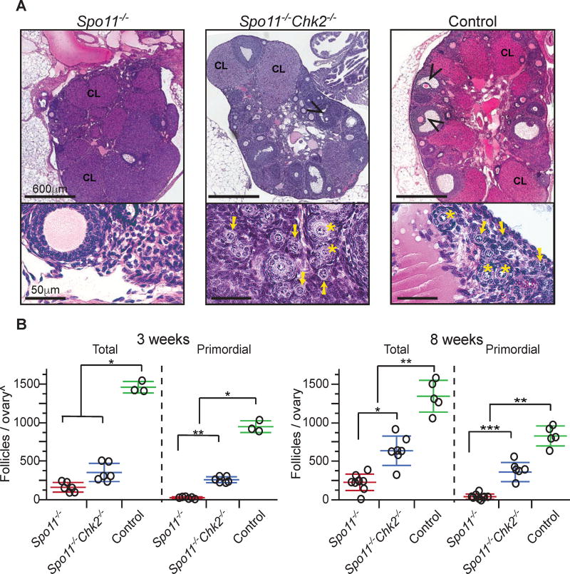
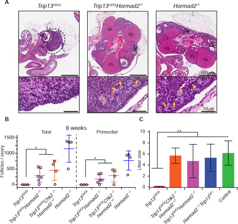
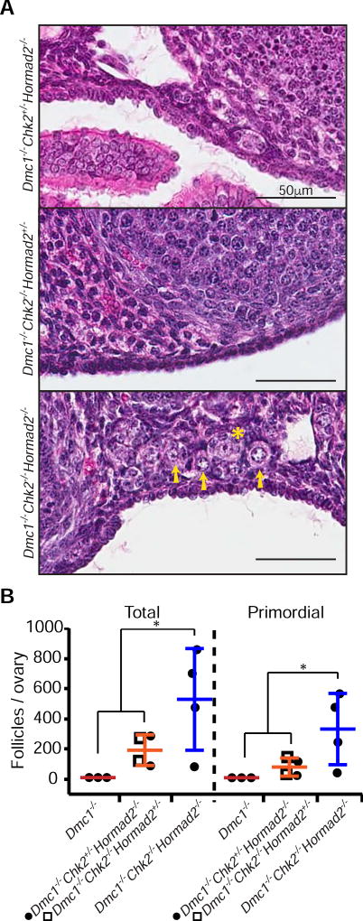
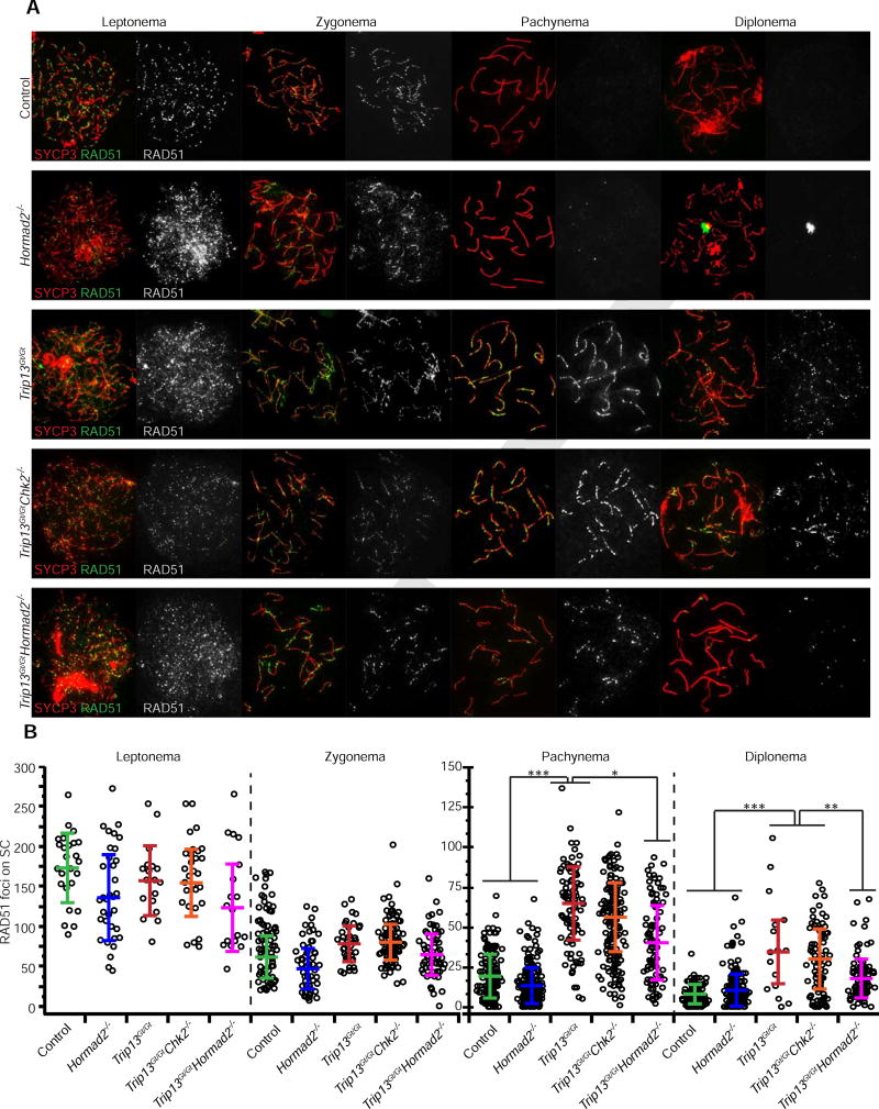
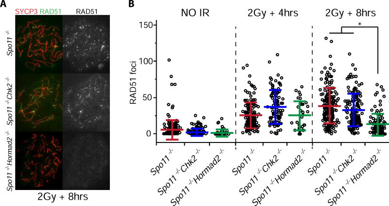
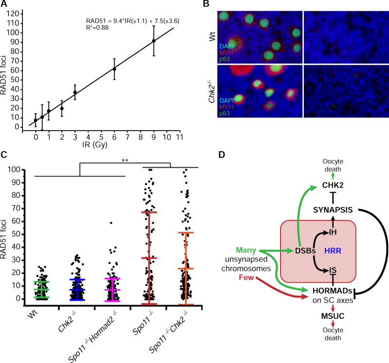
Pairing and synapsis of homologous chromosomes during meiosis is crucial for producing genetically normal gametes and is dependent upon repair of SPO11-induced double-strand breaks (DSBs) by homologous recombination. To prevent transmission of genetic defects, diverse organisms have evolved mechanisms to eliminate meiocytes containing unrepaired DSBs or unsynapsed chromosomes. Here we show that the CHK2 (CHEK2)-dependent DNA damage checkpoint culls not only recombination-defective mouse oocytes but also SPO11-deficient oocytes that are severely defective in homolog synapsis. The checkpoint is triggered in oocytes that accumulate a threshold level of spontaneous DSBs (∼10) in late prophase I, the repair of which is inhibited by the presence of HORMAD1/2 on unsynapsed chromosome axes. Furthermore, Hormad2 deletion rescued the fertility of oocytes containing a synapsis-proficient, DSB repair-defective mutation in a gene (Trip13) required for removal of HORMADs from synapsed chromosomes, suggesting that many meiotic DSBs are normally repaired by intersister recombination in mice.


Figures

Figure 1
Figure 1. CHK2 is required for efficient elimination of asynaptic Spo11−/− mouse oocytes
(A) H&E stained histological sections of 8 weeks old ovaries. Black arrowheads indicate antral follicles. CL= Corpus Luteum; the presence of corpora lutea are indicative of prior rounds of ovulation. The lower portion of each panel contains a higher magnification image of an ovarian cortical region, where primordial follicles reside. Yellow arrows and stars indicate primordial and primary follicles, respectively. (B) Follicle counts from ovaries of indicated genotypes at 3 and 8 weeks postpartum, respectively. Each data point is from a single ovary, each being from a different animal. Total = all follicle types. Horizontal hashes denote mean and standard deviation. Littermate controls included animals with the following genotypes: Spo11+/+Chk2+/+, Spo11+/− Chk2+/− and Spo11+/+Chk2+/−. (^) The values obtained for the 3 weeks follicles/ovaries counts are not comparable to the 8 weeks (see methods). Asterisks indicate p-values: (*) 0.005 ≤ p-values ≤ 0.05, (**)0.001 ≤ p-values ≤ 0.005 and (***) p-values ≤ 0.001 derived from a non-parametric, one-way ANOVA test (Kruskal-Wallis).
Figure 2
Figure 2. Synapsis-competent Trip13Gt/Gt oocytes are eliminated in a HORMAD2-dependent manner
(A) H&E stained histological sections of 8 week old ovaries of indicated genotypes. Black arrowheads indicate antral follicles. CL= Corpus Luteum. The lower half of each panel shows a higher magnification of cortical regions of ovaries. Yellow arrows and stars indicate primordial and primary follicles, respectively. (B) Follicle quantification of 8 week old ovaries. Each data point is from a single ovary, each being from a different animal. “Total” = all follicle types. Horizontal hashes denote mean and standard deviation. The statistic used was Kruskal-Wallis. * indicates p-value = 0.002. (C) Graphed are mean litter sizes. N ≥ 3 females tested for fertility per genotypic group. Control matings were between mice with the genotypes Trip13Gt/+ and Trip13Gt/+ Hormad2+/−. Error bars represent standard deviation and ** indicates p-value ≤ 0.005 derived from the Kruskal-Wallis test.
Figure 3
Figure 3. HORMAD2 and CHK2 are not in the same checkpoint pathway
(A) H&E stained histological sections of cortical regions of 8 week old mutant mouse ovaries, where primordial follicles are concentrated. Histology of whole ovaries of these genotypes are represented in Fig. S2. Primordial follicles, which constitute the oocyte reserve, are indicated by yellow arrows, and a primary follicle by a star. Residual Dmc1−/− ovaries are not represented because they are completely devoid of oocytes (Pittman et al., 1998). (B) Follicle counts from ovaries of indicated genotypes at 8 weeks of age. “Total” = all types of follicles. Data points represent follicle counts derived from one ovary, each ovary originating from a different animal. Asterisk indicates p-value ≤ 0.05 (Kruskal-Wallis test).
Figure 4
Figure 4. Depletion of HORMAD2 accelerates DSB-repair during early stages of meiotic prophase I
(A) Representative images of meiotic chromosome spreads from oocytes at different substages of meiotic prophase I, probed with antibodies for SYCP3 (SC axis protein) and the DSB marker RAD51. Oocytes were isolated from female embryos ranging from 15.5 dpc to newborns. See Figure S1 for HORMAD2 localization in meiotic mutants. (B) Numbers of RAD51 foci in specified meiotic prophase I substage of indicated mutants. Only RAD51 foci present on SYCP3 stained axes were scored. Each data point represents one cell. In each genotypic group, at each stage, the counts are derived from at least three animals. Horizontal hashes in summary statistic plots denote mean and standard deviation. Values of the mixed model calculation can be found in Table S1. Colors correspond to genotypes. Asterisks indicate statistical significant differences between groups in terms of the least square means of RAD51 foci. p-values: *** p ≤ 0.001; ** p ≤ 0.005; * p ≤ 0.05 (Tukey HSD). See Table S1 for raw data and statistical calculations associated with (B).
Figure 5
Figure 5. Depletion of HORMAD2 accelerates repair of induced DSBs in oocytes
(A) Immunolabeling of surface spread chromosomes from oocytes after exposure to ionizing radiation (IR). Fetal ovaries were collected at 15.5 dpc, cultured 24 hours, exposed to 2 Gy of IR, then cultured for an additional 4–8 hours. Shown are those recovered 8 hours after IR. See Figure S4 for single Hormad2−/− single mutant results. (B) Quantification of RAD51 foci. Each data point represents one oocyte. The graphs include mean and standard deviation, and are color coded according to genotypic group. The 4 and 8 hrs unirradiated samples were combined. Data were derived from at least two different animals per condition. See Table S2 for raw data and statistical calculations associated with (B).
Figure 6
Figure 6. DNA damage threshold required to trigger oocyte death, and evidence for HORMAD-mediated inhibition of IS repair
(A) Linear regression for conversion of radiation dosages to RAD51 focus counts. Meiotic surface spreads were made from WT neonatal ovaries 2.5 hrs after IR. Plotted are means with standard deviations. Each IR dose has focus counts from ~25 oocytes derived from a total of 18 animals. See Figure S5 for single cell foci counts and numerical values. (B) Chk2−/− oocytes are highly IR resistant. Shown are immunofluorescence images of ovarian sections labeled with nuclear and cytoplasmic germ cell markers (p63 and MVH, respectively). (C) RAD51 focus counts from newborn oocyte spreads. Only oocytes with discrete patterns of RAD51 foci were scored, as defined in Fig. S3. Data points represent individual oocytes, derived from at least five different animals from each genotypic group. Horizontal hashes denote means and standard deviations calculated using a mixed model (see methods). Asterisks indicate statistically significant differences between groups with p-values: *** p ≤ 0.001; ** p ≤ 0.005; * p ≤ 0.05 (Tukey HSD). See Table S3 for raw data and statistical calculations. (D) Model for pachytene checkpoint activation in mouse oocytes. Oocytes with many unsynapsed chromosomes (green) ultimately accumulate DSBs, which cannot be repaired due to block to IS recombination imposed by HORMADs on asynapsed axes. Failure of DSB repair leads to activation of CHK2 and downstream effector proteins (p53/TAp63) that trigger apoptosis. Few asynapsed chromosomes (red) lead to inactivation of essential genes by MSUC thereby causing oocyte death. HRR - Homologous Recombination Repair; IH - Interhomolog; IS - Intersister; MSUC - Meiotic Silencing of Unsynapsed Chromatin.

References

    1. Agoulnik AI, Lu B, Zhu Q, Truong C, s Ty MT, Arango N, Chada KK, Bishop CE. A novel gene, Pog, is necessary for primordial germ cell proliferation in the mouse and underlies the germ cell deficient mutation, gcd. Hum Mol Genet. 2002;11:3047–3053. - PubMed
    1. Baudat F, Manova K, Yuen JP, Jasin M, Keeney S. Chromosome synapsis defects and sexually dimorphic meiotic progression in mice lacking Spo11. Mol Cell. 2000;6:989–998. - PubMed
    1. Bellani MA, Romanienko PJ, Cairatti DA, Camerini-Otero RD. SPO11 is required for sex-body formation, and Spo11 heterozygosity rescues the prophase arrest of Atm−/− spermatocytes. J Cell Sci. 2005;118:3233–3245. - PubMed
    1. Bolcun-Filas E, Rinaldi VD, White ME, Schimenti JC. Reversal of female infertility by Chk2 ablation reveals the oocyte DNA damage checkpoint pathway. Science. 2014;343:533–536. - PMC - PubMed
    1. Burgoyne PS, Baker TG. Perinatal oocyte loss in XO mice and its implications for the aetiology of gonadal dysgenesis in XO women. J Reprod Fertil. 1985;75:633–645. - PubMed

MeSH terms

LinkOut - more resources