Review
. 2018 Jan;1862(1):81-196.
doi: 10.1016/j.bbagen.2017.08.014. Epub 2017 Aug 24.

Marine natural product peptides with therapeutic potential: Chemistry, biosynthesis, and pharmacology

Affiliations
Review

Marine natural product peptides with therapeutic potential: Chemistry, biosynthesis, and pharmacology

Vedanjali Gogineni et al. Biochim Biophys Acta Gen Subj. 2018 Jan.

Abstract

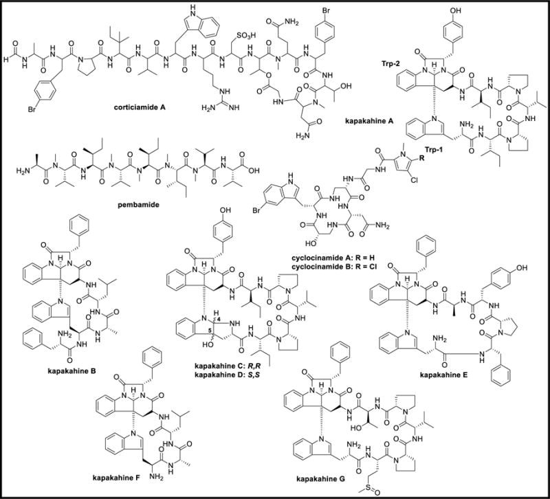
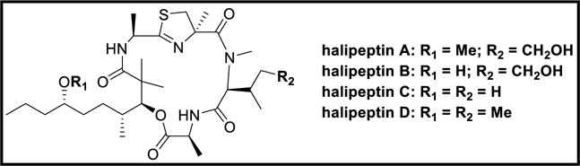
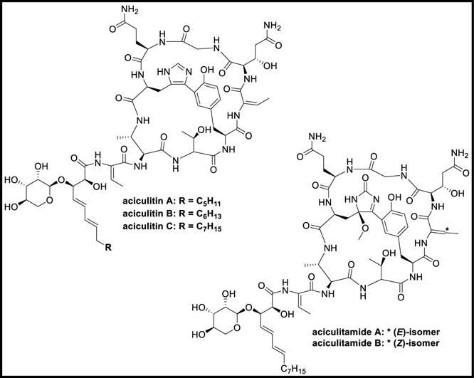
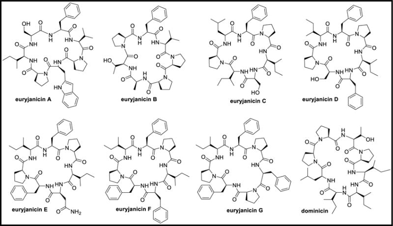
The oceans are a uniquely rich source of bioactive metabolites, of which sponges have been shown to be among the most prolific producers of diverse bioactive secondary metabolites with valuable therapeutic potential. Much attention has been focused on marine bioactive peptides due to their novel chemistry and diverse biological properties. As summarized in this review, marine peptides are known to exhibit various biological activities such as antiviral, anti-proliferative, antioxidant, anti-coagulant, anti-hypertensive, anti-cancer, antidiabetic, antiobesity, and calcium-binding activities. This review focuses on the chemistry and biology of peptides isolated from sponges, bacteria, cyanobacteria, fungi, ascidians, and other marine sources. The role of marine invertebrate microbiomes in natural products biosynthesis is discussed in this review along with the biosynthesis of modified peptides from different marine sources. The status of peptides in various phases of clinical trials is presented, as well as the development of modified peptides including optimization of PK and bioavailability.

Keywords: Bioactive peptides; Biosynthesis; Challenges; Marine organisms; Peptide isolation; Therapeutic peptides.


Conflict of interest statement

Notes

The authors declare no competing financial interest.

Figures

Figure 1
Figure 1
Chemical structures of discodermins A-H and kasumigamide
Figure 2
Figure 2
Chemical structures of phakellistatins 1–12 and isophakellistatin 3
Figure 3
Figure 3
Chemical structures of phakellistatins 13–19
Figure 4
Figure 4
Chemical structures of geodiamolides A-R, geodiamolide-TA, neosiphoniamolide, jasplakinolide, and pipestelides A-C
Figure 5
Figure 5
Chemical structures of milnamides A-G, hemiasterlins A, D, and scleritodermin A
Figure 6
Figure 6
Chemical structures of mirabamides A-H
Figure 7
Figure 7
Chemical structures of celebesides A-C and theopapuamides A-D
Figure 8
Figure 8
Chemical structures of homophymines A-E and A1-E1
Figure 9
Figure 9
Chemical structures of neamphamides A-D
Figure 10
Figure 10
Chemical structures of callipeltins A-D
Figure 11
Figure 11
Chemical structures of callipeltins E-K
Figure 12
Figure 12
Chemical structures of callipeltins L-Q
Figure 13
Figure 13
Chemical structures of microspinosamide and carteritins A-B
Figure 14
Figure 14
Chemical structures of mycothiazole, dysidenin, isodysidenin, dysideaprolines A-F, barbaleucamides A-B, and dysithiazolamide
Figure 15
Figure 15
Chemical structures of microcionamides A-B
Figure 16
Figure 16
Chemical structures of halicylindramides A-C and seco-methyl ester of halicylindramide B
Figure 17
Figure 17
Chemical structures of haligramides A-B, waiakeamide, and sulfone derivative of waiakeamide
Figure 18
Figure 18
Chemical structures of corticiamide A, pembamide, cyclocinamides A-B, and kapakahines A-G
Figure 19
Figure 19
Chemical structures of taumycins A-B
Figure 20
Figure 20
Chemical structures of pipecolidepsins A-B
Figure 21
Figure 21
Chemical structures of halipeptins A-D
Figure 22
Figure 22
Chemical structures of tausalarin C, arenastatin A, and axinastatins 1–3
Figure 23
Figure 23
Chemical structures of hymenamides A-H and J-K
Figure 24
Figure 24
Chemical structures of wainunuamide, axinellins A-C, stylopeptides 1–2, stylostatins 1–2, and cyclonellin
Figure 25
Figure 25
Chemical structures of fenestins A-B and hymenistatin 1
Figure 26
Figure 26
Chemical structures of discobahamins A-B and calyxamides A-B
Figure 27
Figure 27
Chemical structures of microsclerodermins A-K and anhydromicrosclerodermin C
Figure 28
Figure 28
Chemical structures of aciculitins A-C and aciculitamides A-B
Figure 29
Figure 29
Chemical structures of polydiscamides A-D
Figure 30
Figure 30
Chemical structures of criamides A-B and gombamide A
Figure 31
Figure 31
Chemical structures of euryjanicins A-G and dominicin
Figure 32
Figure 32
Chemical structures of neopetrosiamides A-B, jamaicensamide A, sulfolipodiscamides A-C, and lipodiscamides A-C
Figure 33
Figure 33
Structures of stylissamides A-H, X, stylissatins A-D, and stylisins 1–2
Figure 34
Figure 34
Chemical structures of reniochalistatins A-E and yaku’amides A-B
Figure 35
Figure 35
Chemical structures of chujamides A-B and leucamide A
Figure 36
Figure 36
Chemical structures of azumamides A-E and phoriospongins A-B
Figure 37
Figure 37
Chemical structures of callyaerins A-H
Figure 38
Figure 38
Chemical structures of motuporin, theonellapeptolide Id, and nazumazoles A-F
Figure 39
Figure 39
Chemical structures of polytheonamides A-C
Figure 40
Figure 40
Chemical structures of pseudotheonamides A1-A2, B2, C-D, dihydrocyclotheonamide A, nazumamide A, cyclotheonamides A-B, and orbiculamide A
Figure 41
Figure 41
Chemical structures of cyclotheonamides E, E2, and E3
Figure 42
Figure 42
Chemical structures of theonellamides A-G, keramamides A-H, and J-N
Figure 43
Figure 43
Chemical structures of koshikamides A1-A2 and B-H
Figure 44
Figure 44
Chemical structures of papuamides A-F, theonegramide, nagahamide A, and cupolamide A
Figure 45
Figure 45
Chemical structures of barangamides A-D, theonellapeptolides Ia-Ie, and IId-IIe
Figure 46
Figure 46
Chemical structure of theonellapeptolide IIIe [169]
Figure 47
Figure 47
Chemical structures of congeners 1–2 and solomonamides A-B
Figure 48
Figure 48
Chemical structures of cyclolithistide A, revised structure of cyclolithistide A [176], theopalauamide A, isotheopalauamide, and oriamide
Figure 49
Figure 49
Chemical structures of miraziridine A, paltolides A-C, perthamides B-K, and mutremdamide A
Figure 50
Figure 50
Structural similarity of peptides isolated from Theonella sp., and microorganisms
Figure 51
Figure 51
Chemical structures of turnagainolides A-B, solonamides A-B, actinoramides A-C, fijimycins A-C, and brunsvicamides A-C
Figure 52
Figure 52
Chemical structures of malyngamides 2–3, cocosamides A-B, pitiprolamide, and pitipeptolides A-F
Figure 53
Figure 53
Chemical structures of lagunamides A-C, wewakamide A, and guineamides A-G
Figure 54
Figure 54
Chemical structures of wewakazole, wewakazole B, and wewakpeptins A-D
Figure 55
Figure 55
Chemical structures of porpoisamides A-B, bisebromoamide, and norbisebromoamide
Figure 56
Figure 56
Chemical structure of somocystinamide A
Figure 57
Figure 57
Chemical structures of cyclic desmethoxymajusculamide C, linear desmethoxymajusculamide C, majusculamides A-D, dolastatins 11–12, 57-normajusculamide C, lyngbyastatins 1, 3, epilyngbyastatin 1, and deoxymajusculamide D
Figure 58
Figure 58
Chemical structures of apratoxins A-E and dehydroapratoxin A
Figure 59
Figure 59
Chemical structures of dragonamides A-E, herbamides A-B, dragomabin, and carmabins A-B
Figure 60
Figure 60
Chemical structures of almiramides A-C
Figure 61
Figure 61
Chemical structures of grassystatins A-C
Figure 62
Figure 62
Chemical structures of lobocyclamides A-C and obyanamide
Figure 63
Figure 63
Chemical structures of hantupeptins A-C
Figure 64
Figure 64
Chemical structures of trungapeptins A-C and antanapeptins A-D
Figure 65
Figure 65
Chemical structures of palmyramide A, dudawalamides A-E, and mantillamide
Figure 66
Figure 66
Chemical structures of grassypeptolide and carriebowmide
Figure 67
Figure 67
Chemical structure of hoiamide A
Figure 68
Figure 68
Chemical structures of tiglicamides A-C, largamides A-C, and methyl esters of largamides A-C
Figure 69
Figure 69
Chemical structures of itralamides A-B
Figure 70
Figure 70
Chemical structures of lyngbyastatins 4–6 and 8–10
Figure 71
Figure 71
Chemical structures of somamides A-B, lyngbyastatin 7, kempopeptins A-B, and scyptolin A
Figure 72
Figure 72
Chemical structures of lyngbyazothrins A-D, pahayokolides A-B, schizotrin A, and tychonamides A-B
Figure 73
Figure 73
Chemical structures of grassypeptolides A-E and ibu-epidemethoxylyngbyastatin
Figure 74
Figure 74
Chemical structures of grassypeptolides F-G
Figure 75
Figure 75
Chemical structures of lyngbyapeptins A-D and lyngbyabellins A-J
Figure 76
Figure 76
Chemical structures of alotamide A, lyngbyabellins K-N, and 7-epi-lyngbyabellin L
Figure 77
Figure 77
Chemical structures of barbamide and jamaicamides A-C
Figure 78
Figure 78
Chemical structure of hectochlorin
Figure 79
Figure 79
Chemical structures of apramides A-G
Figure 80
Figure 80
Chemical structures of antillatoxin, barbaramide A, aurilide, aurilides B-C, and kalkitoxin
Figure 81
Figure 81
Chemical structures of georgamide and yanucamides A-B
Figure 82
Figure 82
Chemical structures of dysidenamide, pseudodysidenin, nordysidenin, and isodysidenin
Figure 83
Figure 83
Chemical structures of ulongamides A-F
Figure 84
Figure 84
Chemical structures of isomalyngamides A-B, malyngamides A-K, and 8-epi-malyngamide C
Figure 85
Figure 85
Chemical structures of malyngamides L-Y
Figure 86
Figure 86
Chemical structures of hermitamides A-B and laxaphycins A-B
Figure 87
Figure 87
Chemical structures of tasiamide, tasiamides B-F, pepstatin A, and tasipeptins A-B
Figure 88
Figure 88
Chemical structures of symplocamide A, symplocin A, veraguamides A-G, and semisynthetic veraguamide
Figure 89
Figure 89
Chemical structures of symplostatins 1–4
Figure 90
Figure 90
Chemical structures of malevamides A-D, belamide A, and largazole
Figure 91
Figure 91
Chemical structures of mitsoamide and gallinamide
Figure 92
Figure 92
Chemical structures of nostocyclamide, tenuecyclamides A-D, and cryptophycin
Figure 93
Figure 93
Chemical structures of microcystin, raocyclamides A-B, and venturamides A-B
Figure 94
Figure 94
Chemical structures of anabaenopeptins A-J and pompanopeptins A-B
Figure 95
Figure 95
Chemical structures of floridamide, coibamide A, scytonemin, hormothamnin A, and trichamide,
Figure 96
Figure 96
Chemical structures of arenamides A-C and caldoramide
Figure 97
Figure 97
Chemical structures of viequeamides A-F and companeramides A-B
Figure 98
Figure 98
Chemical structures of nodulapeptins A-B, aeruginosins NAL2, NOL6, spumigins A, C-H, and spumigins B1-B2
Figure 99
Figure 99
Chemical structures of kailuins A-H
Figure 100
Figure 100
Chemical structures of ngercheumicins A-I, unnarmicins A and C
Figure 101
Figure 101
Chemical structures of ariakemicins A-B, mollemycin A, thiocoraline, cyclomarins A-D, and cyclomarazines A-B
Figure 102
Figure 102
Chemical structures of surugamides A-F and champacyclin
Figure 103
Figure 103
Chemical structures of tumescenamides A-C and streptocidins A-D
Figure 104
Figure 104
Chemical structures of salinamides A-F
Figure 105
Figure 105
Chemical structures of tauramamide, tupuseleiamides A-B, and bogorols A-E
Figure 106
Figure 106
Chemical structures of loloatins A-D and marthiapeptide A
Figure 107
Figure 107
Chemical structures of loihichelins A-F, marinobactins A-C, D1, D2, and E
Figure 108
Figure 108
Chemical structures of aquachelins A-D, amphibactins B-I, petrobactin, petrobactin sulfate, aerobactin, and amphi-enterobactin
Figure 109
Figure 109
Chemical structures of alterobactins A-B and pseudoalterobactins A-B
Figure 110
Figure 110
Chemical structures of trivanchrobactin, divanchrobactin, vanchrobactin, anguibactin, and vibrioferrin
Figure 111
Figure 111
Chemical structures of ochrobactins A-C and synechobactins A-C
Figure 112
Figure 112
Chemical structures of unguisins A-F
Figure 113
Figure 113
Chemical structures of versicotides A-B, versicoloritides A-C, fellutamides A-D, F, and asperterrestide A
Figure 114
Figure 114
Chemical structures of sansalvamide A, scopularides A-B, and exumolides A-B
Figure 115
Figure 115
Chemical structures of cyclo-(L-Pro-L-Tyr), cyclo-(L-Pro-L-Val), cyclo-(L-Phe-L-Pro), and cis-bis(methylthio)silvatin
Figure 116
Figure 116
Chemical structures of penilumamide, penilumamides B-D, and asperpeptide A
Figure 117
Figure 117
Chemical structures of emericellamides A-B, guangomides A-B, homodestcardin, and azonazine
Figure 118
Figure 118
Chemical structures of endolides A-D and hirsutide
Figure 119
Figure 119
Chemical structures of simplicilliumtides A-H and dictyonamides A-B
Figure 120
Figure 120
Chemical structures of halolitoralins A-C, sclerotides A-B, cyclo(L-Pro-L-Val), cyclo(L-Pro-L-Leu), and cyclo(L-Ile-L-Val)
Figure 121
Figure 121
Chemical structures of RHM1, RHM2, and efrapeptin G
Figure 122
Figure 122
Chemical structures of cordyheptapeptides A-E and oryzamides A-E
Figure 123
Figure 123
Chemical structures of cycloxazoline, diazonamides A-E, revised structure of diazonamide A, and diazonamide A analog
Figure 124
Figure 124
Chemical structure of vitilevuamide
Figure 125
Figure 125
Chemical structures of bistratamides A-J, westiellamide, and didmolamides A-B
Figure 126
Figure 126
Chemical structures of lissoclinamides 1–8, patellamides A-E, ulithiacyclamide A, and ascidiacyclamide
Figure 127
Figure 127
Chemical structures of didemnins A-E, G-H, M-N, X-Y, epididemnin A1, nordidemnin N, and acyclodidemnin A
Figure 128
Figure 128
Chemical structures of eusynstyelamide and eudistomides A-B
Figure 129
Figure 129
Chemical structures of mollamide, mollamides B-C, keenamide A, and cycloforskamide
Figure 130
Figure 130
Chemical structures of virenamides A-C
Figure 131
Figure 131
Chemical structures of cyclodidemnamide, comoramides A-B, mayotamides AB, didmolamides A-B, prepatellamide A, patellamides A-C, and tamandarins A-B
Figure 132
Figure 132
Chemical structures of patellins 1–6 and trunkamide A
Figure 133
Figure 133
Chemical structures of kulolides 1–3, kulokainalide-1, kulomo’opunalides 1–2, and pupukeamide
Figure 134
Figure 134
Chemical structures of ulicyclamide, preulithiacyclamide, ulithiacyclamide, and ulithiacyclamide B
Figure 135
Figure 135
Chemical structures of nobilamides A-H, A-3302 A-B, and N-acetyl-L-phenylalanyl-L-leucinamide
Figure 136
Figure 136
Chemical structure of aplidine
Figure 137
Figure 137
Chemical structures of dolastatins 3, 10–15, homodolastatin 3, kororamide, and epidolastatin 12
Figure 138
Figure 138
Chemical structures of dolastatins 16–18 and homodolastatin 16
Figure 139
Figure 139
Chemical structures of dolastatins C-E, G-I, nordolastatin G, isodolastatin H, lyngbyastatin 2, and norlyngbyastatin 2
Figure 140
Figure 140
Chemical structures of dolabellin and doliculide
Figure 141
Figure 141
Chemical structures of kulokekahilides 1–2 and aurilide
Figure 142
Figure 142
Chemical structures of viridamides A-B, dolastatin 12, lyngbyastatin 1, malyngamide C, malyngamide C acetate, lyngbic acid, janadolide, and microcolins A-B
Figure 143
Figure 143
Chemical structures of tumonoic acids A-B, D-I, and epi-tumonoic acid
Figure 144
Figure 144
Chemical structures of onchidins A-B and nocardiamides A-B
Figure 145
Figure 145
Chemical structures of kahalalides A-H, J, W, norkahalalide A, isokahalalide F, and 5OH-kahalalide F
Figure 146
Figure 146
Chemical structures of kahalalides K, O-Q, R1-R2, S1-S2, V,X, and Y
Figure 147
Figure 147
Chemical structures of cis,cis-ceratospongamide, trans,trans-ceratospongamide, galaxamide, and mebamamides A-B
Figure 148
Figure 148
Chemical structures of echinocandin B, pneumocandins A0, B0, C0, and caspofungin acetate
Figure 149
Figure 149
Biosynthesis of nodularin
Figure 150
Figure 150
Biosynthesis of barbamide & barbaleucamide
Figure 151
Figure 151
Biosynthesis of jamaicamide A
Figure 152
Figure 152
Biosynthesis of lyngbyatoxins [452]
Figure 153
Figure 153
Molecular networking of dolastatin 10 tetrapeptide molecular family
Figure 154
Figure 154
Biosynthesis of anabaenopeptins
Figure 155
Figure 155
Biosyntheses of aeruginosin and spumigin
Figure 156
Figure 156
Biosynthesis of thiocoraline
Figure 157
Figure 157
Biosynthesis of cyclomarin A and cyclomarazine A
Figure 158
Figure 158
Biosynthesis of surugamides
Figure 159
Figure 159
Biosynthesis of salinamides
Figure 160
Figure 160
Biosyntheses of amphibactins and marinobactins
Figure 161
Figure 161
Biosynthesis of amphi-enterobactin
Figure 162
Figure 162
Biosynthetic gene clusters of scopularide and emericellamide
Figure 163
Figure 163
Biosynthesis of Scopularide in Scopulariopsis brevicaulis
Figure 164
Figure 164
Biosynthesis of emericellamide in Aspergillus nidulans
Figure 165
Figure 165
Biosynthesis of N-methyl-3-(3-furyl)alanine
Figure 166
Figure 166
Biosynthesis of kasumigamide
Figure 167
Figure 167
Biosynthesis of polytheonamides
Figure 168
Figure 168
Biosynthesis of keramamides
Figure 169
Figure 169
Biosynthesis of microsclerodermins
Figure 170
Figure 170
Biosynthesis of patellamides
Figure 171
Figure 171
Chemical structure of ziconotide
Figure 172
Figure 172
Chemical structure of leconotide
Figure 173
Figure 173
Chemical structure of Xen-2174
Figure 174
Figure 174
Chemical structures of brentuximab vedotin and monomethylauristatin E
Figure 175
Figure 175
Chemical structures of soblidotin, tasidotin, and cemadotin
Figure 176
Figure 176
Chemical structure of elisidepsin
Figure 177
Figure 177
Chemical structure of HTI-286
Figure 178
Figure 178
Chemical structure of glembatumumab vedotin
Figure 179
Figure 179
Chemical structure of monomethylauristatin F

References

    1. Aneiros A, Garateix A. Bioactive peptides from marine sources: Pharmacological properties and isolation procedures. J Chromatogr B: Anal Technol Biomed Life Sci. 2004;803:41–53. - PubMed
    1. Miijanich GP. Venom peptides as human pharmaceuticals. Sci Med. 1997;4:6–15.
    1. Cragg GM, Newman DJ, Weiss RB. Coral reefs, forests, and thermal vents: The worldwide exploration of nature for novel antitumor agents. Semin Oncol. 1997;24:156–163. - PubMed
    1. Matsunaga S, Fusetani N. Nonribosomal peptides from marine sponges. Curr Org Chem. 2003;7:945–966.
    1. Matsunaga S, Fusetani N, Konosu S. Bioactive marine metabolites VII. Structures of discodermins B, C, and D, antimicrobial peptides from the marine sponge Discodermia kiiensis. Tetrahedron Lett. 1985;26:855–856. - PubMed

Publication types

MeSH terms