. 2017:2017:7375818.
doi: 10.1155/2017/7375818. Epub 2017 Aug 6.

The Lymphotoxin β Receptor Is Essential for Upregulation of IFN-Induced Guanylate-Binding Proteins and Survival after Toxoplasma gondii Infection

Affiliations

The Lymphotoxin β Receptor Is Essential for Upregulation of IFN-Induced Guanylate-Binding Proteins and Survival after Toxoplasma gondii Infection

Kristina Behnke et al. Mediators Inflamm. 2017.

Abstract

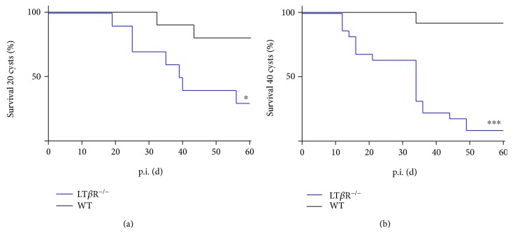
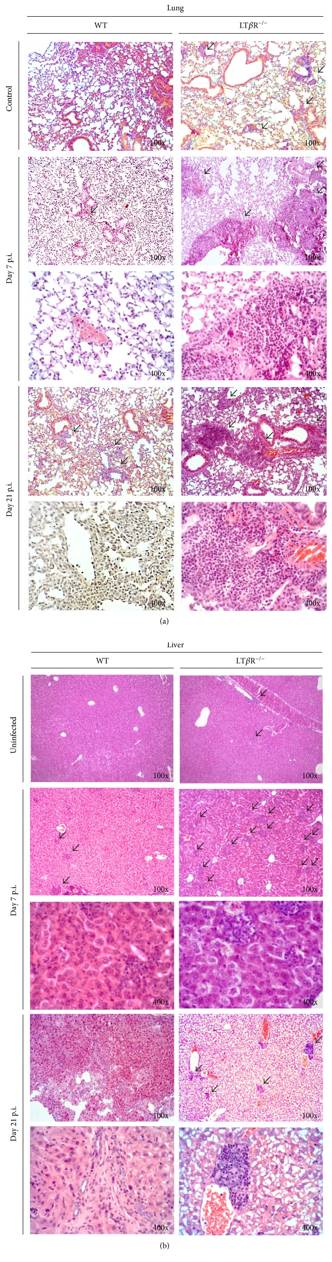
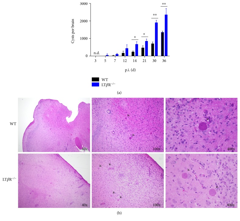
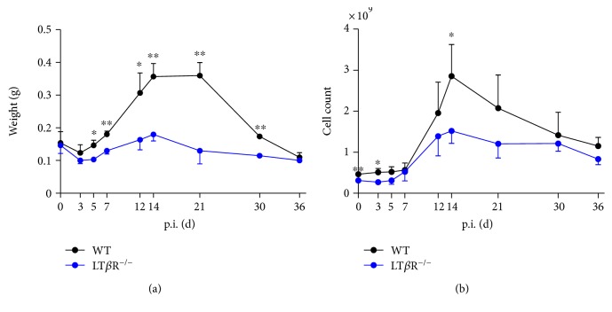
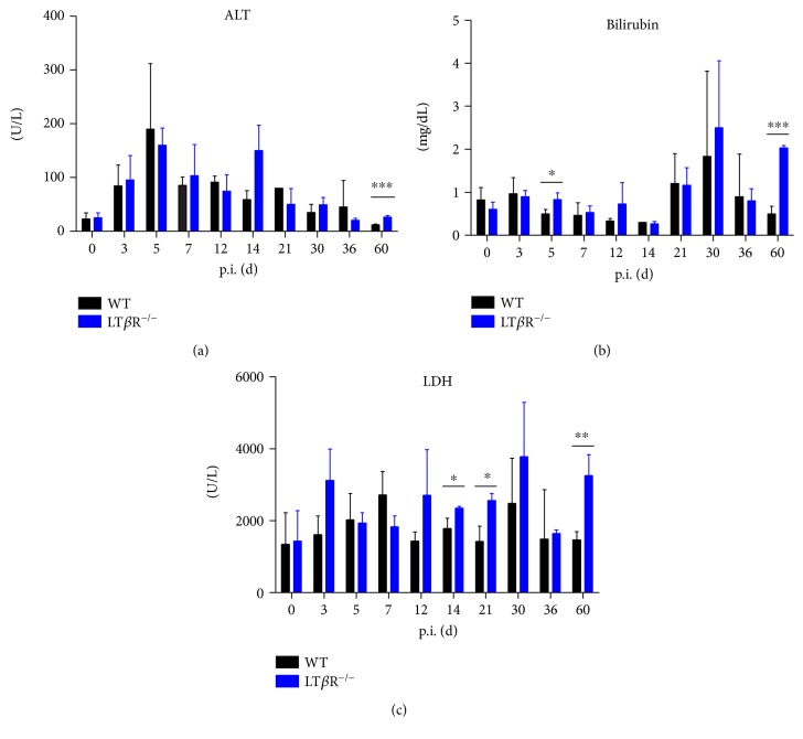
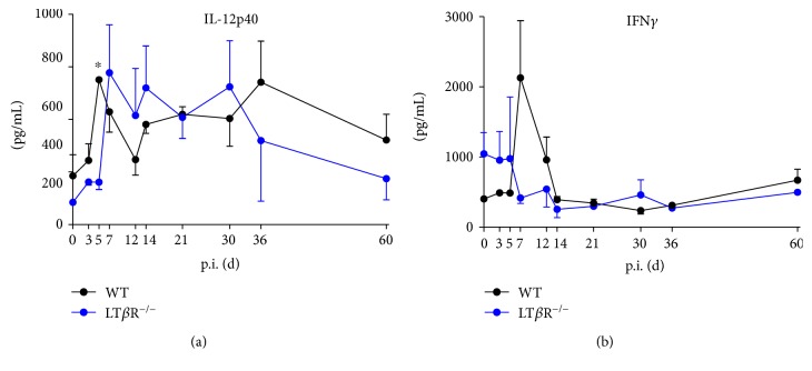
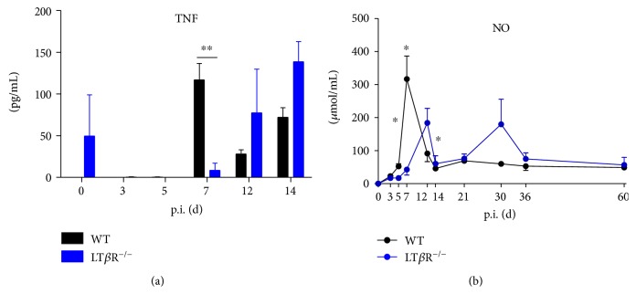
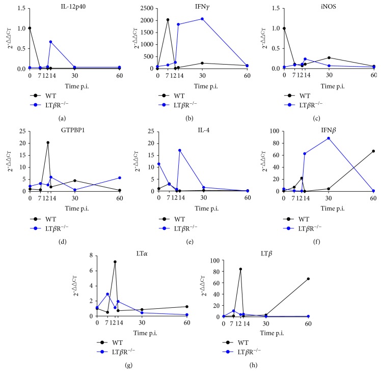
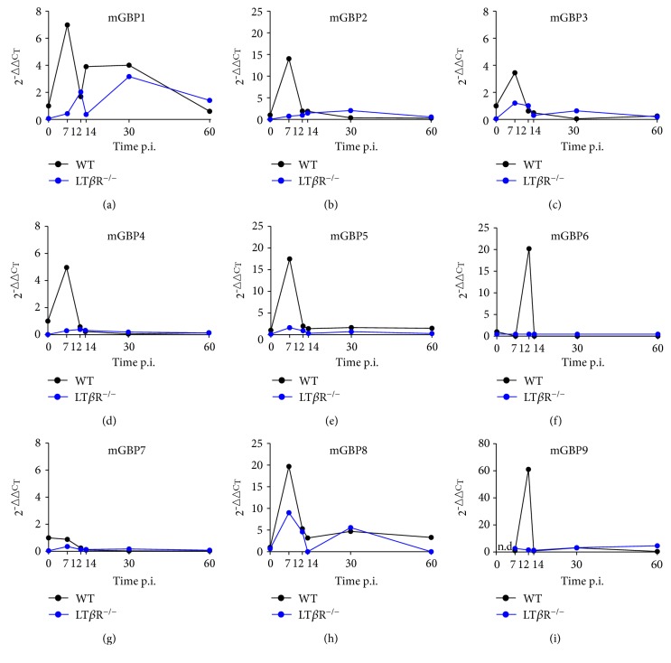
Lymphotoxin β receptor (LTβR) signaling plays an important role in efficient initiation of host responses to a variety of pathogens, encompassing viruses, bacteria, and protozoans via induction of the type I interferon response. The present study reveals that after Toxoplasma gondii infection, LTβR-/- mice show a substantially reduced survival rate when compared to wild-type mice. LTβR-/- mice exhibit an increased parasite load and a more pronounced organ pathology. Also, a delayed increase of serum IL-12p40 and a failure of the protective IFNγ response in LTβR-/- mice were observed. Serum NO levels in LTβR-/- animals rose later and were markedly decreased compared to wild-type animals. At the transcriptional level, LTβR-/- animals exhibited a deregulated expression profile of several cytokines known to play a role in activation of innate immunity in T. gondii infection. Importantly, expression of the IFNγ-regulated murine guanylate-binding protein (mGBP) genes was virtually absent in the lungs of LTβR-/- mice. This demonstrates clearly that the LTβR is essential for the induction of a type II IFN-mediated immune response against T. gondii. The pronounced inability to effectively upregulate host defense effector molecules such as GBPs explains the high mortality rates of LTβR-/- animals after T. gondii infection.


Figures

Figure 1
Figure 1
LTβR−/− animals show significantly reduced survival after infection with T. gondii (ME 49) cysts compared to WT animals. WT and LTβR−/− animals were infected i.p. with (a) 20 cysts (WT: n = 10, LTβR−/−: n = 10) or (b) 40 cysts (WT: n = 12, LTβR−/−: n = 22) of T. gondii (ME49) freshly isolated from the brains of CD1 mice. p < 0.05, ∗∗∗p < 0.001.
Figure 2
Figure 2
LTβR−/− animals show more and larger inflammatory areas in the (a) lung and (b) liver 7 and 21 days after infection with T. gondii (ME49) cysts compared to WT animals. The lung and liver were isolated from uninfected control mice 7 and 21 days after i.p. infection with 40 T. gondii (ME49) cysts and fixed in formalin. Tissues were embedded in paraffin, 10 μm sections were generated, and HE staining was performed. Original magnification as indicated. 3 animals were analyzed for each time point, and a representative section from one organ is shown in each case. Arrows indicate small, dense lymphocyte infiltrates that are considered part of the basal LTβR−/− phenotype. Arrowheads indicate inflammatory infiltrates seen in infected animals.
Figure 3
Figure 3
Analysis of parasite burden in the brain of WT and LTβR−/− animals. Animals were infected i.p. with 40 cysts of T. gondii (ME49), sacrificed on the days indicated, and the brains were prepared. One hemisphere was used for isolation of cysts, which were isolated by mincing the tissue with a scalpel and then passing it through consecutively higher gauge cannulas, followed by two centrifugation steps to first remove pelleted cells and tissue debris and then pellet the cysts. One half of the second hemisphere was used to generate HE stains from paraffin sections after formalin fixing of tissue. (a) Cysts per brain were calculated by multiplying cyst number counted in one hemisphere by two (n = 3 in all cases, except day 30 and day 36 from LTβR−/− animals, where only 2 animals were analyzed). (b) Cysts (arrows) in HE-stained brain sections 60 days after i.p. infection with T. gondii (ME49) are shown. One representative section of brain tissue from one of three animals is shown. Original magnifications as indicated. p < 0.05, ∗∗p < 0.01.
Figure 4
Figure 4
Splenomegaly is observed only in WT but not in LTβR−/− animals after infection with T. gondii (ME49). Mice were infected with 40 cysts and sacrificed on the days indicated. Controls were uninfected animals. (a) Spleens were isolated and weighed. (b) Cell numbers were determined by mincing and homogenizing the spleen, passing the obtained cell supension through a 40 μm cell strainer and counting live cells (n = 3 in all cases except day 30 and day 36 from LTβR−/− animals, where only 2 animals were analyzed). p < 0.05, ∗∗p < 0.01.
Figure 5
Figure 5
Serum parameters in WT and LTβR−/− animals. Mice were infected with 40 cysts of T. gondii (ME49) and sacrificed on the days indicated. Controls were uninfected animals. Serum was obtained by accessing the vena cava inferior, bleeding the animals, and removing cells by centrifugation after allowing a suitable time for clotting. Analysis was performed on a Spotchem 4430. (a) ALT, (b) bilirubin, and (c) LDH (n = 3 in all cases except day 30 and day 36 from LTβR−/− animals, where only 2 animals were analyzed). p < 0.05, ∗∗p < 0.01, and ∗∗∗p < 0.001.
Figure 6
Figure 6
Cytokine production is disturbed in LTβR−/− animals. 50 μL of murine serum was collected from uninfected and infected WT and LTβR−/− animals (T. gondii (ME49), 40 cysts) on the days indicated. (a) IL-12p4 and (b) IFNγ amounts were determined by ELISA. (n = 3 in all cases except day 30 and day 36 from LTβR−/− animals, where only 2 animals were analyzed). p < 0.05.
Figure 7
Figure 7
Compared to WT animals, LTβR−/− animals show delayed increase of TNFα in the serum in the acute phase of infection with T. gondii. 50 μL of murine serum was collected from uninfected and infected WT and LTβR−/− animals, TNFα levels were determined by ELISA (a), and nitric oxide levels were determined by colorimetric detection of nitrite after conversion of nitrate to nitrite (b). (n = 3 in all cases except d 0 (both genotypes) and d 14 (LTβR−/−), where only 2 animals were analyzed). p < 0.05, ∗∗p < 0.01.
Figure 8
Figure 8
LTβR−/− animals show differential expression of immune relevant genes in the lung in comparison to WT animals after infection with T. gondii (ME49). Mice were sacrificed, RNA was isolated from the lungs from uninfected and infected WT and LTβR−/− animals on the days indicated, and expression levels were determined via quantitative RT-PCR. (a) IL-12p40, (b) IFNγ, (c) iNOS, (d) GTPBP1, (e) IL-4, (f) IFNβ, (g) LTα, and (h) LTβ. (n = 3 in all cases except day 30 and day 36 from LTβR−/− animals, where only 2 animals were analyzed).
Figure 9
Figure 9
LTβR−/− animals show abrogated or delayed expression of mGBP genes in comparison to WT animals after infection with T. gondii (ME49). Mice were sacrificed, RNA was isolated from lungs from uninfected and infected WT and LTβR−/− animals on the days indicated, and expression levels were determined via quantitative RT-PCR. (a) mGBP1, (b) mGBP2, (c) mGBP3, (d) mGBP4, (e) mGBP5, (f) mGBP6, (g) mGBP7, (h) mGBP8, and (i) mGBP9 (n = 3 in all cases except day 30 and day 36 from LTβR−/− animals, where only 2 animals were analyzed).

Similar articles

Cited by

References

    1. Hehlgans T., Pfeffer K. The intriguing biology of the tumour necrosis factor/tumour necrosis factor receptor superfamily: players, rules and the games. Immunology. 2005;115(1):1–20. doi: 10.1111/j.1365-2567.2005.02143.x. - DOI - PMC - PubMed
    1. Schluter D., Kwok L. Y., Lütjen S., et al. Both lymphotoxin-alpha and TNF are crucial for control of Toxoplasma gondii in the central nervous system. Journal of Immunology. 2003;170(12):6172–6182. doi: 10.4049/jimmunol.170.12.6172. - DOI - PubMed
    1. Endres R., Luz A., Schulze H., et al. Listeriosis in p47(phox−/−) and TRp55−/− mice: protection despite absence of ROI and susceptibility despite presence of RNI. Immunity. 1997;7(3):419–432. doi: 10.1016/S1074-7613(00)80363-5. - DOI - PubMed
    1. Pfeffer K., Matsuyama T., Kündig T. M., et al. Mice deficient for the 55 kd tumor necrosis factor receptor are resistant to endotoxic shock, yet succumb to L. monocytogenes infection. Cell. 1993;73(3):457–467. doi: 10.1016/0092-8674(93)90134-C. - DOI - PubMed
    1. Ehlers S., Hölscher C., Scheu S., et al. The lymphotoxin beta receptor is critically involved in controlling infections with the intracellular pathogens mycobacterium tuberculosis and Listeria monocytogenes. Journal of Immunology. 2003;170(10):5210–5218. doi: 10.4049/jimmunol.170.10.5210. - DOI - PubMed

LinkOut - more resources