. 2017 Aug 21:3:17054.
doi: 10.1038/cddiscovery.2017.54. eCollection 2017.

Enriched housing promotes post-stroke functional recovery through astrocytic HMGB1-IL-6-mediated angiogenesis

Affiliations

Enriched housing promotes post-stroke functional recovery through astrocytic HMGB1-IL-6-mediated angiogenesis

Jia-Yi Chen et al. Cell Death Discov. .

Abstract

Enriched environment (EE) is shown to promote angiogenesis, neurogenesis and functional recovery after ischemic stroke. However, the underlying mechanisms remain unclear. C57BL/6 mice underwent middle cerebral artery occlusion (60 min) followed by reperfusion, after which mice were housed in either standard environment (SE) or EE. Here we found that post-ischemic EE exhibited decreased depression and anxiety-like behavior, and promoted angiogenesis and functional recovery compared to SE mice. EE mice treated with high-mobility group box-1 (HMGB1) inhibitor glycyrrhizin had an increased post-stroke depression and anxiety-like behavior, and the angiogenesis and functional recovery were decreased. HMGB1 and interleukin-6 (IL-6) expression in astrocyte were increased in EE mice. EE mice treated with glycyrrhizin decreased, whereas EE mice treated with recombinant HMGB1 (rHMGB1) increased the levels of IL-6 and p-AKT. Blockade of IL-6 with anti-IL-6-neutralizing antibody in EE mice attenuated EE-mediated angiogenesis and functional recovery. Furthermore, our in vitro data revealed that in primary astrocyte cultures rHMGB1 promoted the expression of IL-6 in activated astrocytes. PI3K/AKT signaling pathway was involved in HMGB1-mediated expression of astrocytic IL-6. Thus, our results reveal a previously uncharacterized property of HMGB1/IL-6 signaling pathway in EE-mediated angiogenesis and functional recovery after ischemic stroke.


Conflict of interest statement

The authors declare no conflict of interest.

Figures

Figure 1
Figure 1
EE increases the expression of HMGB1 in astrocytes at 21 d.p.i. (a) Representative schematic drawing showing the sites (red dotted line) observed by IF staining. Images were acquired from each slide containing 8 fields view. (b and c) Immunofluorescence imaging and quantitative determinations of HMGB1+ (red) cells coexpressing GFAP (green) in the ischemic striatum at 21 d.p.i. Bar=20 μm. (d) Western blotting for HMGB1 in the ischemic hemisphere at 21 d.p.i. (e) Quantitative determinations of the levels of HMGB1. Data represent mean±S.E.M., n=5; #P<0.05, ##P<0.01.
Figure 2
Figure 2
EE inhibits PSD and anxiety, and promotes angiogenesis in the peri-infarct area and functional recovery in HMGB1-dependent manner during stroke recovery. (a) The influences of EE on depressive and anxiety-like behavior outcome at 21 d.p.i. EE resulted in decreased time of floating in the FST, which is interpreted as decreased depressive-like behavior. EE also led to decreased levels of anxiety-like behavior, as measured by increased central tendency in the open field and the consumption of sucrose in the SCT. Data represent mean±S.E.M., n=12; #P<0.05 and ##P<0.01. (b) Representative schematic drawing showing the sites (red dotted line) observed by IF staining. Images were acquired from each slide containing 8 fields view. (c) CD34 immunostaining in the peri-infarct area for each group. (d) The level of MVD in the brain sections of each group. Data represent mean±S.E.M., n=5; #P<0.05. (e and f) Western blotting and quantitative data for CD34 in the ischemic hemisphere of each group at 21 d.p.i. Data represent mean±S.E.M., n=5; #P<0.05 and ##P<0.01. Behavioral tests were assessed at 21 d.p.i., including pole test (time to turn completely head down (g) and time to reach the floor (h)), the EBST (i) and rotarod test (j). Data represent mean±S.E.M., n=12; **P<0.01 and ***P<0.0001, significantly different between sham-operated and MCAO-operated mice housed in SE; §P<0.05, §§P<0.01 and §§§P<0.0001, significantly different between sham-operated and MCAO-operated mice housed in EE; #P<0.05 and ##P<0.01, significantly different between enriched mice and standard housing mice; &P<0.05 and &&P<0.01, significantly different between glycyrrhizin-treated and NS-treated enriched mice.
Figure 3
Figure 3
Reactive astrocytes secrete IL-6 in a HMGB1/AKT signaling-dependent manner. (ac) Western blotting and quantitative data for IL-6 in the ischemic hemisphere for each group at 21 d.p.i. Data represent mean±S.E.M., n=5; #P<0.05 and ##P<0.01. (d) Representative fluorescence images of primary cells stained with IL-6 (red) and GFAP (green) in cultured mouse astrocytes for each group. (e and f) Western blotting and quantitative data for IL-6 in the culture supernatant or in the cell lysates of LPS-activated mouse astrocytes. Data represent mean±S.E.M. from three experiments; #P<0.05 and ##P<0.01. (g and h) Western blotting and quantitative data for IL-6 in the culture supernatant or in the cell lysates of LPS-activated mouse astrocytes treated with rHMGB1 or NS. Data represent mean±S.E.M. from three experiments; #P<0.05 and ##P<0.01. (i and j) Western blotting and quantitative data for IL-6 in the culture supernatant or in the cell lysates of LPS-activated mouse astrocytes treated with rHMGB1 with or without the addition of LY294002. Data represent mean±S.E.M. from three experiments; #P<0.05.
Figure 4
Figure 4
EE promotes angiogenesis in the peri-infarct area and functional recovery through AKT signaling after ischemic stroke. (a and b) Western blotting and quantitative data for p-AKT in stroke mice placed in SE or EE in the ischemic hemisphere at 21 d.p.i. Data represent mean±S.E.M., n=5; #P<0.05 and ##P<0.01. (c) CD34 immunostaining in the peri-infarct area for each group. (d) The level of MVD in the brain sections of each group. Data represent mean±S.E.M., n=5; #P<0.05. (e) Western blotting and quantitative data for CD34 in the ischemic hemisphere at 21 d.p.i. Data represent mean±S.E.M., n=5; #P<0.05. Behavioral tests were assessed at 21 d.p.i, including pole test (time to turn completely head down (f) and time to reach the floor (g)), the EBST (h) and rotarod test (i). Data represent mean±S.E.M., n=12; *P<0.05 and **P<0.01, significantly different between LY294002-treated and DMSO-treated enriched mice; #P<0.05 and ##P<0.01, significantly different between LY294002-treated and LY294002 plus rIL-6-treated enriched mice.
Figure 5
Figure 5
EE promotes angiogenesis in the peri-infarct area and functional recovery through HMGB1/AKT signaling-mediated IL-6 after ischemic stroke. (a and b) Western blotting and quantitative data for IL-6 in the ischemic hemisphere at 21 d.p.i. Data represent mean±S.E.M., n=5; #P<0.05. (c) CD34 immunostaining in the peri-infarct area for each group. (d) The level of MVD in the brain sections of each group. Data represent mean±S.E.M., n=5; #P<0.05. (e and f) Western blotting and quantitative data for CD34 in the ischemic hemisphere at 21 d.p.i. Data represent mean±S.E.M., n=5; #P<0.05. Behavioral tests were assessed at 21 d.p.i., including pole test (time to turn completely head down (g) and time to reach the floor (h)), the EBST (i) and rotarod test (j). Data represent mean±S.E.M., n=12; #P<0.05, significantly different between aCSF-treated and anti-IL-6-neutralizing antibody (anti-IL-6)-treated enriched mice.
Figure 6
Figure 6
Main mechanisms of EE in post-stroke neurogenesis and functional recovery. Post-ischemic EE increased the production and secretion of activated astrocytes after ischemic stroke. Astrocytic HMGB1 then enhances the production and secretion of activated astrocytes through PI3K/AKT signaling pathway. Astrocytic IL-6 promotes post-stroke neurogenesis and functional recovery.
Figure 7
Figure 7
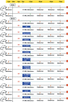
Experimental procedures and animal groups. Focal cerebral ischemia was induced by transient occlusion of the right middle cerebral artery. n=12/group/time point for each functional assay, and n=5/group for western blotting (WB) and n=5/group for IF. Sham, sham operation.

References

    1. Donnan GA, Fisher M, Macleod M, Davis SM. Stroke. Lancet 2008; 371: 1612–1623. - PubMed
    1. Go AS, Mozaffarian D, Roger VL, Benjamin EJ, Berry JD, Blaha MJ et al. Heart disease and stroke statistics—2014 update: a report from the American Heart Association. Circulation 2014; 129: e28–e292. - PMC - PubMed
    1. Will B, Galani R, Kelche C, Rosenzweig MR. Recovery from brain injury in animals: relative efficacy of environmental enrichment, physical exercise or formal training (1990-2002). Prog Neurobiol 2004; 72: 167–182. - PubMed
    1. Keyvani K, Sachser N, Witte OW, Paulus W. Gene expression profiling in the intact and injured brain following environmental enrichment. J Neuropathol Exp Neurol 2004; 63: 598–609. - PubMed
    1. Johansson BB. Functional and cellular effects of environmental enrichment after experimental brain infarcts. Restor Neurol Neurosci 2004; 22: 163–174. - PubMed