

What the young physician should know about May-Thurner syndrome
- PMID: 28845392
- PMCID: PMC4592040
What the young physician should know about May-Thurner syndrome
Abstract
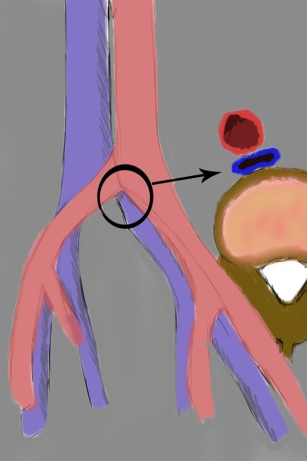
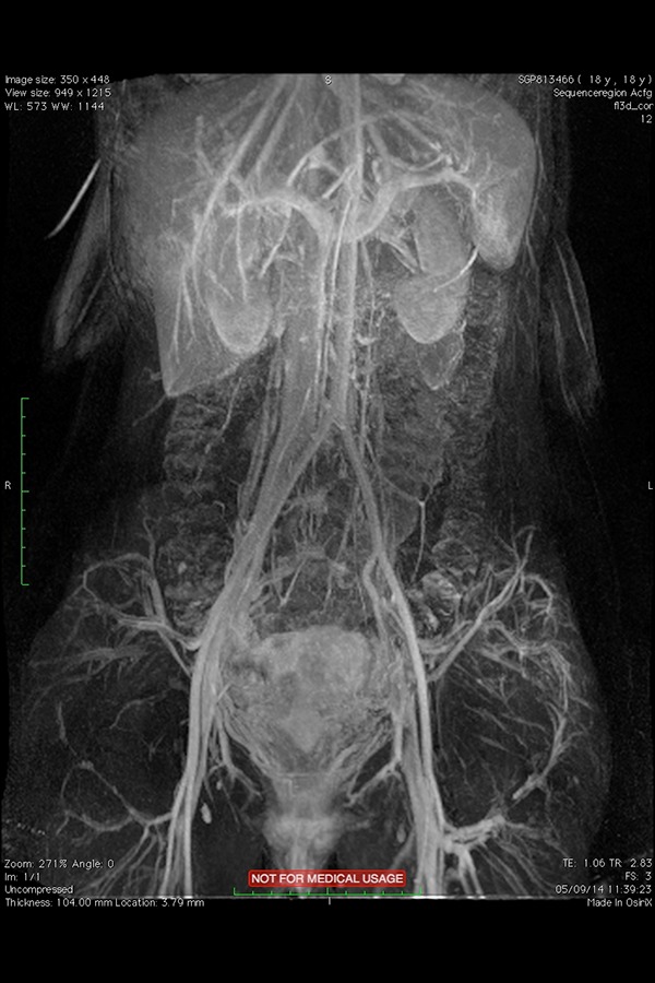
May-Thurner syndrome (MTS) is an anatomically variable condition resulting in compression of the left common iliac vein between the right common iliac artery and the underlying spine with subsequent development of a left deep vein thrombosis (DVT). Although this syndrome is rare, its true prevalence is likely underestimated. Mainly, clinical symptoms and signs include, but are not limited to, pain, swelling, venous stasis ulcers, skin pigmentation changes and post-thrombotic syndrome. Correct treatment is not well established and is based on clinical presentation. Staged thrombolysis with/without prophylactic retrievable inferior vena cava filter placement followed by angioplasty/stenting of the left iliac vein appears to be the best option in MTS patients with extensive DVT. The aim of this review is to present in a simple and didactic form all variable clinical presentations of MTS and to outline possible management within the current guidelines.
Keywords: DVT; May-Thurner Syndrome; endovascular treatment; medical education; thrombectomy.
Figures


References
-
- Cockett FB, Thomas ML. The iliac compression syndrome. Br J Surg. 1965;52(10):816–21. - PubMed
-
- Burke RM, Rayan SS, Kasirajan K, Chaikof EL, Milner R. Unusual case of right-sided May-Thurner syndrome and review of its management. Vascular. 2006;14(1):47–50. - PubMed
-
- Abboud G, Midulla M, Lions C, El Ngheoui Z, Gengler L, Martinelli T, Beregi JP. “Right-sided” May-Thurner syndrome. Cardiovasc Intervent Radiol. 2010;33(5):1056–9. - PubMed
-
- Virchow R. Uber die Erweiterung Kleiner Gefasse. Arch Path Anat. 1851;3:427.
-
- McMurrich JP. The occurrence of congenital adhesions in the common iliac veins, and their relation to thrombosis of the femoral and iliac veins. Am J Med Sci. 1908;135:342–6.
LinkOut - more resources
Full Text Sources