Review
. 2017 Aug 23;88(2):134-142.
doi: 10.23750/abm.v88i2.5558.

Diagnostic Imaging and workup of Malignant Pleural Mesothelioma

Affiliations
Review

Diagnostic Imaging and workup of Malignant Pleural Mesothelioma

Luciano Cardinale et al. Acta Biomed. .

Abstract

Malignant pleural mesothelioma is the most frequent primary neoplasm of the pleura and its incidence is still increasing.This tumor has a strong association with exposure to occupational or environmental asbestos, often after a long latent period of 30-40 years.Plain chest radiography (CXR) is usually the first-line radiologic examination, but the radiographic findings are nonspecific due to its limited contrast resolution and they need to be complemented by other imaging modalities such as computed tomography (CT), magnetic resonance Imaging (MRI), Positron emission tomography-computed tomography (PET-CT) and ultrasound (US).The aim of this paper is to describe the imaging features of this malignancy, underlining the peculiarity of CXR, CT, MRI, PET-CT and US and also focusing on diagnostic workup, based on the literature evidence and according to our experience.

Keywords: Malignant Pleural Mesothelioma, Computed Tomography, Video assisted Thoracic Surgery, Magnetic Resonance Imaging, Thoracic Ultrasound, Positron-emission tomography.


Figures

Figure 1
Figure 1
Malignant pleural mesothelioma. Standard posteroanterior chest X-ray (a) demonstrates a large lobulated pleural mass invading the chest wall (note rib destruction). No pleural effusion is seen. Standard posteroanterior chest X-ray (b) showing right pleural effusion and calcified pleural plaques (white arrows) secondary to long-standing asbestos exposure
Figure 2
Figure 2
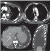
Malignant pleural mesothelioma. Axial contrast-enhanced CT images in arterial (a) and portal phases (b). This example shows that the pleural ticknening is less evident in a more arterial phase than with a 70-80 seconds scan delay. The enhancement of pleural thickening is maximum in the portal phase
Figure 3
Figure 3
Malignant pleural mesothelioma. Axial contrast-enhanced CT image (a) demonstrates circumferential irregular pleural thickening (white arrow) and mediastinal lymphadenopathy. Axial contrast-enhanced CT image (b) demonstrates right subtle circumferential pleural thickening (white arrow) and ipsilateral volume loss. Axial (c) and sagittal reformatted (d) contrast-enhanced CT images showing extensive nodular pleural thickening (white arrows) and a large-sized pleural effusion
Figure 4
Figure 4
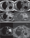
Malignant pleural mesothelioma. Axial T1-weighted MR images showing a iso/hypointense (in phase,a) a large pleural mass involving the chest wall without signal loss (out phase, b), demonstrating absence of fat tissue. Axial T2-weighted (HASTE) MR image (c) shows a pleural mass (black arrow) with irregular hyperintense signal because of presence of fibrous tissue inside tumor lesion. A small posterior pleural nodule is also seen (white arrow). Axial T2-Weighted Fat Saturated MR image (d) shows a hyperintense lesion (white arrowhead) without signal drop because of absence of fat tissue inside. Note also bilateral axillary linphoadenopathy (black arrowhead) and pleural thickening. Axial diffusion-weighted MR image (b value = 750 s/mm2) showing pleural tumor (e) and thickened left pleura with higher signal intensity than adjacent skeletal muscle, with restricted diffusion with low ADC values, more frequent in neoplastic disease (f) (1-1.5)
Figure 5
Figure 5
Malignant pleural mesothelioma. Axial fused well-collimated PET/CT image (a) shows two small FDG-avid nodules in the inferior right hemithorax. Axial fused well-collimated PET/CT image (b) shows extensive FDG-avid pleural thickening in the inferior right hemithorax
Figure 6
Figure 6
Malignant pleural mesothelioma. Axial US scan (a) through the right upper abdominal quadrant allows visualization of the liver and diaphragm as well as the supradiaphragmatic hypoechoic regular and subtle thickening of the diaphragmatic pleura. As well is present a fibrinous septaeted pleural effusion. Axial contrast-enhanced CT image (b) showing diaphragmatic pleural thickening and a large pleural effusion. Note the absence of septations in the pleural fluid. Intraoperative (video-assisted thoracic surgery) photograph (c) of the same patient.
Figure 7
Figure 7
Malignant pleural mesothelioma diagnostic flowchart

References

    1. Nickell LT Jr, Lichtenberger JP 3rd, Khorashadi L, Abbott GF, Carter BW. Multimodality imaging for characterization, classification, and staging of malignant pleural mesothelioma. Radiographics. 2014 Oct;34(6):1692–706. doi: 10.1148/rg.346130089. - PubMed
    1. Rusch VW. Diffuse malignant mesothelioma. General thoracic surgery. In: Shields TW, LoCicero J III, Reed CE, Feins RH, editors. Philadelphia: Wolters Kluwer/Lippincott Williams & Wilkins; 2009. pp. 847–59.
    1. Raj V, Kirke R, Bankart MJ, Entwisle JJ. Multidetector CT imaging of pleura: comparison of two contrast infusion protocols. The British Journal of Radiology. 2011;84(1005):796–9. doi:10.1259/bjr/55980445. - PMC - PubMed
    1. Boutin C, Rey F. Thoracoscopy in pleural malignant mesothelioma: a prospective study of 188 consecutive patients. Part 1: diagnosis. Cancer. 1993;72(2):389–93. - PubMed
    1. Flores RM, et al. The impact of lymph node station on survival in 348 patients with surgically resected malignant pleural mesothelioma: implications for revision of the American Joint Committee on Cancer staging system. J Thorac Cardiovasc Surg. 2008;136(3):605–10. - PubMed

MeSH terms

LinkOut - more resources