. 2017 Oct 2;127(10):3652-3656.
doi: 10.1172/JCI94031. Epub 2017 Aug 28.

Mice expressing KrasG12D in hematopoietic multipotent progenitor cells develop neonatal myeloid leukemia

Affiliations

Mice expressing KrasG12D in hematopoietic multipotent progenitor cells develop neonatal myeloid leukemia

Stefan P Tarnawsky et al. J Clin Invest. .

Abstract

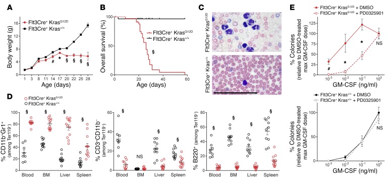
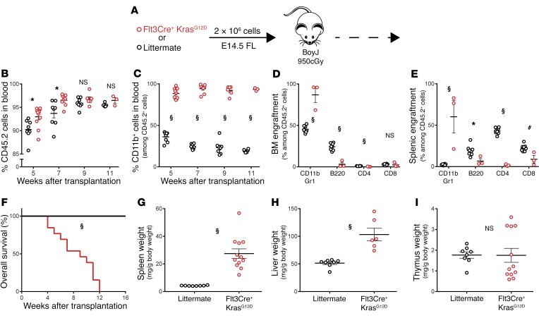
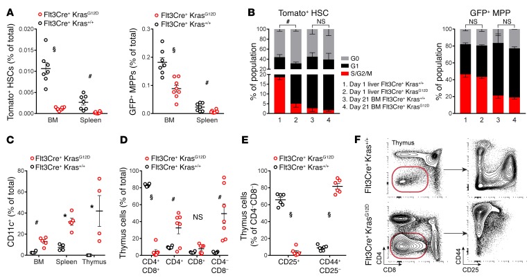
Juvenile myelomonocytic leukemia (JMML) is a pediatric myeloproliferative neoplasm that bears distinct characteristics associated with abnormal fetal development. JMML has been extensively modeled in mice expressing the oncogenic KrasG12D mutation. However, these models have struggled to recapitulate the defining features of JMML due to in utero lethality, nonhematopoietic expression, and the pervasive emergence of T cell acute lymphoblastic leukemia. Here, we have developed a model of JMML using mice that express KrasG12D in multipotent progenitor cells (Flt3Cre+ KrasG12D mice). These mice express KrasG12D in utero, are born at normal Mendelian ratios, develop hepatosplenomegaly, anemia, and thrombocytopenia, and succumb to a rapidly progressing and fully penetrant neonatal myeloid disease. Mutant mice have altered hematopoietic stem and progenitor cell populations in the BM and spleen that are hypersensitive to granulocyte macrophage-CSF due to hyperactive RAS/ERK signaling. Biased differentiation in these progenitors results in an expansion of neutrophils and DCs and a concomitant decrease in T lymphocytes. Flt3Cre+ KrasG12D fetal liver hematopoietic progenitors give rise to a myeloid disease upon transplantation. In summary, we describe a KrasG12D mouse model that reproducibly develops JMML-like disease. This model will prove useful for preclinical drug studies and for elucidating the developmental origins of pediatric neoplasms.


Conflict of interest statement

Conflict of interest: The authors have declared that no conflict of interest exists.

Figures

Figure 1
Figure 1. Flt3Cre+ KrasG12D mice develop a JMML-like disease.
(A) Weight gain from birth (n = 12 mutants and 19 controls). (B) Overall survival (statistical analysis by Mantel-Cox test). (C) Peripheral blood smear (n = 5). Scale bar: 100 μm. (D) Flow cytometric quantification of tissue leukocytes. (E) Seven-day BM colony formation with 100 nM PD0325901 or 0.1% DMSO (n = 3 biological replicates/group). All analyses were performed on 3- to 4-week-old moribund Flt3Cre+ KrasG12D mice and age-matched littermates. *P < 0.05, #P < 0.01, and §P < 0.001, by unpaired, 2-tailed Student’s t test (A, D, and E).
Figure 2
Figure 2. Fetal Flt3Cre+ KrasG12D progenitors initiate a JMML-like disease upon transplantation.
(A) Schematic of FL transplants (n = 12 mutant and 8 control recipients). Analysis of donor cell (B) engraftment and (C) myeloid contribution in peripheral blood. (D and E) Flow cytometric quantification of donor leukocytes in BM and spleens of moribund mutant recipients and control recipients 16 weeks after transplantation. max, maximum. (F) Overall survival following transplantation (statistical analysis by Mantel-Cox test). (GI) Normalized tissue weights of analyzed animals. *P < 0.05, #P < 0.01, and §P < 0.001, by unpaired, 2-tailed Student’s t test (BE and GI).
Figure 3
Figure 3. Analysis of Flt3Cre+ KrasG12D progenitor frequency and differentiation.
(A) Frequency of HSCs and MPPs in the BM and spleens of mutants and littermates. (B) Cell-cycle analysis of HSCs and MPPs from 1-day-old liver (n = 3 WT and 4 mutants) and 21-day-old BM (n = 2/group); representative gating is shown in Supplemental Figure 9. (C) Flow cytometric quantification of tissue DCs. (DF) Flow cytometric quantification and representative gating of thymic cells. Error bars represent the SEM. Cell-cycle statistical analyses were performed using a χ2 test, and other analyses were performed using an unpaired, 2-tailed Student’s t test. *P < 0.05, #P < 0.01, and §P < 0.001.

References

    1. Chan RJ, Cooper T, Kratz CP, Weiss B, Loh ML. Juvenile myelomonocytic leukemia: a report from the 2nd International JMML Symposium. Leuk Res. 2009;33(3):355–362. doi: 10.1016/j.leukres.2008.08.022. - DOI - PMC - PubMed
    1. Ng CS, et al. Juvenile chronic myeloid leukemia. A malignancy of S-100 protein-positive histiocytes. Am J Clin Pathol. 1988;90(5):575–582. doi: 10.1093/ajcp/90.5.575. - DOI - PubMed
    1. Locatelli F, Niemeyer CM. How I treat juvenile myelomonocytic leukemia. Blood. 2015;125(7):1083–1090. doi: 10.1182/blood-2014-08-550483. - DOI - PubMed
    1. Loh ML. Recent advances in the pathogenesis and treatment of juvenile myelomonocytic leukaemia. Br J Haematol. 2011;152(6):677–687. doi: 10.1111/j.1365-2141.2010.08525.x. - DOI - PubMed
    1. Locatelli F, et al. Analysis of risk factors influencing outcomes after cord blood transplantation in children with juvenile myelomonocytic leukemia: a EUROCORD, EBMT, EWOG-MDS, CIBMTR study. Blood. 2013;122(12):2135–2141. doi: 10.1182/blood-2013-03-491589. - DOI - PMC - PubMed

MeSH terms

Substances