

Microbial Lineages in Sarcoidosis. A Metagenomic Analysis Tailored for Low-Microbial Content Samples
- PMID: 28846439
- PMCID: PMC5768903
- DOI: 10.1164/rccm.201705-0891OC
Microbial Lineages in Sarcoidosis. A Metagenomic Analysis Tailored for Low-Microbial Content Samples
Abstract
Rationale: The etiology of sarcoidosis is unknown, but microbial agents are suspected as triggers.
Objectives: We sought to identify bacterial, fungal, or viral lineages in specimens from patients with sarcoidosis enriched relative to control subjects using metagenomic DNA sequencing. Because DNA from environmental contamination contributes disproportionately to samples with low authentic microbial content, we developed improved methods for filtering environmental contamination.
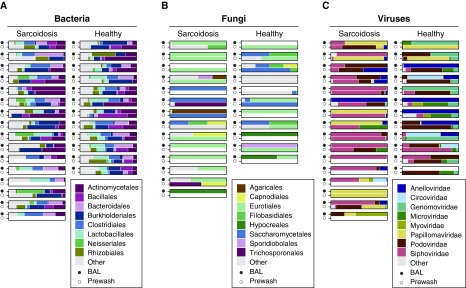
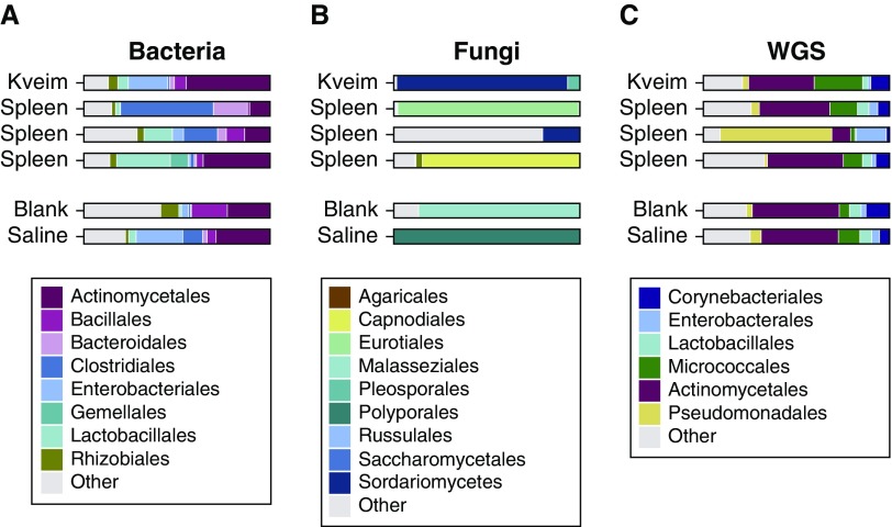
Methods: We analyzed specimens from subjects with sarcoidosis (n = 93), control subjects without sarcoidosis (n = 72), and various environmental controls (n = 150). Sarcoidosis specimens consisted of two independent sets of formalin-fixed, paraffin-embedded lymph node biopsies, BAL, Kveim reagent, and fresh granulomatous spleen from a patient with sarcoidosis. All specimens were analyzed by bacterial 16S and fungal internal transcribed spacer ribosomal RNA gene sequencing. In addition, BAL was analyzed by shotgun sequencing of fractions enriched for viral particles, and Kveim and spleen were subjected to whole-genome shotgun sequencing.
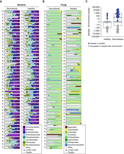
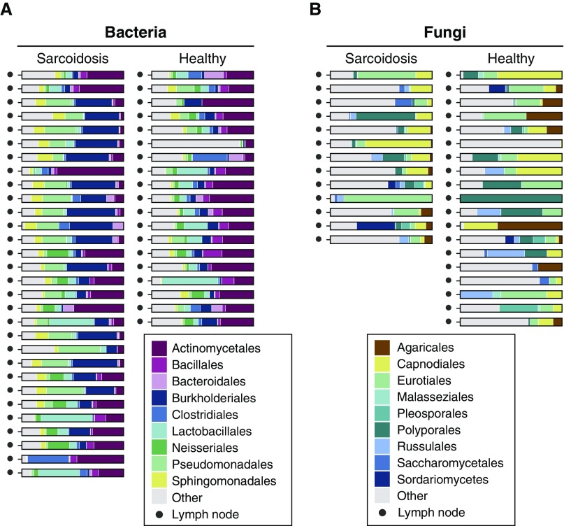
Measurements and main results: In one tissue set, fungi in the Cladosporiaceae family were enriched in sarcoidosis compared with nonsarcoidosis tissues; in the other tissue set, we detected enrichment of several bacterial lineages in sarcoidosis but not Cladosporiaceae. BAL showed limited enrichment of Aspergillus fungi. Several microbial lineages were detected in Kveim and spleen, including Cladosporium. No microbial lineage was enriched in more than one sample type after correction for multiple comparisons.
Conclusions: Metagenomic sequencing revealed enrichment of microbes in single types of sarcoidosis samples but limited concordance across sample types. Statistical analysis accounting for environmental contamination was essential to avoiding false positives.
Keywords: bacterial 16S ribosomal RNA; fungal internal transcribed spacer ribosomal RNA; metagenomic; microbiome; sarcoidosis.
Figures
Comment in
-
Turning "Sarkoid" into "Dropsy". A Valiant, Next-Generation Attempt.Am J Respir Crit Care Med. 2018 Jan 15;197(2):154-155. doi: 10.1164/rccm.201709-1843ED. Am J Respir Crit Care Med. 2018. PMID: 28930643 Free PMC article. No abstract available.
References
-
- Chen ES, Moller DR. Etiologic role of infectious agents. Semin Respir Crit Care Med. 2014;35:285–295. - PubMed
-
- Chen ES, Moller DR. Etiologies of sarcoidosis. Clin Rev Allergy Immunol. 2015;49:6–18. - PubMed
-
- Dubaniewicz A. Microbial and human heat shock proteins as ‘danger signals’ in sarcoidosis. Hum Immunol. 2013;74:1550–1558. - PubMed
-
- Fischer A, Grunewald J, Spagnolo P, Nebel A, Schreiber S, Müller-Quernheim J. Genetics of sarcoidosis. Semin Respir Crit Care Med. 2014;35:296–306. - PubMed
-
- Fingerlin TE, Hamzeh N, Maier LA. Genetics of sarcoidosis. Clin Chest Med. 2015;36:569–584. - PubMed
Publication types
MeSH terms
Substances
Grants and funding
LinkOut - more resources
Full Text Sources
Other Literature Sources
Medical
