. 2017 Nov/Dec;9(8):1337-1348.
doi: 10.1080/19420862.2017.1366395. Epub 2017 Aug 28.

Identification of multiple serine to asparagine sequence variation sites in an intended copy product of LUCENTIS® by mass spectrometry

Affiliations

Identification of multiple serine to asparagine sequence variation sites in an intended copy product of LUCENTIS® by mass spectrometry

François Griaud et al. MAbs. 2017 Nov/Dec.

Abstract

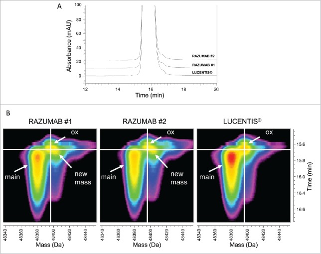
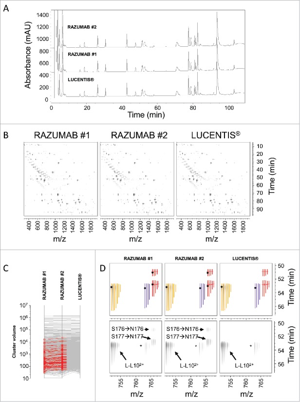
Patent expiration of first-generation biologics and the high cost of innovative biologics are 2 drivers for the development of biosimilar products. There are, however, technical challenges to the production of exact copies of such large molecules. In this study, we performed a head-to-head comparison between the originator anti-VEGF-A Fab product LUCENTIS® (ranibizumab) and an intended copy product using an integrated analytical approach. While no differences could be observed using size-exclusion chromatography, capillary electrophoresis-sodium dodecyl sulfate and potency assays, different acidic peaks were identified with cation ion exchange chromatography and capillary zone electrophoresis. Further investigation of the intact Fab, subunits and primary sequence with mass spectrometry demonstrated the presence of a modified light chain variant in the intended copy product batches. This variant was characterized with a mass increase of 27.01 Da compared to the originator sequence and its abundance was estimated in the range of 6-9% of the intended copy product light chain. MS/MS spectra interrogation confirmed that this modification relates to a serine to asparagine sequence variant found in the intended copy product light chain. We demonstrated that the integration of high-resolution and sensitive orthogonal technologies was beneficial to assess the similarity of an originator and an intended copy product.

Keywords: LUCENTIS®; RAZUMAB; amino acid substitution; biosimilar; error-tolerant search; intended copy product; mass spectrometry; misincorporation; sequence variant; time-resolved deconvolution.


Figures

Figure 1.
Figure 1.
Analysis of size and charge variants of RAZUMAB batches and LUCENTIS® by separation techniques. (A) UV chromatogram overlay of SEC analysis. Absorbance was recorded at 280 nm. High molecular weight species (HMW) and the intact Fab peak (main peak) are shown. (B) Overlay of CE-SDS electropherograms. Fragments, the intact Fab peak (main peak) and HMW species are annotated. (C) Analysis of charge variants by CEX. Main peak, acidic and basic variants are displayed on the overlay of UV chromatograms at 280 nm. RAZUMAB and LUCENTIS® exhibit different acidic variant profiles. (D) Charge variant profiles with CZE. UV electropherograms at 214 nm are shown for RAZUMAB batches and LUCENTIS®. Main peak, acidic and basic variants are displayed. Different acidic variants are present in RAZUMAB and LUCENTIS®. *: main peak shoulder in the acidic region of RAZUMAB batches. Insert: zoomed-in view from 10 to 11 min. For all chromatograms, a signal offset of 10% has been applied between samples.
Figure 2.
Figure 2.
Intact Fab LC-UV/ESI-MS analysis. (A) Overlay of UV chromatograms of intact LUCENTIS® and RAZUMAB batches 1 and 2. Absorbance was recorded at 280 nm. Signal offset: 10%. (B) Time-resolved deconvolution of intact LUCENTIS® and intact RAZUMAB MS data for batches 1 and 2. Heat maps were generated from time-resolved deconvolution performed in parallel for all samples. Intensity is color-coded ranging from less intense (black) to most intense (red). The unmodified Fab (main peak) and an oxidized variant (ox) are annotated. A new mass (+ ∼25 Da) is found exclusively in RAZUMAB samples.
Figure 3.
Figure 3.
Fab subunit LC-UV/ESI-MS analysis. (A) Overlay of UV chromatograms at 214 nm of light and heavy chains of LUCENTIS® and RAZUMAB batches 1 and 2 after reduction and carbamidomethylation. Signal offset: 10%. (B) Time-resolved deconvolution for the light chain (LC). LC, and LC + 27 Da (in RAZUMAB samples), are annotated. ║: In-source dehydration, ◊: Guanidine adduct, §: Possible LC + (2 × 27 Da) in RAZUMAB batches, ‡: Sodium adduct. (C) Time-resolved deconvolution for the heavy chain (HC) species. HC, oxidized HC (HCox) and N-terminal pyroglutamate formation (HC(pE)) are annotated. *major sample preparation artifact is overalkylation with iodoacetamide as shown by the addition of +57 Da. Heat maps were generated from time-resolved deconvolution performed in parallel for all samples. Intensity is color-coded ranging from less intense (black) to most intense (red).
Figure 4.
Figure 4.
Differential analysis of RAZUMAB and LUCENTIS® peptide mapping signals. (A) Overlay of UV chromatograms of Lys-C peptide mapping of LUCENTIS® and RAZUMAB batches 1 and 2 after reduction and carbamidomethylation. Absorbance was acquired at 214 nm. No major differences were observed using UV chromatogram analysis. Signal offset: 10%. (B) Differential analysis of Lys-C peptide mapping MS data. A chromatogram plane (RT-m/z) of the MS and MS/MS data is built for each sample, in which MS peaks detected in one or more samples are clustered according to their monoisotopic mass and RT. The volume of each cluster is calculated and compared across all samples. (C) Identification of RAZUMAB-specific MS signals. Differential cluster analysis retrieved 57 clusters (shown in red) present in both RAZUMAB batches and absent in LUCENTIS®. (D) Mapping RAZUMAB-specific MS signals with a mass increment of 27.01 Da. An example is shown for L-L102+ peptide using the chromatogram planes of RAZUMAB batches and LUCENTIS®. Top panels: Clusters with isotope peak boundaries for all isotopes are shown for L-L102+ species across samples. Native L-L102+ peptide (yellow cluster) was detected in RAZUMAB batches along with variant species (red clusters), absent from LUCENTIS® sample. These variant peptides were detected with a mass increment of 27.01 Da and were assigned by MS/MS interpretation to S176→N176 and S177→N177 substitutions in RAZUMAB. A black dot represents the acquisition of MS/MS data for the specific cluster in the respective sample. The purple cluster corresponds to L-L102+ sodium adduct. Bottom panels: same chromatogram planes as in top panels without cluster isotopic peak boundaries.* L-L102+ sodium adduct.
Figure 5.
Figure 5.
MS/MS spectra of native and variant L-L10 peptides. (A) MS/MS spectrum of L-L102+ at 765.39 m/z and retention time 50.9 min with serine to asparagine substitution localized at position 176 (marked in red). Signature ions used for Ser → Asn substitution localization are shown in red. (B) MS/MS spectrum of L-L102+ at 765.39 m/z and retention time 52.8 min with serine to asparagine substitution localized at position 177. Position and signature ions are shown in red. (C) MS/MS spectrum of native L-L102+ peptide at 751.88 m/z and retention time 53.4 min.

References

    1. Reichert JM. Antibodies to watch in 2016. Mabs. 2016;8:197-204. doi: 10.1080/19420862.2015.1125583. PMID:26651519 - DOI - PMC - PubMed
    1. Reichert JM. Antibodies to watch in 2015. Mabs. 2015;7:1-8. doi: 10.4161/19420862.2015.988944. PMID:25484055 - DOI - PMC - PubMed
    1. Ecker DM, Jones SD, Levine HL. The therapeutic monoclonal antibody market. Mabs. 2015;7:9-14. doi: 10.4161/19420862.2015.989042. PMID:25529996 - DOI - PMC - PubMed
    1. Reichert JM. Antibodies to watch in 2017. MAbs. 2017;9:167-81. doi: 10.1080/19420862.2016.1269580 - DOI - PMC - PubMed
    1. Holzmann J, Balser S, Windisch J. Totality of the evidence at work: The first US biosimilar. Expert Opin Biol Ther. 2016;16:137-42. doi: 10.1517/14712598.2016.1128410. PMID:26634611 - DOI - PubMed

Publication types