. 2017 Nov/Dec;9(8):1282-1296.
doi: 10.1080/19420862.2017.1371383. Epub 2017 Aug 28.

Rare, high-affinity anti-pathogen antibodies from human repertoires, discovered using microfluidics and molecular genomics

Affiliations

Rare, high-affinity anti-pathogen antibodies from human repertoires, discovered using microfluidics and molecular genomics

Adam S Adler et al. MAbs. 2017 Nov/Dec.

Abstract

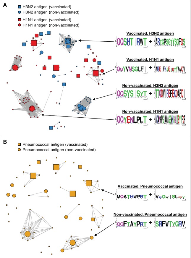
Affinity-matured, functional anti-pathogen antibodies are present at low frequencies in natural human repertoires. These antibodies are often excellent candidates for therapeutic monoclonal antibodies. However, mining natural human antibody repertoires is a challenge. In this study, we demonstrate a new method that uses microfluidics, yeast display, and deep sequencing to identify 247 natively paired anti-pathogen single-chain variable fragments (scFvs), which were initially as rare as 1 in 100,000 in the human repertoires. Influenza A vaccination increased the frequency of influenza A antigen-binding scFv within the peripheral B cell repertoire from <0.1% in non-vaccinated donors to 0.3-0.4% in vaccinated donors, whereas pneumococcus vaccination did not increase the frequency of antigen-binding scFv. However, the pneumococcus scFv binders from the vaccinated library had higher heavy and light chain Replacement/Silent mutation (R/S) ratios, a measure of affinity maturation, than the pneumococcus binders from the corresponding non-vaccinated library. Thus, pneumococcus vaccination may increase the frequency of affinity-matured antibodies in human repertoires. We synthesized 10 anti-influenza A and nine anti-pneumococcus full-length antibodies that were highly abundant among antigen-binding scFv. All 10 anti-influenza A antibodies bound the appropriate antigen at KD<10 nM and neutralized virus in cellular assays. All nine anti-pneumococcus full-length antibodies bound at least one polysaccharide serotype, and 71% of the anti-pneumococcus antibodies that we tested were functional in cell killing assays. Our approach has future application in a variety of fields, including the development of therapeutic antibodies for emerging viral diseases, autoimmune disorders, and cancer.

Keywords: Influenza A; antibody repertoire; deep sequencing; microfluidics; pneumococcus.


Figures

Figure 1.
Figure 1.
Overview of the workflow used to generate the scFv libraries from B cells isolated from human donors. Peripheral B cells are isolated from either vaccinated or non-vaccinated human donors. B cells are then encapsulated into droplets with oligo-dT beads and a lysis solution. The throughput of microfluidic cell encapsulation is ∼ 3 million cells per hour. mRNA-bound beads are purified from the droplets, and then injected into a second emulsion with an OE-RT-PCR amplification mix that generates DNA amplicons that encode scFv with native pairing of heavy and light chain Ig. Libraries of natively paired amplicons are then electroporated into yeast for scFv display. FACS is used to identify high affinity scFv. Finally, deep antibody sequencing is used to identify all clones in the pre- and post-sort scFv libraries. The complete workflow, from isolated B cells to sequenced scFv candidates, requires only three weeks.
Figure 2.
Figure 2.
scFv libraries subjected to FACS for influenza A virus antigens. Staining for c-Myc (AF488) is used to differentiate yeast cells that express scFv from yeast cells that do not express scFv (x-axis). Staining for biotinylated antigen (PE) is used to identify yeast that express scFv that bind to the antigen (y-axis). The Fc negative control is used to set gates that are used to capture yeast cells that express scFv and bind antigen (upper right corner of the FACS plot). Gates for yeast selection are indicated by the quadrangle in the upper right corner of each FACS plot. The percentage in each quadrangle (red text) indicates the proportion of c-Myc-positive yeast that fell within the gate. A vertical dotted line (black) indicates the gate used to determine the number of yeast that express scFv (c-Myc+). (A) 1st and 2nd sort FACS data for H3N2 and H1N1, using an scFv library generated from B cells from donors vaccinated with the influenza A vaccine Fluvirin. (B) 1st and 2nd sort FACS data for H3N2 and H1N1, using an scFv library generated from B cells from non-vaccinated donors.
Figure 3.
Figure 3.
scFv libraries subjected to FACS for pneumococcus polysaccharides. Staining for c-Myc (AF488) is used to differentiate yeast cells that express scFv from yeast cells that do not express scFv (x-axis). Staining for biotinylated antigen (PE) is used to identify yeast that express scFv that bind to the antigen (y-axis). The Fc negative control is used to set gates that are used to capture yeast cells that express scFv and bind antigen (upper right corner of the FACS plot). Gates for yeast selection are indicated by the quadrangle in the upper right corner of each FACS plot. The percentage in each quadrangle (red text) indicates the proportion of c-Myc positive yeast that fell within the gate. A vertical dotted line (black) indicates the gate used to determine the number of yeast that express scFv (c-Myc+). (A) 1st, 2nd, and 3rd sort FACS data for a pool of pneumococcus polysaccharides, using an scFv library generated from B cells from donors vaccinated with the pneumococcus vaccine Pneumovax-23. (B) 1st, 2nd, and 3rd sort FACS data for a pool of pneumococcus polysaccharides, using an scFv library generated from B cells from non-vaccinated donors.
Figure 4.
Figure 4.
Clonal cluster analysis for FACS-sorted scFv binders. We computed the total number of amino acid differences between each pairwise alignment of FACS-sorted scFv. Edges were drawn only for pairwise alignments with ≤9 amino acid differences. The node for each scFv sequence was sized based on frequency in the FACS-sorted population: small (<1% frequency), medium (1-10% frequency), and large (>10% frequency). Web logos of the CDR3K + CDR3H amino acid sequences of the clusters used in Figure 5 are on the right. (A) Clonal clusters for anti-H3N2 scFv (blue) and anti-H1N1 scFv (red). scFv isolated from vaccinated donors are indicated with squares, and scFv isolated from non-vaccinated donors are indicated with circles. Note that two of the 19 sequences in the Vaccinated, H3N2 antigen logo were from non-vaccinated donors. (B) Clonal clusters for anti-pneumococcal antigen scFv. scFv isolated from vaccinated donors are indicated with squares, and scFv isolated from non-vaccinated donors are indicated with circles.
Figure 5.
Figure 5.
Amino acid sequence logos for groups of evolutionarily related clones. Though 1–4 such groups were present after the 2nd (influenza A) or 3rd (pneumococcus) FACS sort, we displayed a single representative dominant clonal expansion from each. Full V(D)J amino acid sequences are shown. Variant amino acids are emphasized with red arrows (the first seven amino acids are omitted due to the high probability of mis-priming during OE-RT-PCR). (A) Clonal groups for vaccinated donors: H3N2 antigen (top), H1N1 antigen (middle), and pneumococcal polysaccharides (bottom). Note that two of the 19 sequences in the H3N2 logo were from non-vaccinated donors. (B) Clonal groups for non-vaccinated donors: H3N2 antigen (top), H1N1 antigen (middle), and pneumococcal polysaccharides (bottom).

References

    1. Flyak AI, Shen X, Murin CD, Turner HL, David JA, Fusco ML, Lampley R, Kose N, Ilinykh PA, Kuzmina N, et al.. Cross-Reactive and Potent Neutralizing Antibody Responses in Human Survivors of Natural Ebolavirus Infection. Cell. 2016;164(3):392-405. doi:10.1016/j.cell.2015.12.022. PMID:26806128 - DOI - PMC - PubMed
    1. Gilchuk I, Gilchuk P, Sapparapu G, Lampley R, Singh V, Kose N, Blum DL, Hughes LJ, Satheshkumar PS, Townsend MB, et al.. Cross-Neutralizing and Protective Human Antibody Specificities to Poxvirus Infections. Cell. 2016;167(3):684-694.e9. doi:10.1016/j.cell.2016.09.049. PMID:27768891 - DOI - PMC - PubMed
    1. Beerli RR, Rader C. Mining human antibody repertoires. MAbs. 2010;2(4):365-78. doi:10.4161/mabs.12187. PMID:20505349 - DOI - PMC - PubMed
    1. Smith SA, Crowe JE Jr. Use of Human Hybridoma Technology To Isolate Human Monoclonal Antibodies. Microbiol Spectr. 2015;3(1):AID-0027-2014. doi:10.1128/microbiolspec.AID-0027-2014. PMID:26104564 - DOI - PMC - PubMed
    1. Köhler G, Milstein C. Continuous cultures of fused cells secreting antibody of predefined specificity. Nature. 1975;256(5517):495-7. doi:10.1038/256495a0. PubMed PMID:1172191 - DOI - PubMed

Publication types

MeSH terms