

In Vitro Innervation as an Experimental Model to Study the Expression and Functions of Acetylcholinesterase and Agrin in Human Skeletal Muscle
- PMID: 28846617
- PMCID: PMC6151842
- DOI: 10.3390/molecules22091418
In Vitro Innervation as an Experimental Model to Study the Expression and Functions of Acetylcholinesterase and Agrin in Human Skeletal Muscle
Abstract
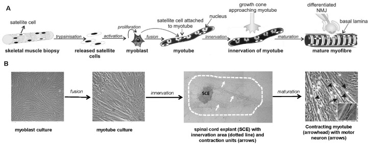
Acetylcholinesterase (AChE) and agrin, a heparan-sulfate proteoglycan, reside in the basal lamina of the neuromuscular junction (NMJ) and play key roles in cholinergic transmission and synaptogenesis. Unlike most NMJ components, AChE and agrin are expressed in skeletal muscle and α-motor neurons. AChE and agrin are also expressed in various other types of cells, where they have important alternative functions that are not related to their classical roles in NMJ. In this review, we first focus on co-cultures of embryonic rat spinal cord explants with human skeletal muscle cells as an experimental model to study functional innervation in vitro. We describe how this heterologous rat-human model, which enables experimentation on highly developed contracting human myotubes, offers unique opportunities for AChE and agrin research. We then highlight innovative approaches that were used to address salient questions regarding expression and alternative functions of AChE and agrin in developing human skeletal muscle. Results obtained in co-cultures are compared with those obtained in other models in the context of general advances in the field of AChE and agrin neurobiology.
Keywords: acetylcholinesterase; agrin; apoptosis; co-cultures; human muscle; in vitro innervation; neuromuscular junction.
Conflict of interest statement
The authors declare no conflict of interest.
Figures
References
Publication types
MeSH terms
Substances
LinkOut - more resources
Full Text Sources
Other Literature Sources
