Review
. 2017 Aug 28;5(3):51.
doi: 10.3390/microorganisms5030051.

The Sea as a Rich Source of Structurally Unique Glycosaminoglycans and Mimetics

Affiliations
Review

The Sea as a Rich Source of Structurally Unique Glycosaminoglycans and Mimetics

Ariana A Vasconcelos et al. Microorganisms. .

Abstract

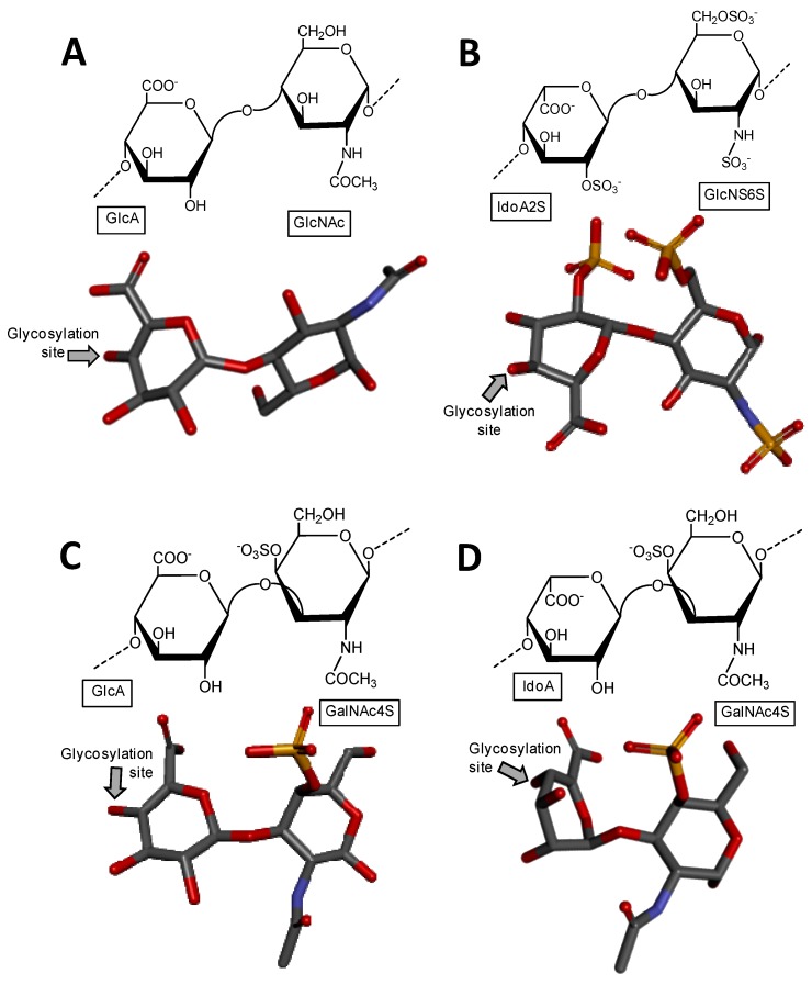
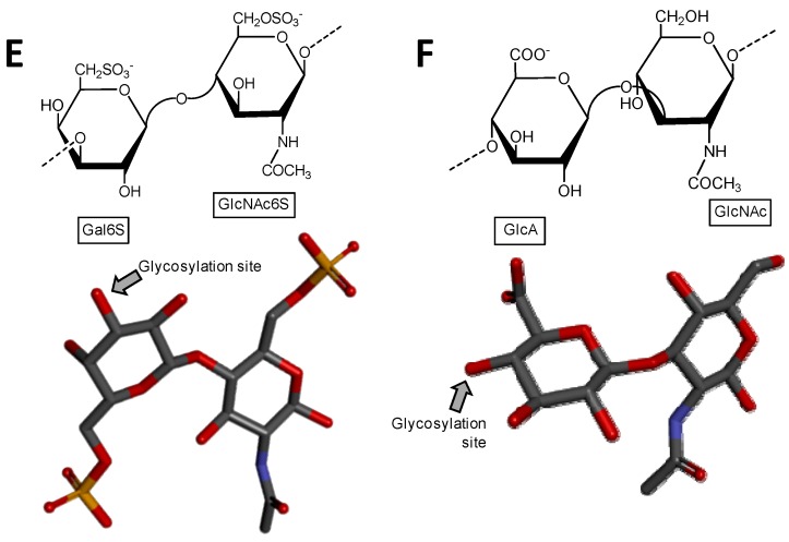
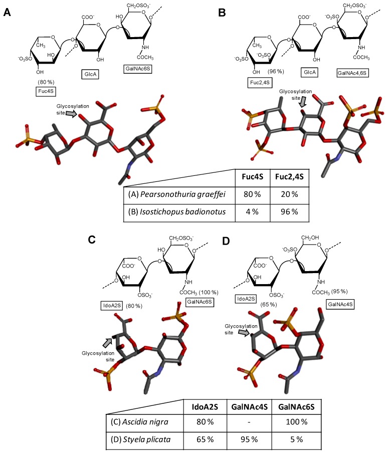
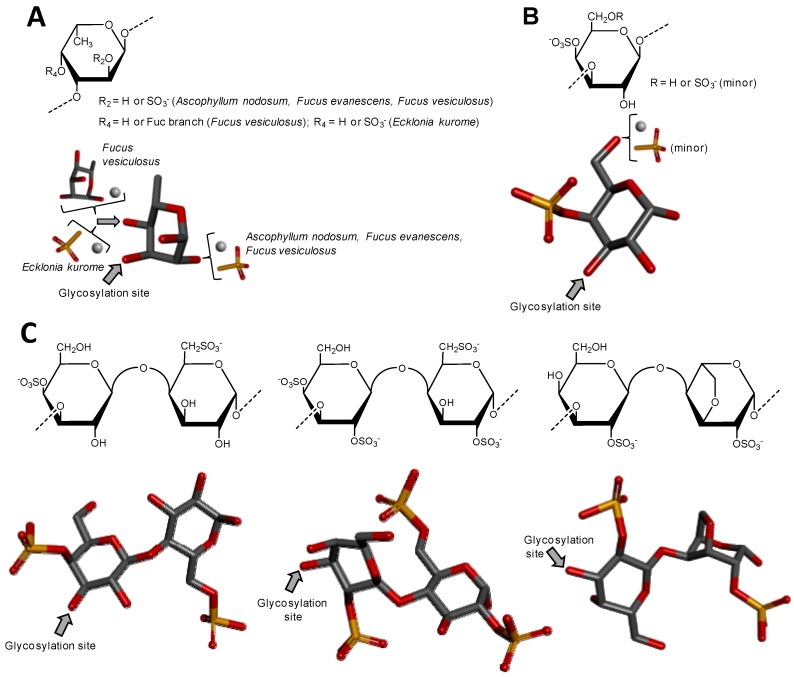
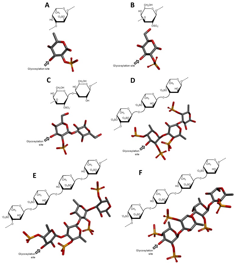
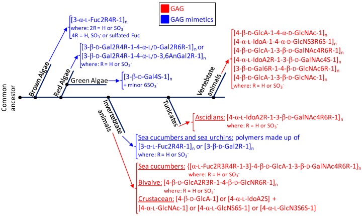
Glycosaminoglycans (GAGs) are sulfated glycans capable of regulating various biological and medical functions. Heparin, heparan sulfate, chondroitin sulfate, dermatan sulfate, keratan sulfate and hyaluronan are the principal classes of GAGs found in animals. Although GAGs are all composed of disaccharide repeating building blocks, the sulfation patterns and the composing alternating monosaccharides vary among classes. Interestingly, GAGs from marine organisms can present structures clearly distinct from terrestrial animals even considering the same class of GAG. The holothurian fucosylated chondroitin sulfate, the dermatan sulfates with distinct sulfation patterns extracted from ascidian species, the sulfated glucuronic acid-containing heparan sulfate isolated from the gastropode Nodipecten nodosum, and the hybrid heparin/heparan sulfate molecule obtained from the shrimp Litopenaeus vannamei are some typical examples. Besides being a rich source of structurally unique GAGs, the sea is also a wealthy environment of GAG-resembling sulfated glycans. Examples of these mimetics are the sulfated fucans and sulfated galactans found in brown, red and green algae, sea urchins and sea cucumbers. For adequate visualization, representations of all discussed molecules are given in both Haworth projections and 3D models.

Keywords: chondroitin sulfate; glycosaminoglycans; heparan sulfate; heparin; sulfated fucans; sulfated galactans.


Conflict of interest statement

The authors declare no conflict of interest.

Figures

Figure 1
Figure 1
Haworth projections and stick model representations of the commonest structures in (A) heparan sulfate [GlcA-(β1→4)-GlcNAc] whose GlcNAc is α(1→4)-linked to the GlcA of the next disaccharide unit (extracted from PDB ID 3E7J) [35]; (B) heparin: [IdoA2S-(α1→4)-GlcNS6S] whose GlcNS6S is α(1→4)-linked to the IdoA2S of the next disaccharide unit (extracted from PDB ID 1HPN) [36]; (C) chondroitin 4-sulfate [GlcA-(β1→3)-GalNAc4S] whose GalNAc is β(1→4)-linked to the GlcA of next disaccharide unit (extracted from PDB ID 1OFM) [37]; (D) dermatan sulfate [IdoA-(α1→3)-GalNAc4S] whose GalNAc4S is β(1→4)-linked to the IdoA of the next disaccharide unit (extracted from PDB ID 1OFL) [37]; (E) keratan sulfate [Gal6S-(β1→4)-GlcNAc6S] whose GlcNAc6S is (β1→3)-linked to the Gal6S of the next disaccharide unit (extracted from PDB ID 1KES) [38]; and (F) hyaluronan [GlcA-(β1→3)-GlcNAc] whose GlcNAc is (β1→4)-linked to the GlcA of the next disaccharide unit (extracted from PDB ID 2BVK) [39]. The unsaturated uronic acid (∆4,5UroA) of the original structures 3E7J, 1OFM and 1OFL were converted, respectively, to GlcA, GlcA and IdoA. The monosaccharide nomenclatures are IdoA2S for 2-sulfated iduronic acid; GlcNS6S for N,6-disulfated glucosamine; GlcA for glucuronic acid; GlcNAc for N-acetylglucosamine; GalNAc4S for 4-sulfated N-acetylgalactosamine; IdoA for iduronic acid; Gal6S for 6-sulfated galactose; GlcNAc6S for 6-sulfated N-acetylglucosamine. The colors of the atoms used in the representations are grey for carbon, blue for nitrogen, red for oxygen and yellow for sulfur. The hydrogen atoms were omitted for visual simplification. The arrows labeled as “Glycosylation site” indicate the position of the glycosidic bond. Structures were created using ChemDraw Ultra 8.0 for Haworth projections and Discovery Studio Visualizer v.4.5 software (BIOVIA, Dassault Systèmes, San Diego, CA, USA) for 3D representations. The monosaccharides are displayed in their commonest chair configurations: 4C1 for GlcA, GalNAc, GlcNAc and 1C4 for IdoA.
Figure 1
Figure 1
Haworth projections and stick model representations of the commonest structures in (A) heparan sulfate [GlcA-(β1→4)-GlcNAc] whose GlcNAc is α(1→4)-linked to the GlcA of the next disaccharide unit (extracted from PDB ID 3E7J) [35]; (B) heparin: [IdoA2S-(α1→4)-GlcNS6S] whose GlcNS6S is α(1→4)-linked to the IdoA2S of the next disaccharide unit (extracted from PDB ID 1HPN) [36]; (C) chondroitin 4-sulfate [GlcA-(β1→3)-GalNAc4S] whose GalNAc is β(1→4)-linked to the GlcA of next disaccharide unit (extracted from PDB ID 1OFM) [37]; (D) dermatan sulfate [IdoA-(α1→3)-GalNAc4S] whose GalNAc4S is β(1→4)-linked to the IdoA of the next disaccharide unit (extracted from PDB ID 1OFL) [37]; (E) keratan sulfate [Gal6S-(β1→4)-GlcNAc6S] whose GlcNAc6S is (β1→3)-linked to the Gal6S of the next disaccharide unit (extracted from PDB ID 1KES) [38]; and (F) hyaluronan [GlcA-(β1→3)-GlcNAc] whose GlcNAc is (β1→4)-linked to the GlcA of the next disaccharide unit (extracted from PDB ID 2BVK) [39]. The unsaturated uronic acid (∆4,5UroA) of the original structures 3E7J, 1OFM and 1OFL were converted, respectively, to GlcA, GlcA and IdoA. The monosaccharide nomenclatures are IdoA2S for 2-sulfated iduronic acid; GlcNS6S for N,6-disulfated glucosamine; GlcA for glucuronic acid; GlcNAc for N-acetylglucosamine; GalNAc4S for 4-sulfated N-acetylgalactosamine; IdoA for iduronic acid; Gal6S for 6-sulfated galactose; GlcNAc6S for 6-sulfated N-acetylglucosamine. The colors of the atoms used in the representations are grey for carbon, blue for nitrogen, red for oxygen and yellow for sulfur. The hydrogen atoms were omitted for visual simplification. The arrows labeled as “Glycosylation site” indicate the position of the glycosidic bond. Structures were created using ChemDraw Ultra 8.0 for Haworth projections and Discovery Studio Visualizer v.4.5 software (BIOVIA, Dassault Systèmes, San Diego, CA, USA) for 3D representations. The monosaccharides are displayed in their commonest chair configurations: 4C1 for GlcA, GalNAc, GlcNAc and 1C4 for IdoA.
Figure 2
Figure 2
Haworth projections and stick model representations of the galactosaminoglycan structures from marine invertebrates. (A,B) Fucosylated chondroitin sulfates from two holothurian species: (A) Pearsonothuria graeffei mostly composed of {[Fuc4S-α(1→3)]-GlcA-(β1→3)-GalNAc6S} whose GalNAc6S is β(1→4)-linked to the GlcA of the next unit to make up the backbone (disaccharide of backbone extracted from PDB ID 1OFM as model) [40]; and (B) Isostichopus badionotus mostly composed of {[Fuc2,4S-α(1→3)]-GlcA-(β1→3)-GalNAc4,6S} whose GalNAc4,6S is β(1→4)-linked to the GlcA of the next unit to make up the backbone (disaccharide extracted from PDB ID 1OFM as model) [40]; Dermatan sulfate from two ascidian species: (C) Ascidia nigra mostly composed of [IdoA2S-(α1→3)-GalNAc6S] whose GalNAc6S is β(1→4)-linked to the IdoA2S of the next disaccharide unit (disaccharide extracted from PDB ID 1OFL as model) [42]; and (D) Styela plicata mostly composed of [IdoA2S-(α1→3)-GalNAc4S] whose GalNAc4S is β(1→4)-linked to the IdoA2S of the next disaccharide unit (disaccharide of backbone extracted from PDB ID 1OFL as model) [43]. The unsaturated uronic acid (∆4,5UroA) of the original structures 1OFM and 1OFL were converted respectively to GlcA and IdoA. The monosaccharide nomenclatures are Fuc2,4S for 2,4-di-sulfated fucose; Fuc4S for 4-sulfated fucose; GlcA for glucuronic acid; GlcNAc for N-acetylglucosamine; GalNAc4S for 4-sulfated N-acetylgalactosamine, GalNAc6S for 6-sulfated N-acetylgalactosamine, GalNAc4,6S for 4,6-di-sulfated N-acetylgalactosamine and IdoA2S for 2-sulfated iduronic acid. The colors of the atoms used in the representations are grey for carbon, blue for nitrogen, red for oxygen and yellow for sulfur. The hydrogen atoms were omitted for visual simplification. The arrows labeled as “Glycosylation site” indicate the position of the glycosidic bond. Structures were created using ChemDraw Ultra 8.0 for Haworth projections and Discovery Studio Visualizer v.4.5 software (BIOVIA, Dassault Systèmes) for 3D representations. The monosaccharides are displayed in their commonest chair configurations: 4C1 for GlcA and GalNAc(6S, 4,6S or 4S) and 1C4 for IdoA2S and Fuc(4S and 2,4S). The percentage of sulfation patterns on the lateral Fuc residues of each holothurian fucosylated chondroitin sulfate and on the IdoA and GalNAc units of the ascidian dermatan sulfates are shown as inserts at the bottom of the panels and close to the respective units.
Figure 3
Figure 3
Haworth projections and stick model representations of the GAG structures from marine invertebrates. (A) Heparan sulfate from the bivalve Nodipecten nodosus composed of [GlcAR2R3-(β1→4)-GlcNRNR6] whose GlcN is β(1→4)-linked to the GlcA of the next disaccharide unit (disaccharide extracted from PDB ID 3E7J as model). R2, R3, R6 can be either hydrogen or sulfate, and RN can be either acetyl or sulfate [49]; (B) The hybrid heparin/heparan sulfate from the shrimp Litopenaeus vannamei composed mostly of [GlcA-(β1→4)-GlcNS6S] and other monosaccharides whose 3D models were extracted from PDB ID 1PHN and 3E7J) [50]. Percentages of the lateral chemical groups (A) and composing monosaccharide types (B) are indicated accordingly in the panels. The unsaturated uronic acid (∆4,5UroA) of the original structure 3E7J was converted to GlcA. The monosaccharide nomenclatures are GlcA for glucuronic acid; GlcN for glucosamine; IdoA2S for 2-sulfated iduronic acid; GlcNAc for N-acetylglucosamine; GlcNS6S for N,6-disulfated glucosamine; and GlcNS3S6S for N,3,6-trisulfated glucosamine. The colors of the atoms used in the representations are grey for carbon, blue for nitrogen, red for oxygen and yellow for sulfur. The hydrogen atoms were omitted on the structures for visual simplification but are represented in light grey discs as possible substituents. The arrows labeled as “Glycosylation site” indicate the position of the glycosidic bond. Structures were created using ChemDraw Ultra 8.0 for Haworth projections and Discovery Studio Visualizer v.4.5 software (BIOVIA, Dassault Systèmes) for 3D representations. The monosaccharides are displayed in their commonest chair configurations: 4C1 for GlcA, GlcN, GlcNAc, GlcNS6S, GlcNS3S6S and 1C4 for IdoA2S.
Figure 4
Figure 4
Haworth projections and stick model representations of the major components in sulfated fucans and sulfated galactans from seaweeds. (A) The sulfated fucans from the well-known brown alga species Ascophyllum nodosum, Fucus evanescens, Fucus vesiculosus and Ecklonia kurome are mostly composed of α(1→3)-linked α-l-Fucose (Fuc) units highly substituted by sulfation at C2 and C4 positions [55,62]. Branches of Fuc units can also occur at the C4 position through α-linkages [55,63]; (B) The sulfated galactan from green algae are dominantly composed of α(1→3)-linked galactose (Gal) units sulfated at position C4 [51]; (C) The sulfated galactans from red algae are mostly composed of regular disaccharide units alternating 3-linked β-Gal and 4-linked α-Gal residues as illustrated with the different types of carrageenans with different substitutions of sulfation and occurrence of the 3,6-anhydro-galactose units such as μ (mu) left structure, λ (lambda) middle structure and θ (theta) right structure [64]. The colors of the atoms used in the representations are grey for carbon, blue for nitrogen, red for oxygen and yellow for sulfur. The hydrogen atoms were omitted on the structures for visual simplification but are represented in light grey discs as possible substituents. The arrows labeled as “Glycosylation site” indicate the position of the glycosidic bond. Structures were created using ChemDraw Ultra 8.0 for Haworth projections and submitted to MM2 force-field. Then, Discovery Studio Visualizer v.4.5 software (BIOVIA, Dassault Systèmes) was used for 3D representations after energy minimization. The monosaccharides are displayed in their commonest chair configurations: 1C4 for Fuc and 4C1 for Gal.
Figure 5
Figure 5
Haworth projections and stick model representations of the repetitive oligosaccharide units of sulfated fucans and sulfated galactans from echinoderms (sea urchins). While (A) Strongylocentrotus franciscanus expresses a sulfated fucan composed of 3-linked 2-sulfated α-fucose (Fuc) units [65]; (B) Echinometra lucunter synthesizes a sulfated galactan composed of 3-linked 2-sulfated α-galactose (Gal) units [66]; (C) The sulfated galactans isolated from Glyptocidaris crenularis is composed of [Gal2S-(α1→3)-Gal] whose non-sulfated Gal unit is also (α1→3)-linked to the Gal2S of the next disaccharide unit [67]; (D) The sulfated fucan-II from Strongylocentrotus purpuratus is composed of [Fuc2,4S-(α1→3)-Fuc4S-(α1→3)-Fuc4S] whose 4-sulfated Fuc unit of the reducing end is also (α1→3)-linked to the Fuc2,4S of the next trisaccharide unit [68]; (E) The sulfated fucan isolated from Strongylocentrotus pallidus is composed of [Fuc4S-(α1→3)-Fuc4S-(α1→3)-Fuc2S-(α1→3)-Fuc2S] whose 2-sulfated Fuc unit of the reducing end is also (α1→3)-linked to the Fuc4S of the next tetrasaccharide unit [69]; (F) The sulfated fucan isolated from Lytechinus variegatus is composed of [Fuc2,4S-(α1→3)-Fuc2S-(α1→3)-Fuc2S-(α1→3)-Fuc4S] whose 4-sulfated Fuc unit is also (α1→3)-linked to the Fuc2,4S of the next tetrasaccharide unit [70]. Hence, these marine sulfated glycans are regularly composed of monosaccharide (A,B); disaccharide (C); trisaccharide (D) and tetrasaccharide (E,F) building blocks. The colors of the atoms used in the representations are grey for carbon, blue for nitrogen, red for oxygen and yellow for sulfur. The hydrogen atoms were omitted for visual simplification. The arrows labeled as “Glycosylation site” indicate the position of the glycosidic bond. Structures were created using ChemDraw Ultra 8.0 for Haworth projections and submitted to MM2 force-field. Then, Discovery Studio Visualizer v.4.5 software (BIOVIA, Dassault Systèmes) was used for 3D representations after energy minimization. The monosaccharides are displayed in their commonest chair configurations: 1C4 for Fuc and 4C1 for Gal.
Figure 6
Figure 6
Phylogenetic relationship of glycosaminoglycans (GAGs) (red) and mimetics (blue) of seaweeds (brown, red and green algae), marine invertebrate animals, marine tunicates and vertebrate animals (from marine or terrestrial environments). The structural abbreviations are S for sulfation, R for radical, Fuc for fucose, Gal for galactose, GlcA for glucuronic acid, GlcNAc for N-acetylglucosamine, GalNAc for N-acetylgalactosamine, IdoA for iduronic acid and GlcN for glucosamine. R and S stand for radicals and sulfation, respectively. The major goal for showing this simplified scheme is to just illustrate the principal structures of the sulfated glycans expressed in the classes of organisms discussed herein and not the accurate evolutionary relationship between the cited organisms in terms of sulfated glycan-related genotypes or phenotypes or through the evolution history.

Similar articles

Cited by

References

    1. Kuettner K., Lindenbaum A. Analysis of mucopolysaccharides in partially aqueous media. Biochim. Biophys. Acta. 1965;101:223–225. doi: 10.1016/0926-6534(65)90055-2. - DOI - PubMed
    1. Sasisekharan R., Venkataraman G. Heparin and heparan sulfate: Biosynthesis, Structure and Function. Curr. Opin. Chem. Biol. 2000;4:626–663. doi: 10.1016/S1367-5931(00)00145-9. - DOI - PubMed
    1. Rabenstein D.L. Heparin and heparan sulfate: Structure and Function. Nat. Prod. Rep. 2002;19:312–333. doi: 10.1039/b100916h. - DOI - PubMed
    1. Sugahara K., Kitagawa H. Heparin and Heparan Sulfate Biosynthesis. IUBMB Life. 2002;54:163–175. doi: 10.1080/15216540214928. - DOI - PubMed
    1. Sugahara K., Tadahisa Mikami T., Uyama T., Mizuguchi S., Nomura K., Kitagawa H. Recent advances in the structural biology of chondroitin sulfate and dermatan sulfate. Curr. Opin. Struct. Biol. 2003;13:612–620. doi: 10.1016/j.sbi.2003.09.011. - DOI - PubMed

LinkOut - more resources