

Gyri of the human parietal lobe: Volumes, spatial extents, automatic labelling, and probabilistic atlases
- PMID: 28846692
- PMCID: PMC5573296
- DOI: 10.1371/journal.pone.0180866
Gyri of the human parietal lobe: Volumes, spatial extents, automatic labelling, and probabilistic atlases
Abstract
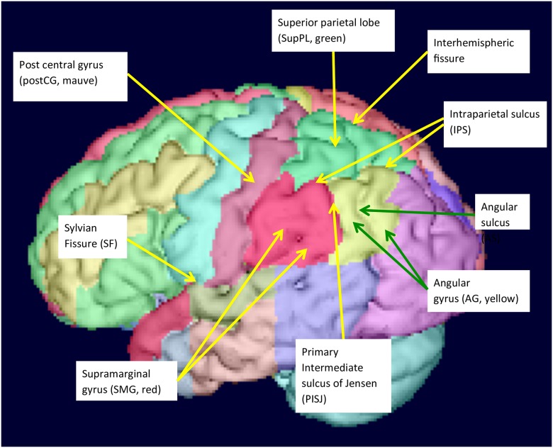
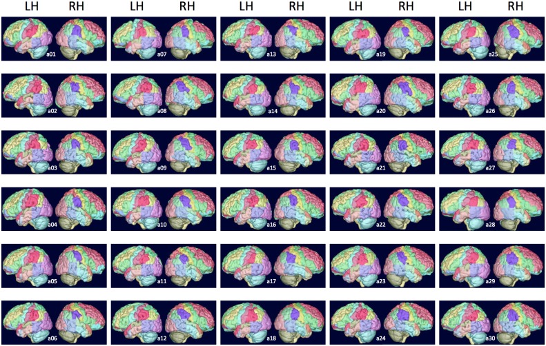
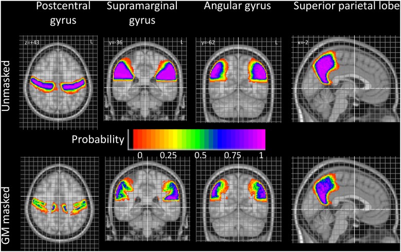
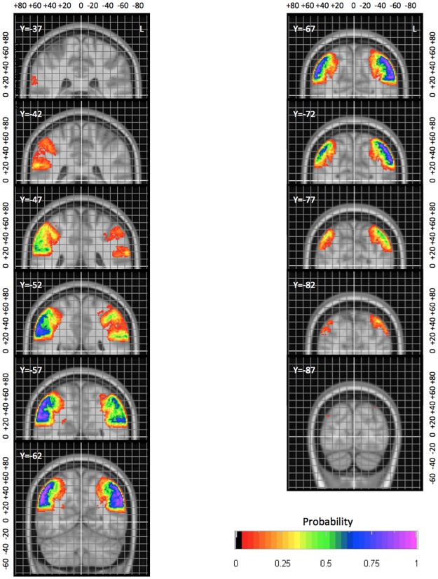
Accurately describing the anatomy of individual brains enables interlaboratory communication of functional and developmental studies and is crucial for possible surgical interventions. The human parietal lobe participates in multimodal sensory integration including language processing and also contains the primary somatosensory area. We describe detailed protocols to subdivide the parietal lobe, analyze morphological and volumetric characteristics, and create probabilistic atlases in MNI152 stereotaxic space. The parietal lobe was manually delineated on 3D T1 MR images of 30 healthy subjects and divided into four regions: supramarginal gyrus (SMG), angular gyrus (AG), superior parietal lobe (supPL) and postcentral gyrus (postCG). There was the expected correlation of male gender with larger brain and intracranial volume. We examined a wide range of anatomical features of the gyri and the sulci separating them. At least a rudimentary primary intermediate sulcus of Jensen (PISJ) separating SMG and AG was identified in nearly all (59/60) hemispheres. Presence of additional gyri in SMG and AG was related to sulcal features and volumetric characteristics. The parietal lobe was slightly (2%) larger on the left, driven by leftward asymmetries of the postCG and SMG. Intersubject variability was highest for SMG and AG, and lowest for postCG. Overall the morphological characteristics tended to be symmetrical, and volumes also tended to covary between hemispheres. This may reflect developmental as well as maturation factors. To assess the accuracy with which the labels can be used to segment newly acquired (unlabelled) T1-weighted brain images, we applied multi-atlas label propagation software (MAPER) in a leave-one-out experiment and compared the resulting automatic labels with the manually prepared ones. The results showed strong agreement (mean Jaccard index 0.69, corresponding to a mean Dice index of 0.82, average mean volume error of 0.6%). Stereotaxic probabilistic atlases of each subregion were obtained. They illustrate the physiological brain torque, with structures in the right hemisphere positioned more anteriorly than in the left, and right/left positional differences of up to 10 mm. They also allow an assessment of sulcal variability, e.g. low variability for parietooccipital fissure and cingulate sulcus. Illustrated protocols, individual label sets, probabilistic atlases, and a maximum-probability atlas which takes into account surrounding structures are available for free download under academic licences.
Conflict of interest statement
Figures
References
-
- Brodmann K. Vergleichende Lokalisationslehre der Großhirnrinde. Leipzig: Barth; 1909.
-
- Duvernoy HM. The Human Brain: Surface, Three-dimensional Sectional Anatomy with MRI, and Blood Supply., 2nd ed New York: Springer; 1999.
-
- Ono M, Kubik S, Abernathey CD. Atlas of the cerebral sulci. Stuttgart New York: Georg Thieme Verlag; 1990.
-
- Bajcsy R, Lieberson R, Reivich M. A computerized system for the elastic matching of deformed radiographic images to idealized atlas images. J Comput Assist Tomogr. 1983;7: 618–625. - PubMed
MeSH terms
Grants and funding
LinkOut - more resources
Full Text Sources
Other Literature Sources
