. 2017 Aug 28;13(8):e1006985.
doi: 10.1371/journal.pgen.1006985. eCollection 2017 Aug.

CTCF counter-regulates cardiomyocyte development and maturation programs in the embryonic heart

Affiliations

CTCF counter-regulates cardiomyocyte development and maturation programs in the embryonic heart

Melisa Gomez-Velazquez et al. PLoS Genet. .

Abstract

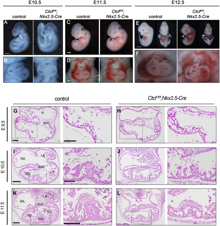
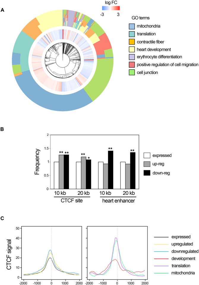
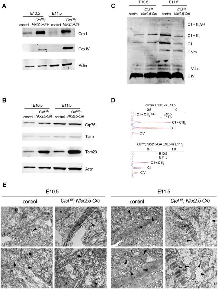
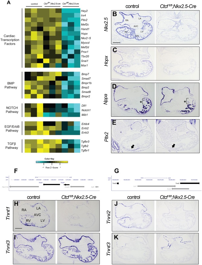
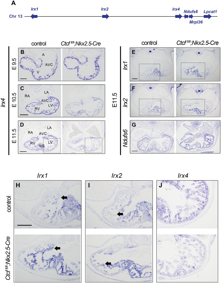
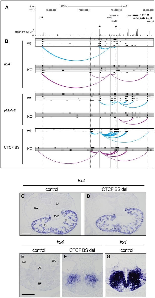
Cardiac progenitors are specified early in development and progressively differentiate and mature into fully functional cardiomyocytes. This process is controlled by an extensively studied transcriptional program. However, the regulatory events coordinating the progression of such program from development to maturation are largely unknown. Here, we show that the genome organizer CTCF is essential for cardiogenesis and that it mediates genomic interactions to coordinate cardiomyocyte differentiation and maturation in the developing heart. Inactivation of Ctcf in cardiac progenitor cells and their derivatives in vivo during development caused severe cardiac defects and death at embryonic day 12.5. Genome wide expression analysis in Ctcf mutant hearts revealed that genes controlling mitochondrial function and protein production, required for cardiomyocyte maturation, were upregulated. However, mitochondria from mutant cardiomyocytes do not mature properly. In contrast, multiple development regulatory genes near predicted heart enhancers, including genes in the IrxA cluster, were downregulated in Ctcf mutants, suggesting that CTCF promotes cardiomyocyte differentiation by facilitating enhancer-promoter interactions. Accordingly, loss of CTCF disrupts gene expression and chromatin interactions as shown by chromatin conformation capture followed by deep sequencing. Furthermore, CRISPR-mediated deletion of an intergenic CTCF site within the IrxA cluster alters gene expression in the developing heart. Thus, CTCF mediates local regulatory interactions to coordinate transcriptional programs controlling transitions in morphology and function during heart development.


Conflict of interest statement

The authors have declared that no competing interests exist.

Figures

Fig 1
Fig 1. Morphological defects in Ctcf mutant embryonic hearts.
Whole mount control and mutant (Ctcffl/fl;Nkx2-5-Cre) embryos at E10.5 (A, B), E11.5 (C, D) and E12.5 (E, F). Arrows in (E) point to the pericardial edema present in mutant embryos. Higher magnification of the heart show morphological defects in Ctcf mutant hearts becoming manifest at E12.5 (F). (G-L) Sections from control and mutant embryos stained with hematoxylin and eosin; higher magnifications (black dashed boxes) for each are shown on the right of each section. Disorganization of the interventricular septum and thinning of the myocardial wall is apparent in Ctcf mutant hearts at E11.5. A, atria; V, ventricle; RA, right atria; LA, left atria; RV, right ventricle; LV, left ventricle; AVC, atrioventricular canal; IVS, interventricular septum. Scale bars, 750 μm (A), 200 μm (B, D), 1 mm (C), 2 mm (E), 800 μm (F), 100 μm (G, H), 200 μm (I-L), and 100 μm in all higher magnifications.
Fig 2
Fig 2. Transcriptional changes in Ctcf mutant hearts.
(A) Iris plot showing enriched GO terms in differentially expressed genes between control and mutant E10.5 hearts (outside circle), together with changes in expression of individual genes (inner circle; red, upregulated in mutants; blue, downregulated). (B) Genes that are up- or downregulated in Ctcf mutant hearts are more likely to have a CTCF binding site in their vicinity (10 or 20 kb surrounding the transcriptional start site) than genes that are expressed in the embryonic heart but do not change. However, only downregulated genes are located closer to heart-specific enhancers. Frequency is expressed relative to that of expressed but unchanged genes. *, p<0.01; **, p<0.0005; Mann-Whitney test. (C) Mean-plots of the distribution of CTCF binding, as determined by ChIP-seq [10], relative to the TSS (0 on the x-axis) of different groups of genes (legend on the right). Note that the peak of the distributions is located slightly upstream of the TSS, indicative of binding to proximal promoter sequences.
Fig 3
Fig 3. Defective mitochondrial biogenesis upon CTCF loss.
Western blot of Cox I, Cox IV (A), Grp75, Tfam and Tom20 (B) of control and mutant (Ctcffl/fl;Nkx2-5-Cre) hearts at E10.5 and E11.5. (C) Blue native gel of control and mutant hearts at E10.5 and E11.5 showing abundance and distribution of mitochondrial respiratory complexes (CI, CIV, CVm) and supercomplexes (CI+CIII). (D) Densitometry of blue native gel quantifying changes between E10.5 and E11.5 in complexes and supercomplexes, in control and mutant hearts. (E) Transmission Electron Microscopy at E10.5 and E11.5 in control and mutant cardiomyocytes. n, nucleus; sm, sarcomere. Arrowheads point to mitochondria. Scale bar, 1 μm top row, 0.5 μm bottom row.
Fig 4
Fig 4. The cardiac developmental program is abrogated in Ctcf mutant hearts.
(A) Deletion of Ctcf leads to downregulation of genes encoding key cardiac developmental transcription factors and signaling-pathway components. The heatmap shows normalized gene expression values from the RNA-seq data for controls (Ctcffl/+), heterozygotes (Ctcffl/+;Nkx2.5-Cre) and homozygotes (Ctcffl/fl;Nkx2.5-Cre) across three biological replicates each. (B-E) In situ hybridization in sections of E10.5 control and mutant (Ctcffl/fl;Nkx2-5-Cre) hearts for Nkx2.5 (B), Hopx (C), which are strongly downregulated; and Nppa (D) and Pitx2 (E), which lose expression in the left ventricle (brackets and asterisks in D) or right ventricle (arrow in E), respectively. (F, G) Genomic region containing Tnnt1 and Tnni3 (F; mm9, chr7:4,433,080–4,493,651) or Tnni2 and Tnnt3 (G; mm9, chr7:149,623,608–149,703,181). Troponin genes are highlighted in black. (H-K) In situ hybridization in sections of E10.5 control and mutant hearts for Tnnt1 (H), Tnni3 (I), Tnni2 (J) and Tnnt3 (K). RA, right atria; LA, left atria; RV, right ventricle; LV, left ventricle; AVC, atrioventricular canal; IVS, interventricular septum. Scale bar, 200 μm.
Fig 5
Fig 5. Ctcf deletion causes global changes in the expression of the IrxA cluster in the developing heart.
(A) Diagram of the extended IrxA cluster showing the location of Irx1, Irx2, Irx4 and the neighboring genes Ndufs6, Mrpl36 and Lpcat1 (not to scale). (B-J) In situ hybridization in sections of control and mutant hearts for Irx4 at E9.5 (B), E10.5 (C) and E11.5 (D); and Irx1 (E), Irx2 (F) and Ndufs6 (G) at E11.5. (H-I) Higher magnifications (black dashed boxes) of Irx1 (H), Irx2 (I) and Irx4 (J) expression in control and mutant hearts at E11.5. Arrows indicate extension in the expression territories of Irx1 (H) and Irx2 (I). A,atria; V, ventricle, RA; right atria; LA, left atria; RV, right ventricle; LV, left ventricle; AVC, atrioventricular canal; IVS, interventricular septum. Scale bars, 100 μm (B), 200 μm (C-J).
Fig 6
Fig 6. CTCF is necessary for the correct chromatin organization of the Irx4-Ndufs6 locus.
(A) ENCODE data for CTCF binding in 8 weeks mouse heart (mm9 chr13:72700492–73802866). Asterisks indicate specific intergenic CTCF binding sites. (B) Chromatin interaction profiles as determined by 4C-seq from the Irx4 (top) and Ndufs6 (middle) promoters and the Irx2/Irx4 intergenic CTCF binding site (BS, bottom) in control (blue) and mutant (KO, purple) E11.5 hearts. Heat map of z-scores > 2 of replicates (grayscale) are shown on top of the interaction plots. (C-F) Irx4 in situ hybridization in E10.5 embryos from a transgenic mouse line where the Irx2/Irx4 intergenic CTCF BS (left asterisk in A) has been deleted. (C, D) Irx4 expression in the heart; (E, F) ectopic Irx4 expression in the oral-esophageal region of foregut. (G) Irx1 expression in the oral-esophageal region of foregut in a wild-type E10.5 embryo. RA, right atria; LA, left atria; RV, right ventricle; LV, left ventricle; AVC, atrioventricular canal; DA, dorsal aorta; TR, trachea; OE, oral-esophageal region. Scale bar, 200 μm (C, D), and 100 μm (E-G).

References

    1. Olson EN. Gene regulatory networks in the evolution and development of the heart. Science. 2006;313(5795):1922–7. doi: 10.1126/science.1132292 - DOI - PMC - PubMed
    1. Hirschy A, Schatzmann F, Ehler E, Perriard JC. Establishment of cardiac cytoarchitecture in the developing mouse heart. Dev Biol. 2006;289(2):430–41. doi: 10.1016/j.ydbio.2005.10.046 - DOI - PubMed
    1. Hom JR, Quintanilla RA, Hoffman DL, de Mesy Bentley KL, Molkentin JD, Sheu SS, et al. The permeability transition pore controls cardiac mitochondrial maturation and myocyte differentiation. Dev Cell. 2011;21(3):469–78. doi: 10.1016/j.devcel.2011.08.008 - DOI - PMC - PubMed
    1. Beutner G, Eliseev RA, Porter GA Jr. Initiation of electron transport chain activity in the embryonic heart coincides with the activation of mitochondrial complex 1 and the formation of supercomplexes. PLoS One. 2014;9(11):e113330 doi: 10.1371/journal.pone.0113330 - DOI - PMC - PubMed
    1. Soonpaa MH, Kim KK, Pajak L, Franklin M, Field LJ. Cardiomyocyte DNA synthesis and binucleation during murine development. Am J Physiol. 1996;271(5 Pt 2):H2183–9. - PubMed

MeSH terms