Review
. 2017 Oct:58:29-37.
doi: 10.1016/j.dnarep.2017.08.003. Epub 2017 Aug 18.

XLF/Cernunnos: An important but puzzling participant in the nonhomologous end joining DNA repair pathway

Affiliations
Review

XLF/Cernunnos: An important but puzzling participant in the nonhomologous end joining DNA repair pathway

Vijay Menon et al. DNA Repair (Amst). 2017 Oct.

Abstract

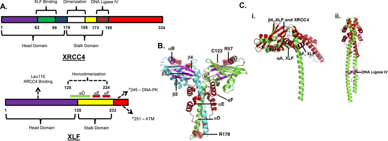
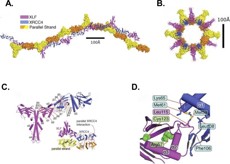
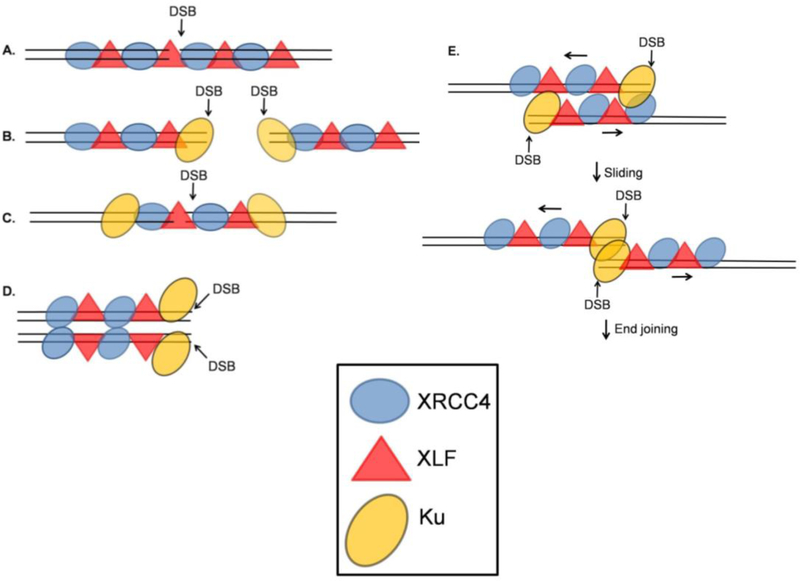
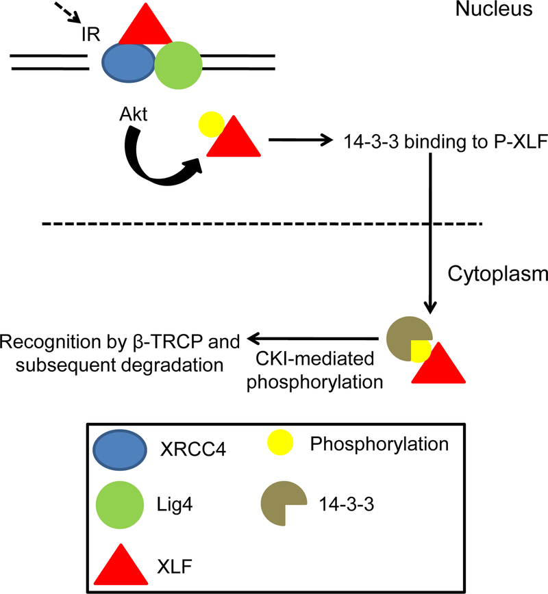
DNA double strand breaks (DSBs) are one of the most deleterious DNA lesions that promote cell death, genomic instability and carcinogenesis. The two major cellular mechanisms that repair DSBs are Nonhomologous End-Joining (NHEJ) and Homologous Recombination Repair (HRR). NHEJ is the predominant pathway, in which XLF (also called Cernunnos) is a key player. Patients with XLF mutation exhibit microcephaly, lymphopenia, and growth retardation, and are immunodeficient and radiosensitive. During NHEJ, XLF interacts with XRCC4-Ligase IV, stimulates its ligase activity, and forms DNA-binding filaments of alternating XLF and XRCC4 dimers that may serve to align broken DNA and promote ligation of noncomplementary ends. Despite its central role in NHEJ, the effects of XLF deficiency are surprisingly variable in different biological contexts, and different individual cell lines. This review summarizes the role of XLF in NHEJ, and the unexpected complexity of its interplay with other repair factors in supporting radiosurvival and V(D)J recombination.

Keywords: Nonhomologous End-Joining (NHEJ); V(D)J recombination; XLF/Cernunnos; XRCC4.


Figures

Figure 1
Figure 1
XLF structure and comparison with XRCC4. A. Domain structures of NHEJ proteins showing select binding sites. XRCC4: The head domain comprises amino acids 1–119 and the stalk domain 120–180 [16]. XLF: The head domain comprises amino acids 1–135 and the stalk domain 136–233. α-Helices D, E and F constituting the stalk are highlighted. The Leu115 residue is important for XRCC4 binding. DNA-PK phosphorylates Ser245 and ATM phosphorylates Ser251. Adapted from [13]. B. Structure of the XLF dimer as determined from crystallography of the 1–233 fragment. Cyan and green α-helices distinguish the two monomers. Segments that would be deleted as a result of a disease-associated nonsense mutation at R178 are shown in red. R57 and C123 are sites of disease-associated point mutations. The region spanning A25 to R57 is shown in magenta. (Image reproduced with permission from [17]). C. (i). Comparison of XLF (red) and XRCC4 (green) structures. α-Helices and antiparallel β-sheets are nearly superimposable in the head domains, but the stalk domains diverge. The angle between the head and stalk is larger in XLF than in XRCC4 because of the insertion of helices αF and αA. (ii). The coiled-coil in XLF is much shorter than that in XRCC4, and does not contain an equivalent region to the LIG4 binding site of XRCC4. LIG4 fragment bound to XRCC4 is shown in magenta. (Image reproduced with permission from [17].)
Figure 2
Figure 2
Structure of the XLF-XRCC4 filament as determined by crystallography. A. Two orthogonal views of the biological unit (one helical turn) of the filament formed by XLF(1–224) (magenta) and XRCC4(1–140) (blue), as seen in the crystal lattice. Parallel strands (yellow/orange) of a second filament are in surface representation. B. View of the biological unit rotated by 90°. C. Overall architecture of the XLF(1–224)-XRCC4(1–140) complex, colored magenta and blue, respectively. Helices α4-α6 correspond to helices αD-αF in Figure 1. Inset shows interaction between XRCC4 dimers in parallel filaments. D. Interface between head domains of XLF(1–224) (magenta) and XRCC4(1–140) (blue). Leu115 of XLF occupies a hydrophobic pocket formed by XRCC4 residues Leu108, Phe106, Met59, Met61, and Lys65. (Image reproduced with permission from [24].)
Figure 3
Figure 3
Different possible arrangements of XRCC4/XLF filaments with respect to DSBs. A. An XRCC4/XLF filament spans a DSB, aligning the DNA ends on either side of the break. This model does not clearly explain how DNA-PKcs and other processing enzymes gain access to the DNA DSB. B. XRCC4/XLF filaments form adjacent to terminally bound Ku. C. An XRCC4/XLF filament spans a DSB after translocation of Ku away from the DSB end. D. XRCC4/XLF filaments aligned side-by-side position two DNA molecules such that processing and ligation can occur at the exposed DSB ends. E. XRCC4/XLF filaments, each binding a DNA segment near a DSB, align side-by-side, then slide to bring the DSB ends into juxtaposition. Adapted from [45, 46, 47].
Figure 4
Figure 4
Akt-mediated phosphorylation and degradation of XLF. Phosphorylation at Thr181 leads to dissociation from the XRCC4-DNA Ligase IV complex. Phosphorylated XLF is then recognized by 14–3-3 protein and translocated to the cytoplasm where it could be further phosphorylated by CKI. It is then recognized by β-TRCP for ubiquitination and subsequent degradation. Adapted from [59].
Figure 5
Figure 5
Comparison of XRCC4 superfamily members. A. Phylogenetic tree of the three proteins, constructed using Clustal Omega. B. Superposition of the crystal structures of PAXX (cyan), XRCC4 (silver), XLF (magenta), and SAS6, a spindle assembly protein (green). (Image reproduced with permission from [73]).

Similar articles

Cited by

References

    1. Povirk LF DNA damage and mutagenesis by radiomimetic DNA-cleaving agents: Bleomycin, neocarzinostatin and other enediynes, Mutat. Res 355 (1996) 71–89. - PubMed
    1. Hutchinson F Chemical changes induced in DNA by ionizing radiation, Prog. Nucleic Acid Res. Mol. Biol 32 (1985) 115–154. - PubMed
    1. Cadet J, Ravanat JL, TavernaPorro M, Menoni H, Angelov D Oxidatively generated complex DNA damage: Tandem and clustered lesions, Cancer Lett 327 (2012) 5–15. - PubMed
    1. Valerie K, Povirk LF Regulation and mechanisms of mammalian double-strand break repair, Oncogene 22 (2003) 5792–5812. - PubMed
    1. Walker JR, Corpina RA, Goldberg J Structure of the Ku heterodimer bound to DNA and its implications for double-strand break repair, Nature 412 (2001) 607–614. - PubMed

Publication types

LinkOut - more resources