. 2017 Aug 31;548(7669):597-601.
doi: 10.1038/nature23670. Epub 2017 Aug 23.

Public antibodies to malaria antigens generated by two LAIR1 insertion modalities

Affiliations

Public antibodies to malaria antigens generated by two LAIR1 insertion modalities

Kathrin Pieper et al. Nature. .

Abstract

In two previously described donors, the extracellular domain of LAIR1, a collagen-binding inhibitory receptor encoded on chromosome 19 (ref. 1), was inserted between the V and DJ segments of an antibody. This insertion generated, through somatic mutations, broadly reactive antibodies against RIFINs, a type of variant antigen expressed on the surface of Plasmodium falciparum-infected erythrocytes. To investigate how frequently such antibodies are produced in response to malaria infection, we screened plasma from two large cohorts of individuals living in malaria-endemic regions. Here we report that 5-10% of malaria-exposed individuals, but none of the European blood donors tested, have high levels of LAIR1-containing antibodies that dominate the response to infected erythrocytes without conferring enhanced protection against febrile malaria. By analysing the antibody-producing B cell clones at the protein, cDNA and gDNA levels, we characterized additional LAIR1 insertions between the V and DJ segments and discovered a second insertion modality whereby the LAIR1 exon encoding the extracellular domain and flanking intronic sequences are inserted into the switch region. By exon shuffling, this mechanism leads to the production of bispecific antibodies in which the LAIR1 domain is precisely positioned at the elbow between the VH and CH1 domains. Additionally, in one donor the genomic DNA encoding the VH and CH1 domains was deleted, leading to the production of a camel-like LAIR1-containing antibody. Sequencing of the switch regions of memory B cells from European blood donors revealed frequent templated inserts originating from transcribed genes that, in rare cases, comprised exons with orientations and frames compatible with expression. These results reveal different modalities of LAIR1 insertion that lead to public and dominant antibodies against infected erythrocytes and suggest that insertion of templated DNA represents an additional mechanism of antibody diversification that can be selected in the immune response against pathogens and exploited for B cell engineering.


Conflict of interest statement

The authors declare competing financial interests. A.L. is the scientific founder and shareholder of Humabs BioMed. F.S. is a shareholder of Humabs BioMed.

Figures
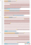
Extended Data Figure 1
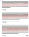
Extended Data Figure 1. Alignment of gDNA and cDNA sequences of LAIR1-containing antibodies.
Shown is one representative antibody from each donor. a, MGE9 (donor E), b, MGF21 (donor F), c, MGO3 (donor O), d, MGQ4 (donor Q).
Extended Data Figure 2
Extended Data Figure 2. Genomic sequences of switch regions containing LAIR1 inserts.
Shown is one representative antibody for each donor; a, MMJ5 (donor J), b, MGM5 (donor M), c, MGB47 (donor B). The chromosome coordinates of the insertion sites are indicated in blue and green.
Extended Data Figure 3
Extended Data Figure 3. Somatically mutated and conserved regions in the LAIR1 domains inserted in the VDJ or in the switch region.
a, Amino acid substitutions in antibodies isolated from different donors and mean R/S ratios at each residue. The mutational analysis takes into consideration the germline LAIR1 alleles found in each donor. In donor C, the P98L substitution is uncolored because it may arise from polymorphism, since the donor is heterozygous at this position. The number of nucleotide mutations and amino acid substitutions are reported in brackets next to the antibody names. b, Graphic representation of mutational hot spots (red) and of most conserved regions (blue).
Extended Data Figure 4
Extended Data Figure 4. Validation of switch region inserts combining Illumina and MinION technologies.
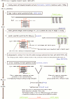
a, Illumina and MinION workflows. Switch regions of polyclonal naïve or IgG/IgA switched B cells were amplified by PCR. For Illumina sequencing, PCR amplicons were fragmented, re-amplified during library preparation and sequenced using the 2x300 bp MiSeq system. The bioinformatic analysis included the assembly of contiguous, chimeric reads. For insert confirmation, independently generated PCR-barcoded primary products were sequenced with MinION technology and analyzed with a different bioinformatic approach for long, error-prone MinION reads. b, Multiple identical switch inserts for donor 6 were confirmed in biological replicate experiments with independent technical and analytical setups. Shown are the experimental designs, shared and unique reads in a Venn diagram and an alignment of Illumina and MinION sequences covering the switch insertion sites for two examples (LCP1, RAVER1). c, Shared and unique switch inserts in technical and biological replicate experiments of donor 5.
Extended Data Figure 5
Extended Data Figure 5. Pipeline for data analysis using the Illumina platform.
Shown is the scheme of the bioinformatics workflow used for the analysis of Illumina sequences.
Extended Data Figure 6
Extended Data Figure 6. Pipeline for data analysis using the MinION technology.
Shown is the scheme of the bioinformatics workflow used for the analysis.
Extended Data Figure 7
Extended Data Figure 7. Examples of genes that donate multiple inserts.
Shown is the original position of the inserts donated by PAX5 and EBF1 as well as a list of genes that donated two or more inserts.
Extended Data Figure 8
Extended Data Figure 8. Examples of potentially functional inserts.
Shown is the alignment of the contig sequence and the genomic region from which the insert was derived, as well as the potential amino acid sequence inserted between the VH and CH1.
Extended Data Figure 9
Extended Data Figure 9. Relationship between LAIR1-IgG or LAIR1-IgM status with protection from febrile malaria.
Shown is the clinical status of 551 members of the Malian cohort, stratified by LAIR1-containing IgG (a) or LAIR1-containing IgM (b) status, over the years 2012 and 2013. Febrile malaria is defined as parasite density ≥2500 asexual parasites per µl of blood and an axillary temperature of ≥37.5°C.
Figure 1
Figure 1. Prevalence and dominance of LAIR1-containing antibodies in malaria-endemic regions.
a, b, Prevalence of LAIR1-containing IgG and IgM in African individuals living in malaria-endemic regions and in European blood donors. Donors from whom LAIR1-containing antibodies were isolated are named and highlighted in red. c, Comparison between LAIR1-containing IgG and IgM values. MFI, median fluorescence intensity. Data points with a delta MFI value below -2000 are not shown. d, Staining of IEs by LAIR1-containing IgG and conventional IgG from three representative donors. e, Dominance of LAIR1-containing B cell clones among memory B cells specific for IEs. Monoclonal antibodies isolated from immortalized memory B cells were classified based on their ability to bind to IEs and the presence of a LAIR1 insert. Bars show number of IgG or IgM monoclonal antibodies isolated from each donor. For gating strategy, see Supplementary Figure 1. nd = not determined.
Figure 2
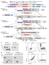
Figure 2. LAIR1-containing antibodies produced by two insertion modalities.
a-d, cDNA and gDNA organization in representative B cell clones of different donors. Donors E and J are Tanzanian, donor M is Malian and donor B is Kenyan. e, Western blot analysis of culture supernatants of B cell clones with LAIR1 insertion in the VDJ (MME2, donor E) or in the switch region (MMJ5, donor J) or a conventional antibody (MME17), (n=4). f, Surface staining of clone MGB47 showing LAIR1/IgG co-expression and lack of light chain (n=3). A positive control and a LAIR1-negative clone (MGB21) are shown for comparison. For gating strategy, see Supplementary Figure 1. g, Western blot analysis of culture supernatant of the camel-like clone MGB47 (n=4). Also shown are supernatants from clones MGO3 (LAIR1 insertion in VDJ), MGB21 (IgG3 control) and MGB4 (IgG1 control). For gel source data, see Supplementary Fig. 2.
Figure 3
Figure 3. The influence of insert position and somatic mutations on antibody specificity.
a, Schematic representation of LAIR1-containing antibodies produced by different insertion modalities. b, Scheme of the constructs (C1-C10) containing LAIR1 or other Ig-like domains in different positions which were tested for binding to a set of antigens or anti-domain antibodies by ELISA or FACS. The construct domains and their binding values to the cognate ligands are color coded as depicted in the figure. The V and J segments that do not contribute to any binding are not colored. BKC3, in grey, is a negative control. LAIR1D21, LAIR1M1 and LAIR1J5 are the exons from MGD21, MGM1 and MGJ5 antibodies. LAIR1gen is the unmutated genomic sequence of LAIR1 encoded on chromosome 19. MGDUCA = unmutated common ancestor of donor D antibodies; GL = germline. Data are from one experiment out of two. c, Binding of LAIR1-containing antibodies to erythrocytes infected with nine parasite isolates and to human collagen (n=1). Values refer to binding at a concentration of 1 μg ml-1. The MGDUCA was also tested at 100 μg ml-1. The number of amino acid substitutions is reported in brackets next to the antibody names.
Figure 4
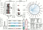
Figure 4. Frequent occurrence of templated inserts in the switch region.
a, Inserts in the switch region were detected by amplification and Illumina MiSeq sequencing (left) of 6 polyclonal samples of primary B cells from European donors and of the MGB47 monoclonal cell line as a control. Biological replicates were analyzed with MinION technology (right). Shown are the size distribution of all detected inserts and the estimated number of B cells that need to be analysed to detect one insert. For each technology, two independent experiments with 2-3 donors were performed. Red lines show mean values. b, Circos plot showing the origin of the inserts from different chromosomes. c, Insert frequencies in switch-µ regions of naïve B cells and in µ-γ joint regions of IgG memory B cells. d, Frequency of intergenic and different types of genic inserts. The red bar shows the number of exon-containing inserts with preserved splice sites in anti-sense and sense orientation. Inserts with the correct orientation and frame of the coding sequence (CDS) are highlighted in pink. e, EnrichR analysis on the Human Gene Atlas reveals significant enrichment for B cell genes. f, List of B cell-specific genes donating inserts.

Comment in

References

    1. Meyaard L. The inhibitory collagen receptor LAIR-1 (CD305) J Leukoc Biol. 2008;83:799–803. - PubMed
    1. Tan J, et al. A LAIR1 insertion generates broadly reactive antibodies against malaria variant antigens. Nature. 2016;529:105–109. - PMC - PubMed
    1. Mwakasungula S, et al. Red blood cell indices and prevalence of hemoglobinopathies and glucose 6 phosphate dehydrogenase deficiencies in male Tanzanian residents of Dar es Salaam. Int J Mol Epidemiol Gen. 2014;5:185–194. - PMC - PubMed
    1. Tran TM, et al. An intensive longitudinal cohort study of Malian children and adults reveals no evidence of acquired immunity to Plasmodium falciparum infection. Clin Infect Dis. 2013;57:40–47. - PMC - PubMed
    1. Muyldermans S. Nanobodies: natural single-domain antibodies. Annu Rev Biochem. 2013;82:775–797. - PubMed

Publication types

MeSH terms

Substances