

Optical Coherence Tomography Predictors of Risk for Progression to Non-Neovascular Atrophic Age-Related Macular Degeneration
- PMID: 28847641
- PMCID: PMC5768932
- DOI: 10.1016/j.ophtha.2017.06.032
Optical Coherence Tomography Predictors of Risk for Progression to Non-Neovascular Atrophic Age-Related Macular Degeneration
Abstract
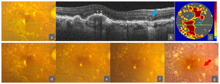
Purpose: Appearance of geographic atrophy (GA) on color photography (CP) is preceded by specific features on spectral-domain optical coherence tomography (SD OCT). We aimed to build SD OCT-based risk assessment models for 5-year new onset of GA and central GA on CP.
Design: Prospective, longitudinal study.
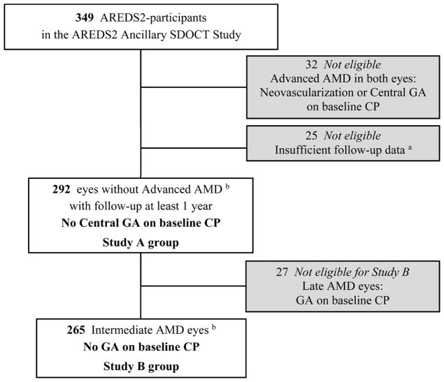
Participants: Age-Related Eye Disease Study 2 Ancillary SD OCT study participants with age-related macular degeneration (AMD) with bilateral large drusen or noncentral GA and at least 1 eye without advanced disease (n = 317).
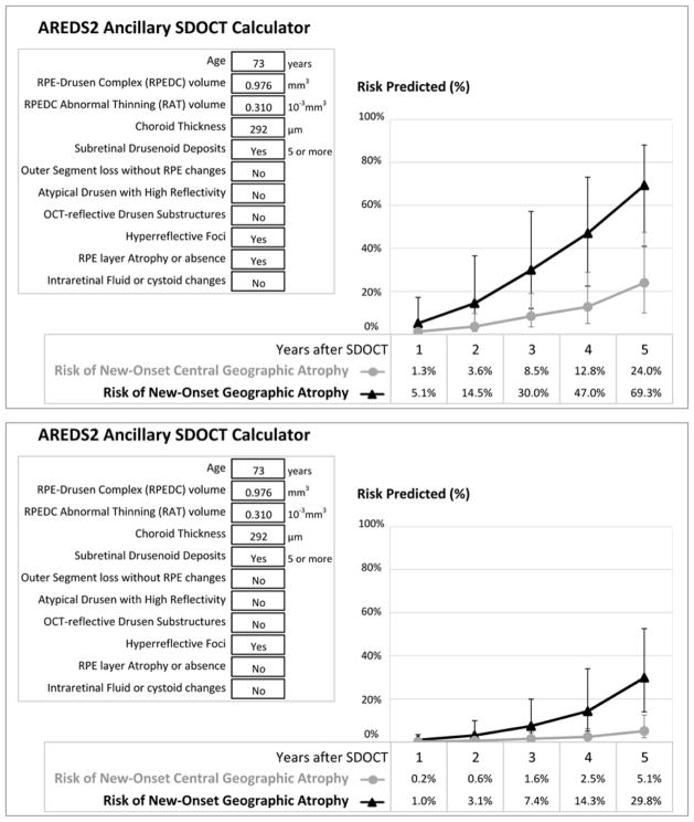
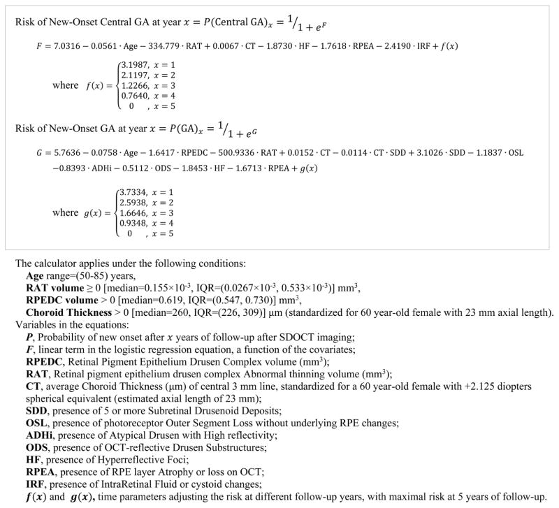
Methods: For 1 eye per participant, qualitative and quantitative SD OCT variables were derived from standardized grading and semiautomated segmentation, respectively, at baseline. Up to 7 years later, annual outcomes were extracted and analyzed to fit multivariate logistic regression models and build a risk calculator.
Main outcome measures: New onset of CP-visible GA and central GA.
Results: Over a follow-up median of 4.0 years and among 292 AMD eyes (without advanced disease at baseline) with complete outcome data, 46 (15.8%) developed central GA. Among 265 eyes without any GA on baseline CP, 70 (26.4%) developed CP-visible GA. Final multivariate models were adjusted for age. In the model for GA, the independent predicting SD OCT factors (P < 0.001-0.03) were: hyperreflective foci and retinal pigment epithelium (RPE) layer atrophy or absence, followed by choroid thickness in absence of subretinal drusenoid deposits, photoreceptor outer segment loss, RPE drusen complex volume, and RPE drusen complex abnormal thinning volume. For central GA, the factors (P < 0.001) were RPE drusen complex abnormal thinning volume, intraretinal fluid or cystoid spaces, hyperreflective foci, and RPE layer atrophy or absence. The models yielded a calculator that computes the probabilities of CP-visible, new-onset GA and central GA after 1 to 5 years.
Conclusions: For AMD eyes with large drusen and no advanced disease, we built a novel risk assessment model-based on age and SD OCT segmentation, drusen characteristics, and retinal pathology-for progression to CP-visible GA over up to 5 years. This calculator may simplify SD OCT grading and with future validation has a promising role as a clinical prognostic tool.
Trial registration: ClinicalTrials.gov NCT00734487.
Copyright © 2017 American Academy of Ophthalmology. All rights reserved.
Figures
References
-
- Klein R, Davis MD, Magli YL, Segal P, Klein BE, Hubbard L. The Wisconsin age-related maculopathy grading system. Ophthalmology. 1991;98(7):1128–1134. - PubMed
-
- The Age-Related Eye Disease Study Research G. The age-related eye disease study system for classifying age-related macular degeneration from stereoscopic color fundus photographs: the age-related eye disease study report number 6. American Journal of Ophthalmology. 2001;132(5):668–681. - PubMed
-
- Joachim N, Mitchell P, Burlutsky G, Kifley A, Wang JJ. The Incidence and Progression of Age-Related Macular Degeneration over 15 Years: The Blue Mountains Eye Study. Ophthalmology. 2015;122(12):2482–2489. - PubMed
Publication types
MeSH terms
Associated data
Grants and funding
LinkOut - more resources
Full Text Sources
Other Literature Sources
Medical
Molecular Biology Databases
Miscellaneous
