. 2017 Oct 18;85(11):e00475-17.
doi: 10.1128/IAI.00475-17. Print 2017 Nov.

Candida albicans and Pseudomonas aeruginosa Interact To Enhance Virulence of Mucosal Infection in Transparent Zebrafish

Affiliations

Candida albicans and Pseudomonas aeruginosa Interact To Enhance Virulence of Mucosal Infection in Transparent Zebrafish

Audrey C Bergeron et al. Infect Immun. .

Abstract

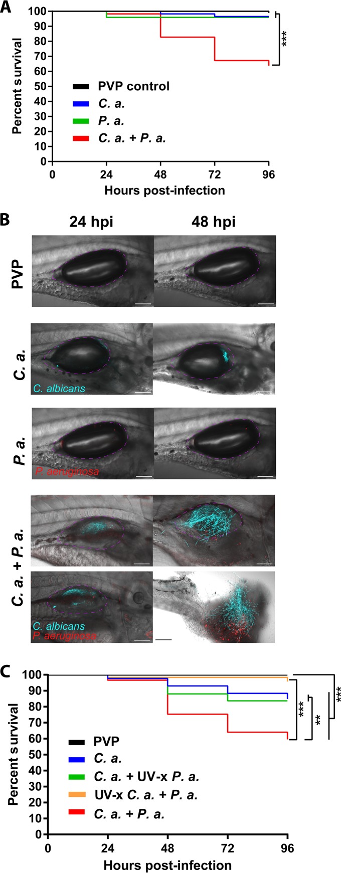
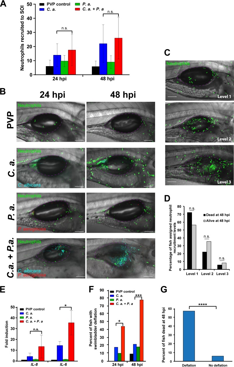
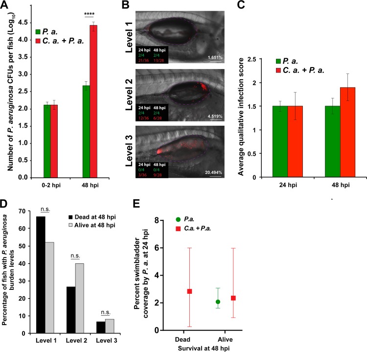
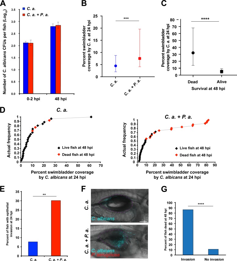
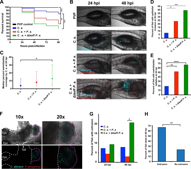
Polymicrobial infections often include both fungi and bacteria and can complicate patient treatment and resolution of infection. Cross-kingdom interactions among bacteria, fungi, and/or the immune system during infection can enhance or block virulence mechanisms and influence disease progression. The fungus Candida albicans and the bacterium Pseudomonas aeruginosa are coisolated in the context of polymicrobial infection at a variety of sites throughout the body, including mucosal tissues such as the lung. In vitro, C. albicans and P. aeruginosa have a bidirectional and largely antagonistic relationship. Their interactions in vivo remain poorly understood, specifically regarding host responses in mediating infection. In this study, we examine trikingdom interactions using a transparent juvenile zebrafish to model mucosal lung infection and show that C. albicans and P. aeruginosa are synergistically virulent. We find that high C. albicans burden, fungal epithelial invasion, swimbladder edema, and epithelial extrusion events serve as predictive factors for mortality in our infection model. Longitudinal analyses of fungal, bacterial, and immune dynamics during coinfection suggest that enhanced morbidity is associated with exacerbated C. albicans pathogenesis and elevated inflammation. The P. aeruginosa quorum-sensing-deficient ΔlasR mutant also enhances C. albicans pathogenicity in coinfection and induces extrusion of the swimbladder. Together, these observations suggest that C. albicans-P. aeruginosa cross talk in vivo can benefit both organisms to the detriment of the host.

Keywords: Candida albicans; Pseudomonas aeruginosa; infection; mucosal; polymicrobial; zebrafish.


Figures

FIG 1
FIG 1
C. albicans and P. aeruginosa demonstrate synergistic virulence in mucosal infection of the swimbladder. (A and B) Wild-type zebrafish larvae at 4 days postfertilization were separated into 4 groups and microinjected into the swimbladder with 5 nl of PVP (control), C. albicans (C. a.) at 2.5 × 10⁷ CFU/ml, P. aeruginosa (P. a.) at 2.5 × 10⁸ CFU/ml, or C. albicans and P. aeruginosa (C. a. + P. a.) at 2.5 × 10⁷ CFU/ml and 2.5 × 10⁸ CFU/ml, respectively. Fish were screened immediately postinjection to select for consistent inocula, and mortality was recorded every 24 h out to 96 h postinjection. Confocal images were acquired at ×10 and ×20 magnification, with scale bars at 100 μm and 200 μm. Data are representative of 4 pooled, independent experiments. Pooled numbers of individual fish are the following: n = 98, 58, 49, and 58 for PVP, C. albicans, P. aeruginosa, and C. albicans plus P. aeruginosa, respectively. A Kaplan-Meier survival analysis and log-rank (Mantel-Cox) test with Bonferroni correction demonstrated a significant reduction in survival. (C) Larvae were injected and monitored as described above with the following groups: PVP control, C. albicans (2.5 × 10⁷ CFU/ml) plus UV-inactivated (UV-x) P. aeruginosa (2.5 × 10⁸ CFU/ml), P. aeruginosa (2.5 × 10⁸ CFU/ml) plus UV-x C. albicans (2.5 × 10⁷ CFU/ml), or C. albicans plus P. aeruginosa at 2.5 × 10⁷ CFU/ml and 2.5 × 10⁸ CFU/ml, respectively. Data are representative of six pooled, independent experiments. Pooled numbers of individual fish are the following: n = 142, 86, 92, 33, and 96 for PVP, C. albicans, C. albicans plus UV-x P. aeruginosa, UV-x C. albicans plus P. aeruginosa, and C. albicans plus P. aeruginosa, respectively. A log-rank (Mantel-Cox) test with Bonferroni correction determined significant differences as indicated. Statistical significance was assigned based on GraphPad Prism conventions (not significant [n.s.], P > 0.05; *, P ≤ 0.05; **, P ≤ 0.01; ***, P ≤ 0.001; ****, P ≤ 0.0001; adjusted in panels A and C with Bonferroni correction). The swimbladder is outlined in a dotted magenta line for clarity.
FIG 2
FIG 2
Coinfection stimulates immune infiltration. Tg(Mpx:EGFP) zebrafish larvae at 4 days postfertilization were separated into 4 groups and microinjected into the swimbladder with 5 nl of PVP (control), C. albicans at 2.5 × 10⁷ CFU/ml, P. aeruginosa at 2.5 × 10⁸ CFU/ml, or C. albicans plus P. aeruginosa at 2.5 × 10⁷ CFU/ml and 2.5 × 10⁸ CFU/ml, respectively. Fish were screened immediately postinjection to select for neutrophil fluorescence and consistent inocula. (A and B) Neutrophils at the site of injection (SOI) were qualitatively scored and blinded, and confocal images of representative fish at ×20 magnification were taken at 24 and 48 h postinjection (hpi). Scale bar, 100 μm. Z-stack animations of the 24-hpi and 48-hpi images of a fish infected with C. albicans only are included in the supplemental material as Movies S1 (24 hpi) and S2 (48 hpi), and animations of a fish infected with both C. albicans and P. aeruginosa are included as Movies S3 (24 hpi) and S4 (48 hpi). Data are pooled from 3 independent experiments. Total numbers of individual fish are the following: n = 21, 23, 27, and 18 for PVP, C. albicans, P. aeruginosa, and C. albicans plus P. aeruginosa, respectively. Analysis was conducted using one-way analysis of variance (ANOVA) with Tukey's multiple-comparisons posttest. (C and D) Fish were imaged at 24 hpi and individuals were monitored to 48 hpi to measure survival. (C) Confocal images acquired at 24 hpi were used to stratify fish based on neutrophil recruitment (level 1, low; level 2, medium; level 3, high; as demonstrated by representative confocal images). (D) No significant differences in 24-hpi neutrophil recruitment phenotype were found between fish that survived or died using Fisher's exact test. (E) Representative fish were homogenized at 48 hpi for isolation of total RNA followed by cDNA synthesis for qPCR analysis. Gene expression of IL-6 and IL-8 was normalized to that of gapdh, with PVP control used for the reference (ΔΔCT). Fold induction (2ΔΔCT) is represented. Total RNA was extracted from 3 independent experiments; total numbers were 22, 24, 28, and 21 larvae for PVP, C. albicans, P. aeruginosa, and C. albicans plus P. aeruginosa groups, respectively. A one-way ANOVA with Tukey's multiple-comparisons posttest revealed significantly higher IL-6 expression in the coinfection than the C. albicans monoinfection (P = 0.011). qPCR for both genes was replicated in triplicate. (F) Percentage of fish with swimbladder deflation at 24 and 48 hpi. A Fisher's exact test revealed a significantly higher percentage of fish with swimbladder deflation in the coinfection compared to the C. albicans monoinfection, as indicated; n = 28 fish for C. albicans infection and n = 27 fish for coinfection. (G) Confocal images acquired at 24 hpi were stratified based on the survival of individual fish at 48 hpi relative to swimbladder deflation, with n = 28 with deflation and n = 34 without deflation. According to a Fisher's exact test, fish that died by 48 hpi had significantly higher incidences of swimbladder deflation than those that survived, as indicated. Statistical significance was assigned based on GraphPad Prism convention (n.s., P > 0.05; *, P ≤ 0.05; **, P ≤ 0.01; ***, P ≤ 0.001; ****, P ≤ 0.0001). The swimbladder is outlined in a dotted magenta line for clarity.
FIG 3
FIG 3
P. aeruginosa burden does not directly contribute to mortality. Tg(Mpx:EGFP) zebrafish larvae were infected and screened as previously described. Three to 4 representative fish per group were subsequently homogenized for quantifying CFU of P. aeruginosa by using selective media, with the remaining fish monitored out to 48 hpi. Representative fish were imaged at 24 and 48 hpi via confocal microscopy. (A) Data are representative of 6 pooled, independent experiments. A total of 22 individual fish were homogenized per time point (0 hpi and 48 hpi) for CFU quantification and plotted on a log10 scale. Student's t test demonstrated a significant increase in the number of P. aeruginosa organisms between mono- and coinfection groups at 48 hpi, as indicated. (B, C, and D) Confocal images acquired at 24 hpi were blinded and qualitatively scored based on Pseudomonas burden (level 1, low; level 2, medium; level 3, high), and then individuals were monitored to determine their survival at 48 hpi. Total numbers of fish analyzed were 15 dead and 25 alive at 48 hpi. (B) Representative images of each level of burden. Fractions in the lower left corner indicate the number of fish of a given phenotype that were scored at a given time point postinfection (green, P. aeruginosa monoinfection; red, P. aeruginosa plus C. albicans). Percentage shown at the lower right is from ImageJ quantification of burden, which correlates well with blinded qualitative scoring. (C) No significant differences in average infection level were observed between mono- and coinfected fish based on qualitative scoring of Pseudomonas burden according to Student's t test. (D) No significant differences were found in the percentages of fish with different qualitatively scored levels of P. aeruginosa burden between mono- and coinfected fish, as tested by Fisher's exact test. (E) Percent swimbladder coverage by P. aeruginosa was quantified via ImageJ analysis of microbial fluorescence from confocal images acquired for representative fish at 24 hpi. The calculated percent coverage from this analysis is also shown in panel B. For fish that died at 48 hpi, n = 11 for C. albicans plus P. aeruginosa; for fish that lived at 48 hpi, n = 4 for P. aeruginosa and n = 22 for C. albicans plus P. aeruginosa. No differences were observed using an unpaired Mann-Whitney test. Confocal images were acquired at ×20 magnification. Scale bar, 100 μm. Statistical significance was assigned based on GraphPad Prism convention (n.s., P > 0.05; *, P ≤ 0.05; **, P ≤ 0.01; ***, P ≤ 0.001; ****, P ≤ 0.0001). The swimbladder is outlined in a dotted magenta line for clarity.
FIG 4
FIG 4
Coinfection enhances C. albicans pathogenicity. Tg(Mpx:EGFP) zebrafish larvae infected and screened as previously described and 3 to 4 random fish per group were subsequently homogenized for quantifying CFU of C. albicans by using selective media, with the remaining fish monitored out to 48 hpi. Random fish were imaged at 24 and 48 hpi via confocal microscopy, and another 3 to 4 fish per group at 48 hpi were homogenized to quantify CFU as described above. Data are representative of 6 to 8 pooled, independent experiments. (A) A total of 22 individual fish were homogenized per time point for CFU quantification and plotted on a log10 scale. There was no significant difference in C. albicans CFU between single infection and coinfection according to Student's t test. (B) Median percent coverage of the swimbladder by C. albicans was quantified via ImageJ analysis of confocal images acquired of representative fish at 24 hpi, with n = 55 and 63 for C. albicans and C. albicans plus P. aeruginosa infections, respectively. An unpaired Mann-Whitney test revealed a significant difference between C. albicans single infection and coinfection, as indicated. (C) Confocal images acquired at 24 hpi were stratified based on the survival of individual fish at 48 hpi relative to the median percent swimbladder coverage by C. albicans, with n = 18 dead and n = 44 alive. Scale bar, 100 μm. According to an unpaired Mann-Whitney test, fish that died by 48 hpi had significantly higher swimbladder coverage by C. albicans than those that survived, as indicated. (D) Cumulative frequency distribution plots representing percent swimbladder coverage by C. albicans at 24 hpi. Fish that died at 48 hpi are indicated by red points; for C. albicans, n = 26; for C. albicans plus P. aeruginosa, n = 36. (E) Percentage of fish with epithelial invasion at 24 hpi; for C. albicans, n = 52; for C. albicans plus P. aeruginosa, n = 63. According to a Fisher's exact test, there was a significant difference between C. albicans and C. albicans plus P. aeruginosa infection, as indicated. (F and G) Confocal images acquired at 24 hpi were quantified by ImageJ and stratified based on the survival of individual fish at 48 hpi relative to epithelial invasion by C. albicans, with n = 15 with invasion and n = 45 without invasion. According to a Fisher's exact test, fish that died by 48 hpi had significantly higher incidences of C. albicans invasion than those that survived, as indicated. Statistical significance was assigned based on GraphPad Prism conventions (n.s., P > 0.05; *, P ≤ 0.05; **, P ≤ 0.01; ***, P ≤ 0.001; ****, P ≤ 0.0001). The swimbladder is outlined in a dotted magenta line for clarity.
FIG 5
FIG 5
P. aeruginosa quorum-sensing-deficient ΔlasR mutant enhances C. albicans pathogenicity in coinfection. Tg(Mpx:EGFP) zebrafish larvae at 4 days postfertilization were separated into four groups and microinjected into the swimbladder with 5 nl of PVP (control), C. albicans at 2.5 × 10⁷ CFU/ml, C. albicans plus P. aeruginosa at 2.5 × 10⁷ CFU/ml and 2.5 × 10⁸ CFU/ml, respectively, or C. albicans plus ΔlasR mutant P. aeruginosa at 2.5 × 10⁷ CFU/ml and 2.5 × 10⁸ CFU/ml, respectively. Fish were screened immediately postinjection to select for neutrophil fluorescence and consistent inocula. Mortality was recorded every 24 h out to 96 hpi, and representative fish were imaged at 24 and 48 hpi via confocal microscopy. Data are representative of 5 to 8 pooled, independent experiments. (A and B) Kaplan-Meier survival analysis and representative images. Pooled numbers of individual fish are the following: n = 96, 61, 71, and 66 for PVP, C. albicans, C. albicans plus P. aeruginosa, and C. albicans plus ΔlasR mutant P. aeruginosa, respectively. A log-rank (Mantel-Cox) test with Bonferroni correction demonstrated a significant reduction in survival between C. albicans and C. albicans plus P. aeruginosa and between C. albicans and C. albicans plus ΔlasR mutant P. aeruginosa, as indicated. Images were acquired at ×20 magnification; scale bar, 100 μm. (C) Median percent coverage of the swimbladder by C. albicans was quantified via ImageJ analysis of confocal images acquired of representative fish at 24 hpi, with n = 41, 44, and 44 for C. albicans, C. albicans plus P. aeruginosa, and C. albicans plus ΔlasR mutant P. aeruginosa, respectively. According to a Kruskal-Wallis test, there was a significant difference between C. albicans and C. albicans plus ΔlasR mutant P. aeruginosa infections, as indicated. (D) Percentage of fish with epithelial invasion at 24 hpi: C. albicans, n = 41; C. albicans plus P. aeruginosa, n = 46; C. albicans plus ΔlasR mutant P. aeruginosa, n = 43. According to a Fisher's exact test, there was a significant difference between C. albicans and C. albicans plus P. aeruginosa infections and between C. albicans and C. albicans plus ΔlasR mutant P. aeruginosa, as indicated. (E) Percentage of fish with swimbladder deflation at 24 hpi. According to a Fisher's exact test, there was a significantly higher percentage of fish with swimbladder deflation in the coinfection compared to the C. albicans monoinfection and a significant difference between C. albicans and C. albicans plus ΔlasR mutant P. aeruginosa, as indicated. The numbers of fish were 41, 46, and 43 for C. albicans, C. albicans plus P. aeruginosa, and C. albicans plus ΔlasR mutant P. aeruginosa, respectively. (F and G) Extrusion events were quantified in confocal images. (F) Representative low- and high-power images of an extrusion event. The fish is outlined in black/white, and the extruded infected tissue is outlined in pink. Scale bars are 200 μm for the 10× images (left) and 100 μm for the ×20 images (right) (G) According to a Fisher's exact analysis, extrusion events at 48 hpi are of significantly higher frequency in C. albicans plus ΔlasR mutant P. aeruginosa than in C. albicans infection, as indicated; for 24 hpi, n = 41, 46, and 43 for C. albicans, C. albicans plus P. aeruginosa, and C. albicans plus ΔlasR mutant P. aeruginosa, respectively; for 48 hpi, n = 38, 34, and 29 for C. albicans, C. albicans plus P. aeruginosa, and C. albicans plus ΔlasR mutant P. aeruginosa, respectively. (H) Confocal images acquired at 24 hpi were quantified by ImageJ and stratified based on the survival of individual fish at 48 hpi relative to extrusion events; n = 9 with extrusion and n = 121 without extrusion. According to a Fisher's exact test, fish that died by 48 hpi had significantly higher incidence of extrusion events than those that survived, as indicated. Statistical significance was assigned based on GraphPad Prism conventions (n.s., P > 0.05; *, P ≤ 0.05; **, P ≤ 0.01; ***, P ≤ 0.001; ****, P ≤ 0.0001; adjusted with Bonferroni correction for panel A). The swimbladder is outlined throughout in a dotted magenta line for clarity.

References

    1. Peleg AY, Hogan DA, Mylonakis E. 2010. Medically important bacterial-fungal interactions. Nat Rev Microbiol 8:340–349. doi:10.1038/nrmicro2313. - DOI - PubMed
    1. Leclair LW, Hogan DA. 2010. Mixed bacterial-fungal infections in the CF respiratory tract. Med Mycol 48:S125–S132. doi:10.3109/13693786.2010.521522. - DOI - PubMed
    1. Schwarz C, Hartl D, Eickmeier O, Hector A, Benden C, Durieu I, Sole A, Gartner S, Milla CE, Barry PJ. 31 July 2017. Progress in definition, prevention and treatment of fungal infections in cystic fibrosis. Mycopathologia doi:10.1007/s11046-017-0182-0. - DOI - PubMed
    1. Kim SH, Clark ST, Surendra A, Copeland JK, Wang PW, Ammar R, Collins C, Tullis DE, Nislow C, Hwang DM, Guttman DS, Cowen LE. 2015. Global analysis of the fungal microbiome in cystic fibrosis patients reveals loss of function of the transcriptional repressor Nrg1 as a mechanism of pathogen adaptation. PLoS Pathog 11:e1005308. doi:10.1371/journal.ppat.1005308. - DOI - PMC - PubMed
    1. Valenza G, Tappe D, Turnwald D, Frosch M, König C, Hebestreit H, Abele-Horn M. 2008. Prevalence and antimicrobial susceptibility of microorganisms isolated from sputa of patients with cystic fibrosis. J Cystic Fibrosis 7:123–127. doi:10.1016/j.jcf.2007.06.006. - DOI - PubMed