Review
. 2018 Oct 1;10(10):a028431.
doi: 10.1101/cshperspect.a028431.

The Tumor Necrosis Factor Family: Family Conventions and Private Idiosyncrasies

Affiliations
Review

The Tumor Necrosis Factor Family: Family Conventions and Private Idiosyncrasies

David Wallach. Cold Spring Harb Perspect Biol. .

Abstract

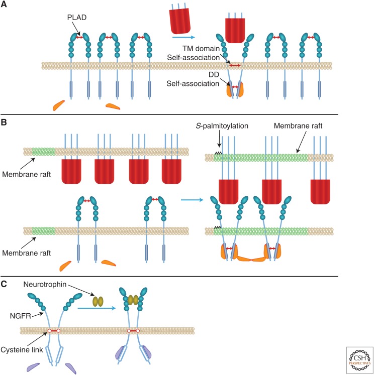
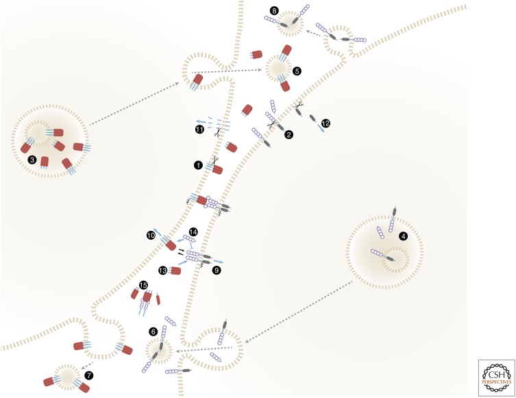
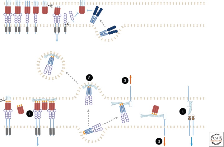
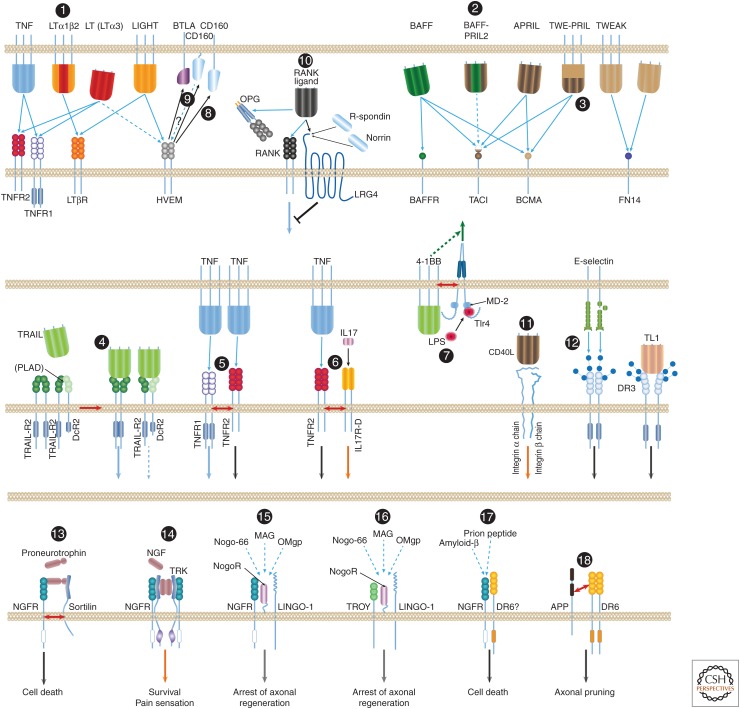
The tumor necrosis factor (TNF) cytokine family and the TNF/nerve growth factor (NGF) family of their cognate receptors together control numerous immune functions, as well as tissue-homeostatic and embryonic-development processes. These diverse functions are dictated by both shared and distinct features of family members, and by interactions of some members with nonfamily ligands and coreceptors. The spectra of their activities are further expanded by the occurrence of the ligands and receptors in both membrane-anchored and soluble forms, by "re-anchoring" of soluble forms to extracellular matrix components, and by signaling initiation via intracellular domains (IDs) of both receptors and ligands. Much has been learned about shared features of the receptors as well as of the ligands; however, we still have only limited knowledge of the mechanistic basis for their functional heterogeneity and for the differences between their functions and those of similarly acting cytokines of other families.


Figures

Figure 1.
Figure 1.
Signaling triggering: proposed mechanisms. (A) Triggering by intracellular domain (ID) juxtaposition imposed by tumor necrosis factor (TNF) family ligands, and its restriction by the pre-ligand assembly domain (PLAD). (B) Triggering potentiation by anchorage of ligands to membranes. (C) Triggering by ID distancing: A fraction of the nerve growth factor receptor (NGFR) molecules occurs as dimers of receptor molecules that are covalently linked through a conserved transmembrane (TM) cysteine residue. In these dimers, the intracellular death domains (DDs) seem to be constitutively associated. Neurotrophin-induced triggering of these dimers to signal for death was suggested to occur by distancing of the IDs, imposed rather like a snail tongue, with the TM cysteine link serving as a fulcrum (Vilar et al. 2009). (See discussion of these findings in the last paragraph of this review.)
Figure 2.
Figure 2.
Membrane-anchored and soluble forms of ligands and receptors of the tumor necrosis factor (TNF) families, and their functional roles. The figure shows an intriguing symmetry of the spectra of soluble and membrane-anchored forms attained by TNF family ligands and TNF/nerve growth factor (NGF) family receptors, and of the functions of these forms. Both ligands (1) and receptors (2) are proteolytically cleaved to yield soluble forms (Kriegler et al. 1988; Engelmann et al. 1990; Nophar et al. 1990; Black et al. 1997; Moss et al. 1997). The cleavage occurs constitutively or inducibly, either on the cell surface as illustrated (Black et al. 1997; Moss et al. 1997; Becker-Pauly and Rose-John 2013) or within the cell (Lopez-Fraga et al. 2001). (3,4) Some of the ligands (Gordon and Galli 1990; Bossi and Griffiths 1999; Koguchi et al. 2007) and receptors (Wang et al. 2003) accumulate within intracellular vesicles from which they are secreted in response to specific stimuli, thus supplementing either the cell-surface-expressed or the soluble pools. Some are released while anchored to membranes that might correspond either to exosomes that have accumulated in intracellular multivesicular bodies (5,6) or to microvesicles exfoliating from the cell surface (7,8) (Albanese et al. 1998; Martinez-Lorenzo et al. 1999; Islam et al. 2007). Binding of membrane-anchored ligands of the TNF family to their receptors, besides triggering receptor signaling (9), also triggers “reverse signaling” by the ligand molecules (10) (Stuber et al. 1995; Arens et al. 2004; Eissner et al. 2004; Grohmann et al. 2007; Kang et al. 2007; Sun et al. 2007; Juhasz et al. 2013). One mechanism contributing to this reverse signaling is intramembrane proteolytic cleavage, yielding ligand intracellular domain (ID) fragments that apparently mediate signaling following their translocation to the nucleus (Domonkos et al. 2001; Fluhrer et al. 2006; Friedmann et al. 2006; Kirkin et al. 2007) (11). Intramembrane cleavage that apparently contributes to signaling has also been reported for two receptors of the TNF/NGF family, nerve growth factor receptor (NGFR) and TNFR1 (Kanning et al. 2003; Kenchappa et al. 2006; Chhibber-Goel et al. 2016) (12). Accumulation of the soluble forms of both receptors and ligands interferes with the binding of membrane-bound forms to the ligands and the receptors, respectively, and can thus block signaling (13,14). To the extent that the soluble forms are incapable of triggering signaling, they may also interfere with signaling activation by their membrane-bound forms. However, the association of soluble receptors with soluble ligands also stabilizes the trimeric structures of the soluble ligands, so that they function not as mere inhibitors but rather as buffering agents. While decreasing the intensity of signaling activation, they also extend its duration (15) (Aderka et al. 1992; Eliaz et al. 1996).
Figure 3.
Figure 3.
“Re-anchoring” of soluble forms of ligands and receptors by their binding to extracellular matrix components. Various reported effects of extracellular matrix components on the functions of soluble ligands and receptors, shown in the lower part of the figure, are compared with the functions of soluble ligands and receptors in the absence of such components, as shown in the upper part. See text for details and for numbering of the illustrated effects. Undulating lines correspond to extracellular matrix components. Orange forms correspond to structural motifs that dictate binding to such components.
Figure 4.
Figure 4.
Noncanonical associations of the tumor necrosis factor (TNF) ligand and the TNF/ nerve growth factor receptor (NGFR) families. Most of the interactions of ligands and receptors of the TNF ligand and TNF/NGFR families are known to occur between homotrimeric molecules of a particular ligand and molecules of a particular receptor. A few exceptions, however, are known: (1) Lymphotoxin β (LTβ) functions only within heterotrimers that it forms with lymphotoxin α (LTα). Whereas homotrimers of LT bind to the TNF receptors, the LTα1β2 heterotrimer binds to the LTβ receptor (LTβR) (Ware 2005). (2) B-cell-activating factor of the tumor necrosis factor family (BAFF) and a proliferation-inducing ligand (APRIL) also form heterotrimers (so far discerned only in patients with autoimmune diseases), and these heterotrimers apparently activate only transmembrane activator and calcium-modulating cyclophilin ligand interactor (TACI) (Roschke et al. 2002; Dillon et al. 2006; Schuepbach-Mallepell et al. 2015). (3) Although APRIL is constitutively shed and its trimers therefore occur only in soluble form, its gene also yields a fused joint splice variant with the neighboring TWEAK gene. The protein encoded by this transcript—TWE-PRIL—is anchored to the cell membrane (Pradet-Balade et al. 2002). (4) Association of molecules of TRAIL-R2 and the membrane-anchored truncated receptor DcR2 through their preligand assembly domain (PLAD) dictate association of TRAIL with the two receptors in mixed complexes wherein TRAIL-R2 signaling is suppressed by DcR2 (Clancy et al. 2005). (5) Molecules of the two receptors for TNF (TNFR1 and TNFR2) reportedly also associate on their binding to ligand. However, this association does not occur through binding of the two receptors to the same ligand molecule. In fact, the two receptor species are incapable of binding simultaneously to the same TNF molecule; molecules of the two receptors rather associate only after binding independently to distinct TNF molecules (Pinckard et al. 1997). (6) Molecules of TNFR2 reportedly also associate with molecules of IL-17 receptor D, a member of an unrelated cytokine-receptor family. This association, which occurs on triggering by the ligands of the two receptors, imposes assembly of aggregates of the two receptors and functional cooperation between them (Yang et al. 2015). (7) Another example of functional interaction with a structurally unrelated receptor is the association of the ligand for 4-1BB with a coexpressed TLR4–MD2 complex. This apparently occurs through the TMs of 4-1BBL and Tlr4 and the consequent potentiation of lipopolysaccharide (LPS)-induced Tlr4 signaling in a way that depends on the function of the intracellular domain (ID) of 4-1BBL but independently of the association of 4-1BBL with 4-1BB (Kang et al. 2007; Ma et al. 2013). (8) A different kind of noncanonical association is observed in the function of herpesvirus entry mediator (HVEM), a receptor of the TNF family. Besides its association with two TNF family ligands (with LIGHT, and [weakly] with LT homotrimers) it also binds to B- and T-lymphocyte attenuator (BTLA) and CD160, two cell-surface proteins of the immunoglobulin superfamily, thereby triggering inhibitory signaling by those two proteins (Shui et al. 2011). (9) Whether this association also triggers signaling by the ID of HVEM is not known. (10) RANKL, besides binding to RANK and to the soluble receptor osteoprotegerin (OPG), also binds to the seven-transmembrane (TM), leucine-rich repeat containing G protein-coupled receptor 4 (LGR4), which also serves as receptor for R-spondins and for Norrin. It thus triggers signaling antagonistic to that initiated by RANK (Luo et al. 2016). (11) Binding of soluble CD40L to integrin αIIbβ3 and to integrin α5β1 triggers signaling by those two membrane–protein complexes (Andre et al. 2002; Leveille et al. 2007). (12) Binding of the cell-adhesion lectin, E-selectin, to DR3-linked sialic-acid-linked sugar chains triggers signaling by DR3 (Porquet et al. 2011). Juxtaposition and activation of the two TRAIL receptors, DR4 and DR4, by TRAIL also depends, for a reason not yet clear, on glycosylation of these receptors (Wagner et al. 2007). The most elaborate known set of noncanonical associations is observed in the function of the NGFR, a receptor of the TNF/NGF family for which no ligand of the TNF family is known. NGFR contributes to signaling for different effects in response to different inducers through association with a series of different coreceptors. (13) It contributes to signaling for death in response to proneurotrophins, to which it binds in association with a sortilin family receptor. (14) When associating with a Trk tyrosine kinase receptor, apparently through the TMs and IDs of both receptors (Esposito et al. 2001), NGFR contributes to high-affinity binding of NGF, and signals for cell survival and for pain sensation. (15) NGFR is also found to form, through extracellular domain (ED) associations, a ternary complex with the glycosylphosphatidylinositol (GPI)-linked Nogo-66 receptor (NogoR) and the TM receptor LINGO-1. In response to myelin-associated inhibitory factors (a 66-amino-acid fragment of the oligodendrocyte-derived growth inhibitory protein Nogo, the oligodendrocyte myelin glycoprotein [OMgp] or the myelin-associated glycoprotein [MAG]), this complex signals for arrest of axonal regeneration following injury (Hempstead 2002; Chao 2003; Ibanez and Simi 2012). (16) In that complex, NGFR can be replaced by the orphan TNF family receptor TROY (Shao et al. 2005). (17) Finally, direct binding of amyloid-β to NGFR (Hempstead 2002; Chao 2003; Ibanez and Simi 2012), and probably also to a complex of NGFR and another orphan receptor of the TNF/NGF family, DR6 (Hu et al. 2013), reportedly triggers signaling for neuronal death. (18) DR6 was found to bind to a carboxyl-terminal region in the ED of the amyloid precursor protein (APP). APP and DR6 cooperate in the induction of axonal pruning. The mechanism underlying this cooperation is not clear, nor is it known whether the cooperation occurs between proteins expressed in the same cell, as illustrated in the figure, or in distinct cells (Olsen et al. 2014). BCMA, B-cell maturation antigen.

References

    1. Adam Klages S, Adam D, Wiegmann K, Struve S, Kolanus W, Schneider Mergener J, Kronke M. 1996. FAN, a novel WD-repeat protein, couples the p55 TNF-receptor to neutral sphingomyelinase. Cell 86: 937–947. - PubMed
    1. Aderka D, Engelmann H, Maor Y, Brakebusch C, Wallach D. 1992. Stabilization of the bioactivity of tumor necrosis factor by its soluble receptors. J Exp Med 175: 323–329. - PMC - PubMed
    1. Akassoglou K, Bauer J, Kassiotis G, Lassmann H, Kollias G, Probert L. 1999. Transgenic models of TNF induced demyelination. Adv Exp Med Biol 468: 245–259. - PubMed
    1. Albanese J, Meterissian S, Kontogiannea M, Dubreuil C, Hand A, Sorba S, Dainiak N. 1998. Biologically active Fas antigen and its cognate ligand are expressed on plasma membrane-derived extracellular vesicles. Blood 91: 3862–3874. - PubMed
    1. Alon R, Cahalon L, Hershkoviz R, Elbaz D, Reizis B, Wallach D, Akiyama SK, Yamada KM, Lider O. 1994. TNF-α binds to the N-terminal domain of fibronectin and augments the β1-integrin-mediated adhesion of CD4+ T lymphocytes to the glycoprotein. J Immunol 152: 1304–1313. - PubMed

MeSH terms

Substances

LinkOut - more resources