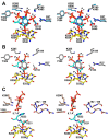
Bacterial PhyA protein-tyrosine phosphatase-like myo-inositol phosphatases in complex with the Ins(1,3,4,5)P4 and Ins(1,4,5)P3 second messengers
- PMID: 28848052
- PMCID: PMC5655508
- DOI: 10.1074/jbc.M117.787853
Bacterial PhyA protein-tyrosine phosphatase-like myo-inositol phosphatases in complex with the Ins(1,3,4,5)P4 and Ins(1,4,5)P3 second messengers
Abstract
myo-Inositol phosphates (IPs) are important bioactive molecules that have multiple activities within eukaryotic cells, including well-known roles as second messengers and cofactors that help regulate diverse biochemical processes such as transcription and hormone receptor activity. Despite the typical absence of IPs in prokaryotes, many of these organisms express IPases (or phytases) that dephosphorylate IPs. Functionally, these enzymes participate in phosphate-scavenging pathways and in plant pathogenesis. Here, we determined the X-ray crystallographic structures of two catalytically inactive mutants of protein-tyrosine phosphatase-like myo-inositol phosphatases (PTPLPs) from the non-pathogenic bacteria Selenomonas ruminantium (PhyAsr) and Mitsuokella multacida (PhyAmm) in complex with the known eukaryotic second messengers Ins(1,3,4,5)P4 and Ins(1,4,5)P3 Both enzymes bound these less-phosphorylated IPs in a catalytically competent manner, suggesting that IP hydrolysis has a role in plant pathogenesis. The less-phosphorylated IP binding differed in both the myo-inositol ring position and orientation when compared with a previously determined complex structure in the presence of myo-inositol-1,2,3,4,5,6-hexakisphosphate (InsP6 or phytate). Further, we have demonstrated that PhyAsr and PhyAmm have different specificities for Ins(1,2,4,5,6)P5, have identified structural features that account for this difference, and have shown that the absence of these features results in a broad specificity toward Ins(1,2,4,5,6)P5 These features are main-chain conformational differences in loops adjacent to the active site that include the extended loop prior to the penultimate helix, the extended Ω-loop, and a β-hairpin turn of the Phy-specific domain.
Keywords: PTP-like; X-ray crystallography; complex; cysteine phosphatase; inositol 1,4,5-trisphosphate (IP3); inositol phosphate; phosphatase; protein complex; second messenger; substrate specificity.
© 2017 by The American Society for Biochemistry and Molecular Biology, Inc.
Conflict of interest statement
The authors declare that they have no conflicts of interest with the contents of this article
Figures


Similar articles
-
Kinetic and structural analysis of a bacterial protein tyrosine phosphatase-like myo-inositol polyphosphatase.Protein Sci. 2007 Jul;16(7):1368-78. doi: 10.1110/ps.062738307. Epub 2007 Jun 13. Protein Sci. 2007. PMID: 17567745 Free PMC article.
-
A protein tyrosine phosphatase-like inositol polyphosphatase from Selenomonas ruminantium subsp. lactilytica has specificity for the 5-phosphate of myo-inositol hexakisphosphate.Int J Biochem Cell Biol. 2008;40(10):2053-64. doi: 10.1016/j.biocel.2008.02.003. Epub 2008 Feb 15. Int J Biochem Cell Biol. 2008. PMID: 18358762
-
Substrate binding in protein-tyrosine phosphatase-like inositol polyphosphatases.J Biol Chem. 2012 Mar 23;287(13):9722-9730. doi: 10.1074/jbc.M111.309872. Epub 2011 Dec 2. J Biol Chem. 2012. PMID: 22139834 Free PMC article.
-
Inositol polyphosphates and intracellular calcium release.Arch Biochem Biophys. 1989 Aug 15;273(1):1-15. doi: 10.1016/0003-9861(89)90156-2. Arch Biochem Biophys. 1989. PMID: 2667466 Review.
-
Control of glomerulosa cell function by angiotensin II: transduction by G-proteins and inositol polyphosphates.Clin Exp Pharmacol Physiol. 1988 Jul;15(7):501-15. doi: 10.1111/j.1440-1681.1988.tb01108.x. Clin Exp Pharmacol Physiol. 1988. PMID: 3152162 Review.
Cited by
-
Functional Metagenomics Reveals a New Catalytic Domain, the Metallo-β-Lactamase Superfamily Domain, Associated with Phytase Activity.mSphere. 2019 Jun 19;4(3):e00167-19. doi: 10.1128/mSphere.00167-19. mSphere. 2019. PMID: 31217298 Free PMC article.
-
Characteristics of the First Protein Tyrosine Phosphatase with Phytase Activity from a Soil Metagenome.Genes (Basel). 2019 Jan 29;10(2):101. doi: 10.3390/genes10020101. Genes (Basel). 2019. PMID: 30700057 Free PMC article.
-
Structural and functional profile of phytases across the domains of life.Curr Res Struct Biol. 2024 Mar 20;7:100139. doi: 10.1016/j.crstbi.2024.100139. eCollection 2024. Curr Res Struct Biol. 2024. PMID: 38562944 Free PMC article. Review.
References
-
- Irvine R. F., and Schell M. J. (2001) Back in the water: The return of the inositol phosphates. Nat. Rev. Mol. Cell Biol. 2, 327–338 - PubMed
-
- York J. D., Odom A. R., Murphy R., Ives E. B., and Wente S. R. (1999) A phospholipase C-dependent inositol polyphosphate kinase pathway required for efficient messenger RNA export. Science 285, 96–100 - PubMed
-
- Hanakahi L. A., Bartlet-Jones M., Chappell C., Pappin D., and West S. C. (2000) Binding of inositol phosphate to DNA-PK and stimulation of double-strand break repair. Cell 102, 721–729 - PubMed
-
- Chatterjee S., Sankaranarayanan R., and Sonti R. V. (2003) PhyA, a secreted protein of Xanthomonas oryzae pv. oryzae, is required for optimum virulence and growth on phytic acid as a sole phosphate source. Mol. Plant Microbe Interact. 16, 973–982 - PubMed
Publication types
MeSH terms
Substances
Associated data
- Actions
- Actions
- Actions
- Actions
- Actions
- Actions
LinkOut - more resources
Full Text Sources
Other Literature Sources

