. 2017 Aug 8:11:227.
doi: 10.3389/fncel.2017.00227. eCollection 2017.

The P2X7 Receptor Primes IL-1β and the NLRP3 Inflammasome in Astrocytes Exposed to Mechanical Strain

Affiliations

The P2X7 Receptor Primes IL-1β and the NLRP3 Inflammasome in Astrocytes Exposed to Mechanical Strain

Farraj Albalawi et al. Front Cell Neurosci. .

Abstract

Inflammatory responses play a key role in many neural pathologies, with localized signaling from the non-immune cells making critical contributions. The NLRP3 inflammasome is an important component of innate immune signaling and can link neural insult to chronic inflammation. The NLRP3 inflammasome requires two stages to contribute: priming and activation. The priming stage involves upregulation of inflammasome components while the activation stage results in the assembly and activation of the inflammasome complex. The priming step can be rate limiting and can connect insult to chronic inflammation, but our knowledge of the signals that regulate NLRP3 inflammasome priming in sterile inflammation is limited. This study examined the link between mechanical strain and inflammasome priming in neural systems. Transient non-ischemic elevation of intraocular pressure increased mRNA for inflammasome components IL-1β, NLRP3, ASC, and CASP1 in rat and mouse retinas. The elevation was greater 1 day after the insult, with the rise in IL-1β most pronounced. The P2X7 receptor was implicated in the mechanosensitive priming of IL-1β mRNA in vivo, as the antagonist Brilliant Blue G (BBG) blocked the increased expression, the agonist BzATP mimicked the pressure-dependent rise in IL-1β, and the rise was absent in P2X7 knockout mice. In vitro measurements from optic nerve head astrocytes demonstrated an increased expression of IL-1β following stretch or swelling. This increase in IL-1β was eliminated by degradation of extracellular ATP with apyrase, or by the block of pannexin hemichannels with carbenoxolone, probenecid, or 10panx1 peptide. The rise in IL-1β expression was also blocked by P2X7 receptor antagonists BBG, A839977 or A740003. The rise in IL-1β was prevented by blocking transcription factor NFκB with Bay 11-7082, while the swelling-dependent fall in NFκB inhibitor IκB-α was reduced by A839977 and in P2X7 knockout mice. In summary, mechanical trauma to the retina primed NLRP3 inflammasome components, but only if there was ATP release through pannexin hemichannels, and autostimulation of the P2X7 receptor. As the P2X7 receptor can also trigger stage two of inflammasome assembly and activation, the P2X7 receptor may have a central role in linking mechanical strain to neuroinflammation.

Keywords: ATP release; IL-1β; NFκB; NLRP3; astrocytes; caspase 1; glaucoma; pannexin.


Figures

FIGURE 1
FIGURE 1
Transient elevation of intraocular pressure primes inflammasome genes in rat and mouse retina. (A) Increased expression of inflammasome-associated genes in rat retina after controlled elevation of IOP (CEI). RNA was extracted from the retina soon after IOP returned to baseline following an elevation to 50–60 mmHg for 4 h. The IOP rise led to increased expression of IL-1β (p = 0.004, n = 10), NLRP3 (p = 0.045, n = 10), CASP1 (p = 0.014, n = 10), and ASC (p = 0.008, n = 5) as compared to contralateral control eye. There was no detectable rise in IL-18 (n = 5). (B) Mouse retina exposed to CEI showed increased expression of IL-1β (p = 0.049, n = 5), NLRP3 (p < 0.001, n = 3), CASP1 (p = 0.021, n = 3) and ASC (p = 0.029, n = 4), but not IL-18 (n = 4). Note the scale difference for IL-1β. RNA from retina was extracted 22 h after returning IOP to baseline from an elevation to 60 mmHg for 4 h.
FIGURE 2
FIGURE 2
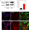
Elevation of IL-1β at the protein level. (A) Representative immunoblots from mouse whole retina lysates probed for pro-IL-1β, at the expected 31 kDa size. Protein levels increased in eyes subject to controlled elevation of IOP (CEI) to 60 mmHg for 4 h and sacrificed 22 h after IOP returned to baseline, as compared to the contralateral non-pressurized control eye. Levels of housekeeping protein GAPDH (37 kDa) were similar between conditions. (B) Summary of relative protein expression in response to IOP elevation, as quantified with densitometry and normalized to GAPDH levels (p = 0.006, n = 5). (C) Immunohistochemistry sections of mouse retina stained for IL-1β (red), GFAP (green) and with the nuclear stain DAPI (blue). The top row shows representative images from the non-pressurized eye, while the bottom row is from a contralateral eye exposed to the CEI procedure as in “(A)”. Increased staining for IL-1β was apparent in the nerve fiber layer, optic nerve and to a lesser extent throughout the retina (left). Higher magnification of the boxed area shows horizontal bands stained for IL-1β throughout the optic nerve head (center). These bands colocalize with GFAP (right), consistent with optic nerve head astrocytes.
FIGURE 3
FIGURE 3
Involvement of the P2X7 receptor in inflammasome priming in vivo. (A) The pressure-dependent rise in IL-1β mRNA in rat retinas exposed to moderate elevation of IOP (CEI) was not present following injection of the P2X7 receptor antagonist BBG. Data are expressed as relative gene expression in the non-pressurized (Control) vs pressurized retina for eyes injected with 0.8% BBG (CEI + BBG) or saline (CEI) 1–3 days before the elevation of IOP to 50 mmHg for 4 h (n = 4–5. p < 0.05 vs. saline pressurized). (B) In C57BL/6J mice, the CEI procedure increased retina levels of IL-1β mRNA relative to contralateral untreated eyes (p = 0.018). In P2X7 knockout mice, the elevation in IOP did not significantly (NS) increase levels of IL-1β. Levels of IL-1β mRNA in pressurized eyes of P2X7 knockout mice were significantly less than in wild type pressurized eyes (∗∗p = 0.036). Data are expressed as gene expression of untreated eyes (Control) relative to pressurized eyes (CEI). Retina including optic nerve head was extracted 22 h after returning IOP to baseline from an elevation to 60 mmHg for 4 h (n = 4). (C) Intravitreal injection of P2X7 agonist BzATP was sufficient to increase levels IL-1β mRNA in mouse retina when extracted 24 h after injection. Data are expressed as relative gene expression of contralateral non-injected eye (Control) vs. injected eye (BzATP; p < 0.01, n = 3).
FIGURE 4
FIGURE 4
Mechanical strain primes IL-1β in optic nerve head astrocytes. (A) Astrocytes plated on silicon substrates and fixed after 4 h in control conditions (left, Control) or after 16% cyclical strain at 0.3 Hz (right, Stretch). Staining for actin with phalloidin (red) showed no obvious changes to the cytoskeleton. (B) Application of 16% cyclical strain for 4 h increased expression of IL-1β mRNA (n = 5, p < 0.03). (C) Astrocytes exposed to moderate swelling induced by 30% hypotonicity (Swell) showed increased expression of IL-1β mRNA relative to untreated cells maintained in isotonic solution (Control; n = 3, p = 0.009).
FIGURE 5
FIGURE 5
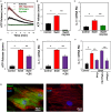
ATP release through pannexin channels required for mechanosensitive priming of IL-1β in astrocytes. (A) Swelling rat astrocytes in hypotonic solution led to a release of ATP into the extracellular medium, as detected by the luciferin/luciferase assay. The ATP hydrolase apyrase (1 U/ml) substantially reduced the response. Symbols represent mean ± SEM, n = 10. (B) Quantification of extracellular ATP levels 18 min after exposure to solutions (Swell vs. Control isotonic, p < 0.05, ∗∗ Swell vs Swell+Apyrase, p < 0.05, n = 10). (C) Swelling astrocytes in the presence of apyrase also prevented the rise in IL-1β mRNA (p = 0.02 Swell vs control, ∗∗p = 0.03 Swell vs Swell+Apyrase, n = 3). (D) The swelling-induced release of ATP was inhibited by pannexin channel blocker carbenoxolone (CBX, 10 μM, p < 0.05, Swell vs. control, ∗∗p < 0.05 Swell vs. Swell+CBX, n = 20, normalized to swell). (E) The swelling-induced rise in IL-1β mRNA was also inhibited by 10 μM carbenoxolone (p < 0.05, Swell vs. control, ∗∗p < 0.05 Swell vs Swell+CBX, n = 7, normalized to swell). (F) Pannexin blocker probenecid (Prob, 1 mM) reduced the swelling-induced rise of IL-1β in astrocytes (p = 0.029). The specific peptide blocker 10Panx1 (100 μM) reduced the expression of IL-1β as compared to the scrambled peptide control (10panxscr, p = 0.003). Swelling alone raised IL-1β (p = 0.03, n = 3 for all). (G) Left: Astrocytes stained for panx1 (green), actin (red) and DAPI (blue). Right: No signal was detected in the absence of panx1 antibody.
FIGURE 6
FIGURE 6
P2X7 receptor involved in priming of IL-1β in astrocytes. (A) Immunocytochemistry showing expression of the P2X7R in cultured optic nerve head astrocytes (left). No signal was detected in the absence of the primary antibody (right). (B) The swelling-induced rise in IL-1β mRNA was inhibited by P2X7R antagonists BBG (10 μM), A839977 (50 nM) and A740003 (5 μM). Cells were pretreated with drugs for 1 h before swelling (p < 0.001 Swell vs. control, ∗∗p < 0.001 Swell vs. Swell+drugs, n = 4). (C) The swelling-induced rise in IL-1β was reduced in astrocytes from P2X7-/- mice as compared to C57BL/6J mice. Data are expressed relative to the matched control group (p < 0.01, ∗∗p = 0.026, n = 6). (D) Application of BzATP (400 μM) for 4 h increased IL-1β expression (p < 0.01, n = 7).
FIGURE 7
FIGURE 7
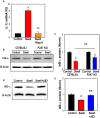
NFκB is involved in inflammasome priming after mechanical strain. (A) NFκB inhibitor Bay11-7082 (Bay11, 4 μM) prevented IL-1β upregulation in rat astrocytes. Bay11-7082 was present for 1 h before and during the 4 h swelling (p < 0.001 Control vs. Swell, ∗∗p < 0.001, Swell vs. Swell+Bay11; n = 4). (B) Representative immunoblots from mouse optic nerve head astrocyte lysates from control C57BL/6J and P2X7-/- mice probed for IκB-α (39 kDa) and housekeeping protein β-actin (42 kDa). Expression of IκB-α was reduced following 4 h of swelling in control astrocytes, consistent with the activation of NFκb. (C) Summary of relative IκB-α protein expression from experiments illustrated in panel B quantified with densitometry. The effect of swelling on IκB-α was significantly less in astrocytes from P2X7-/- mice (p < 0.001 Swell vs. Control C57BL/6J, p = 0.011 Swell vs. Control P2X7-/-, ∗∗p = 0.038 Swell C57BL/6J vs. Swell P2X7-/-; n = 3). (D) Representative immunoblots from mouse optic nerve head astrocyte lysates from control mice probed for IκB-α (39 kDa) and housekeeping protein β-actin (42 kDa). The reduction in IκB-α triggered by swelling was reduced in the presence of P2X7R antagonist A839977 (100 nM). (E) Mean densitometry values for IκB-α protein expression from immunoblots like those in “(D)” (p = 0.002, ∗∗p = 0.004; n = 4).
FIGURE 8
FIGURE 8
Model summarizing the hypothesized role of the P2X7 receptor in the priming of inflammasome genes after mechanical strain, which leads to ATP release through pannexin hemichannels. Extracellular ATP autostimulates P2X7 receptors, leading to NFκB activation and transcriptional elevation of IL-1β and NLRP3 in optic nerve head astrocytes. Swelling may also activate inflammasome genes through other pathways.

References

    1. Albalawi F., Lu W., Lim J., Mitchell C. H. (2016). The role of P2X7R in priming the NLRP3 inflammasome after mechanical strain. FASEB J. 30 745.
    1. Beckel J. M., Argall A. J., Lim J. C., Xia J., Lu W., Coffey E. E., et al. (2014). Mechanosensitive release of adenosine 5′-triphosphate through pannexin channels and mechanosensitive upregulation of pannexin channels in optic nerve head astrocytes: a mechanism for purinergic involvement in chronic strain. Glia 62 1486–1501. 10.1002/glia.22695 - DOI - PMC - PubMed
    1. Bernier L. P. (2012). Purinergic regulation of inflammasome activation after central nervous system injury. J. Gen. Physiol. 140 571–575. 10.1085/jgp.201210875 - DOI - PMC - PubMed
    1. Boaru S. G., Borkham-Kamphorst E., Van de Leur E., Lehnen E., Liedtke C., Weiskirchen R. (2015). NLRP3 inflammasome expression is driven by NF-kappaB in cultured hepatocytes. Biochem. Biophys. Res. Commun. 458 700–706. 10.1016/j.bbrc.2015.02.029 - DOI - PubMed
    1. Bruzzone R., Barbe M. T., Jakob N. J., Monyer H. (2005). Pharmacological properties of homomeric and heteromeric pannexin hemichannels expressed in Xenopus oocytes. J. Neurochem. 92 1033–1043. 10.1111/j.1471-4159.2004.02947.x - DOI - PubMed

LinkOut - more resources