. 2017 Aug 9:8:1517.
doi: 10.3389/fmicb.2017.01517. eCollection 2017.

Curcumin Quantum Dots Mediated Degradation of Bacterial Biofilms

Affiliations

Curcumin Quantum Dots Mediated Degradation of Bacterial Biofilms

Ashish K Singh et al. Front Microbiol. .

Abstract

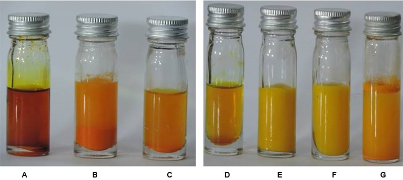
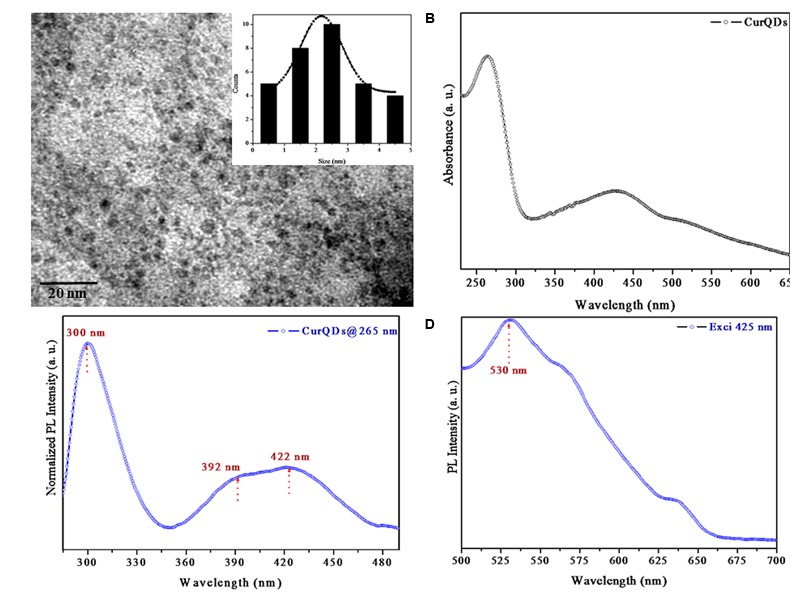
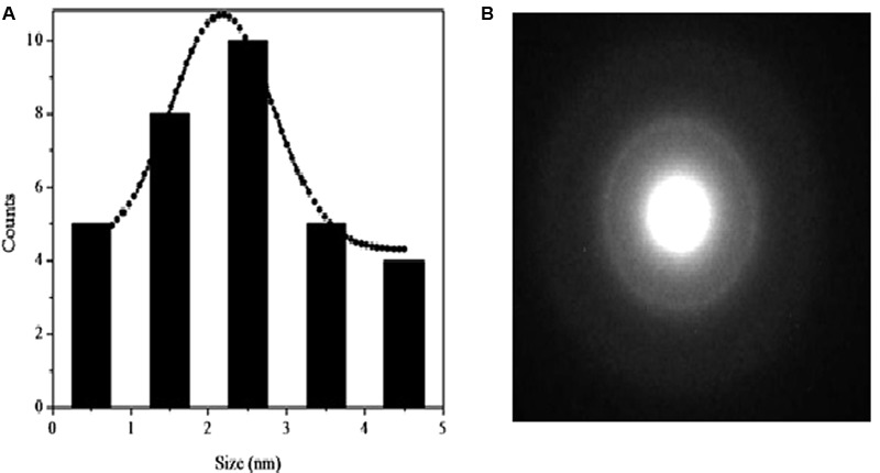
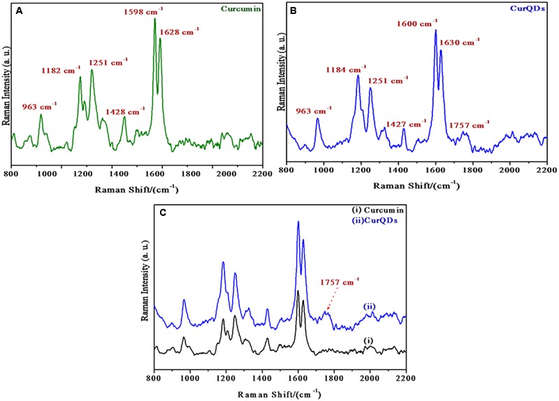
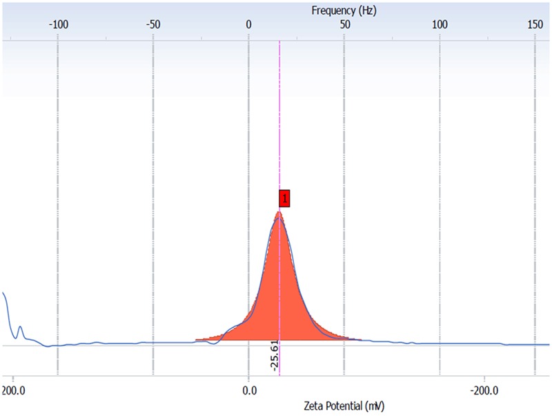
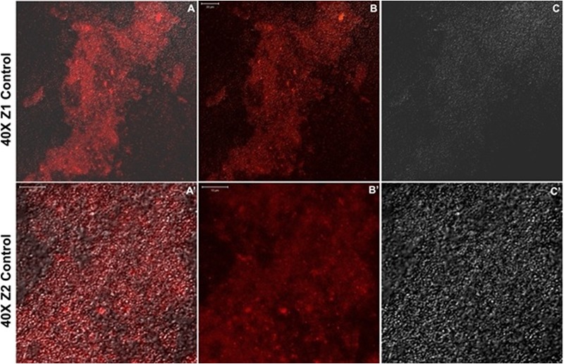
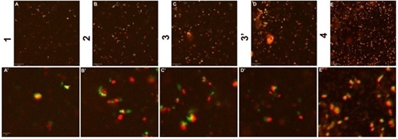
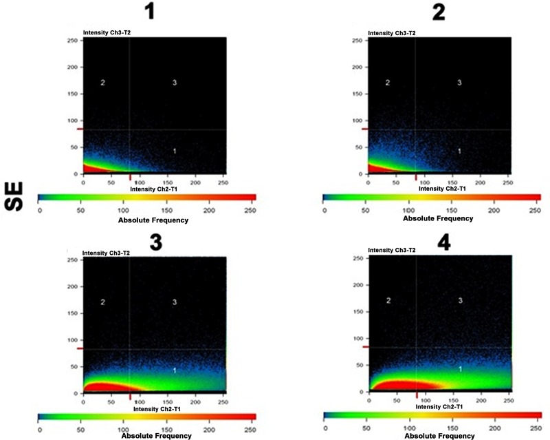
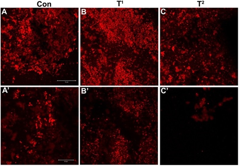
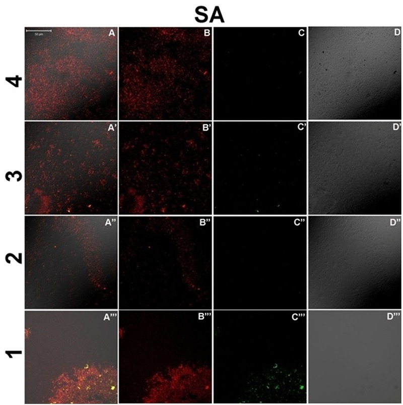
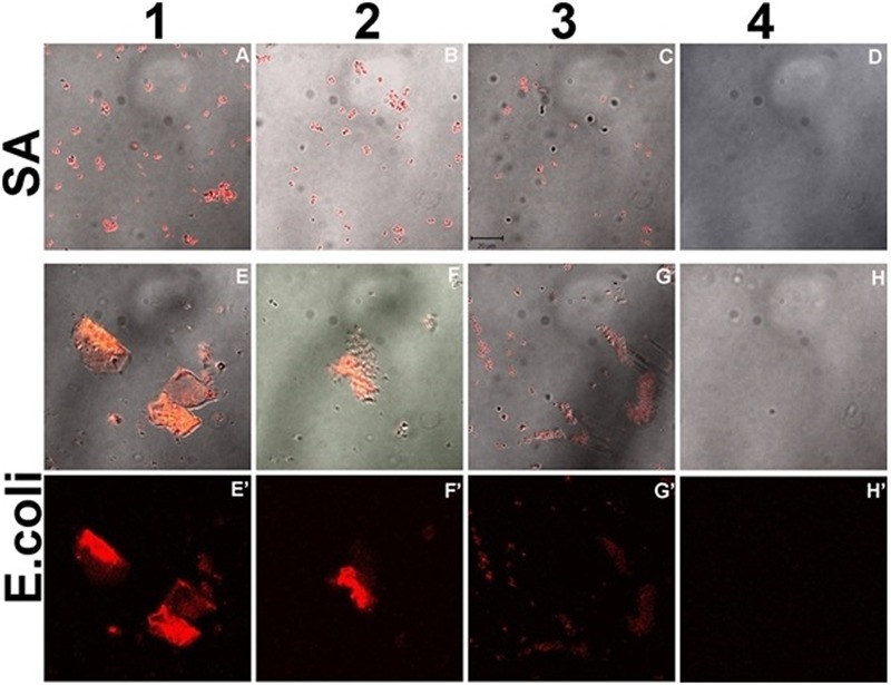
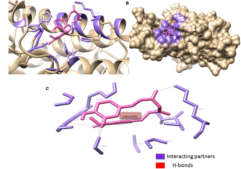
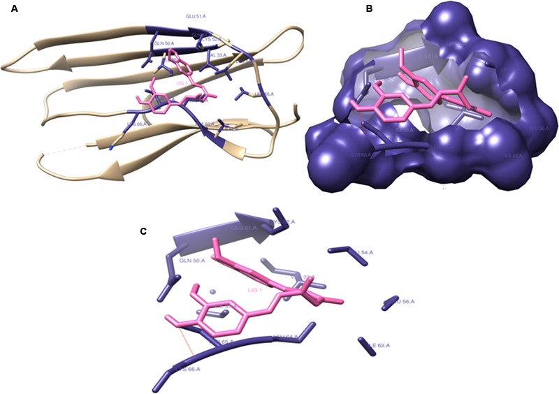
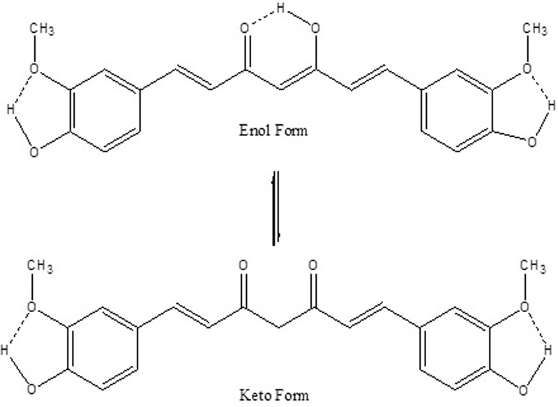
Bacterial biofilm has been reported to be associated with more than 80% of bacterial infections. Curcumin, a hydrophobic polyphenol compound, has anti-quorum sensing activity apart from having antimicrobial action. However, its use is limited by its poor aqueous solubility and rapid degradation. In this study, we attempted to prepare quantum dots of the drug curcumin in order to achieve enhanced solubility and stability and investigated for its antimicrobial and antibiofilm activity. We utilized a newer two-step bottom up wet milling approach to prepare Curcumin Quantum Dots (CurQDs) using acetone as a primary solvent. Minimum inhibitory concentration against select Gram-positive and Gram-negative bacteria was performed. The antibiofilm assay was performed at first using 96-well tissue culture plate and subsequently validated by Confocal Laser Scanning Microscopy. Further, biofilm matrix protein was isolated using formaldehyde sludge and TCA/Acetone precipitation method. Protein extracted was incubated with varying concentration of CurQDs for 4 h and was subjected to SDS-PAGE. Molecular docking study was performed to observe interaction between curcumin and phenol soluble modulins as well as curli proteins. The biophysical evidences obtained from TEM, SEM, UV-VIS, fluorescence, Raman spectroscopy, and zeta potential analysis confirmed the formation of curcumin quantum dots with increased stability and solubility. The MICs of curcumin quantum dots, as observed against both select gram positive and negative bacterial isolates, was observed to be significantly lower than native curcumin particles. On TCP assay, Curcumin observed to be having antibiofilm as well as biofilm degrading activity. Results of SDS-PAGE and molecular docking have shown interaction between biofilm matrix proteins and curcumin. The results indicate that aqueous solubility and stability of Curcumin can be achieved by preparing its quantum dots. The study also demonstrates that by sizing down the particle size has not only enhanced its antimicrobial properties but it has also shown its antibiofilm activities. Further, study is needed to elucidate the exact nature of interaction between curcumin and biofilm matrix proteins.

Keywords: adhesion; antimicrobial agents; bacterial biofilm; curcumin; nano-curcumin; quantum dots.


Figures

FIGURE 1
FIGURE 1
(A) Native curcumin in DMSO. (B) 1st phase curcumin in DMSO with water. (C) 2nd phase curcumin in DMSO with water. (D) Native curcumin in acetone. (E) 1st phase curcumin in acetone with water. (F) 2nd phase curcumin in acetone with water. (G) Naïve curcumin suspended in water. The aqueous insolubility of the native curcumin can be perceived as heterogeneous yellow colored aggregations present at the top of aqueous phase and heterogeneity of suspension seen throughout (G). Curcumin upon dissolution in polar aprotic DMSO and non-polar acetone appears as dark and light mustard yellow solution, respectively (A-G). Phase 1 and 2 of wet milling utilizing DMSO (B,C) and acetone is also shown (E,F).
FIGURE 2
FIGURE 2
(A) Transmission electron microscopy (TEM) images of Curcumin Quantum Dots (CurQDs). (B) UV-Visible absorption spectra of CurQDs. (C) Photoluminescence (PL) spectra of CurQDs 265 nm. (D) PL spectra of CurQDs excited at wavelength 425 nm.
FIGURE 3
FIGURE 3
(A) Gaussian fitted curve for different size CurQDs. (B) Selected area electron diffraction (SAED) pattern of CurQDs.
FIGURE 4
FIGURE 4
Raman spectra of (A) native curcumin, (B) CurQDs, (C) native curcumin and CurQDs represented together.
FIGURE 5
FIGURE 5
The averaged (n D 4) zeta potential distribution for aqueous CurQDs. Sample concentration was 2 mg/mL in MiliQ water with a pH value of 7.4 after six months (day 180th).
FIGURE 6
FIGURE 6
(A–C) Confocal sections of biofilm of Staphylococcus aureus (ATCC 29213) after incubation of 72 h. (A’–C’) Twice magnified view of (A–C), (A) is merged view of DAPI staining and differential interference contrast (DIC) imaging, while (B,C) are DAPI stained and DIC images, respectively.
FIGURE 7
FIGURE 7
Confocal sections of Biofilm layer of S. aureus (ATCC 29213) incubated with 0.125 μg/ml concentration of CurQDs (1–4). (A–E) Depicts the different layers of the corresponding bacterial biofilm. (A’–E’) is the magnified view of the same. Note the stable association of the drug molecule with the bacterium, which suggests its affinity for the organism. Strong co-localization of the DAPI (red) with the drug (green) supports this affinity.
FIGURE 8
FIGURE 8
Co-localization maps revealing the strong physical association of CurQDs with the biofilm matrix in the different layers as seen in Figures 7B’–E’.
FIGURE 9
FIGURE 9
(A) Confocal sections of biofilms of S. epidermidis (ATCC35984), (B,C) confocal sections of biofilms of S. aureus (ATCC 29213) (T1) and Escherichia coli (ATCC 25922) (T2) stained with DAPI, treated with 0.125 μg/ml concentration of curcumin. (A’–C’) Post treatment magnified view of (A–C). Note the diffusion and disintegration of the superficial biofilm at concentration 0.125 μg/ml.
FIGURE 10
FIGURE 10
Confocal sections of bacterial culture of S. aureus (ATCC 29213), treated with 0.0156, 0.0312, 0.0625, and 0.125 μg/ml concentration (present in lanes 4–1, respectively) of CurQDs for 24 h. Cultures have been stained with DAPI (red), and green is the auto-fluorescent CurQDs. (A–A”’) Merged view of biofilm matrix treated with 0.0156–0.125 μg/ml CurQDs respectively. (B–B”’) DAPI stained confocal sections of biofilm matrix treated with 0.0156–0.125 μg/ml CurQDs respectively. (C–C”’) Confocal localization view of CurQDs in increasing concentration (0.0156–0.125 μg/ml). (D–D”’) Differential interference contrast microscopic contours of biofilm matrix after exposure to with 0.0156–0.125 μg/ml CurQDs respectively.
FIGURE 11
FIGURE 11
Confocal sections of biofilm of S. aureus (ATCC 29213) (A–D) and E. coli (ATCC 25922) (E–H’) incubated with increasing concentration of CurQDs for 72 h, representing the corresponding dynamics of bacterial association and biofilm matrix formation in the presence of drug (Resolution 40X).
FIGURE 12
FIGURE 12
Phenol soluble modulins complexed with Curcumin: Curcumin (shown in hot pink) interacting with the Gln82, His83, Phe107:195, Ala114, Ile117:120, Lys118:198, Asp184:190, Ser185, Trp187, and Thr191 residues (shown with forest green) of phenol soluble modulins. Curcumin forms four hydrogen bonds with Lys118:194, Asp84, and Ser85 residues of the PSM’s rendering extra stability to the interaction. Lys118 seems to form two hydrogen bonds with two ortho-positioned hydroxyl residues present on the phenol ring of curcumin. (A) Ribbon image view of the interaction of PSMs with curcumin. (B) Surface topological view of the interaction of PSMs with curcumin. (C) Interacting partners of curcumin and PSM complex.
FIGURE 13
FIGURE 13
Curli protein complexed with Curcumin: Curcumin interacting with (Val33, Gln34:50, Ile35:62, Glu51, Lys52:66, Leu54:56:640, Ala63, and Thr65) residues and forming catalytic pocket in which the snugly fit curcumin is present. (A) Ribbon image view of the interaction of Curli protein with curcumin. (B) Surface topological view of the interaction of Curli protein with curcumin. (C) Interacting partners of curcumin and Curli protein complex.
FIGURE 14
FIGURE 14
A schematic representation of intramolecular H-bonds in enol and keto forms of curcumin. Dotted bonds represent H-bonds.

References

    1. Ali M. S., Pandit V., Jain M., Dhar K. L. (2014). Mucoadhesive microparticulate drug delivery system of curcumin against Helicobacter pylori infection: design, development and optimization. J. Adv. Pharm. Technol. Res. 5 48–56. 10.4103/2231-4040.126996 - DOI - PMC - PubMed
    1. Bales P. M., Renke E. M., May S. L., Shen Y., Nelson D. C. (2013). Purification and characterization of biofilm-associated EPS exopolysaccharides from ESKAPE organisms and other pathogens. PLoS ONE 8:e67950 10.1371/journal.pone.0067950 - DOI - PMC - PubMed
    1. Barik A., Priyadarsini K. I., Mohan H. (2003). Photophysical studies on binding of curcumin to bovine serum albumin. Photochem. Photobiol. 77 597–603. - PubMed
    1. Beloin C., Ghigo J. M. (2005). Finding gene-expression patterns in bacterial biofilms. Trends Microbiol. 13 16–19. 10.1016/j.tim.2004.11.008 - DOI - PubMed
    1. Bhawana, Basniwal R. K., Buttar H. S., Jain V. K., Jain N. (2011). Curcumin nanoparticles: preparation, characterization, and antimicrobial study. J. Agric. Food Chem. 59 2056–2061. 10.1021/jf104402t - DOI - PubMed

LinkOut - more resources