. 2017 Aug 11:8:1520.
doi: 10.3389/fmicb.2017.01520. eCollection 2017.

Biochemical Mechanisms and Microorganisms Involved in Anaerobic Testosterone Metabolism in Estuarine Sediments

Affiliations

Biochemical Mechanisms and Microorganisms Involved in Anaerobic Testosterone Metabolism in Estuarine Sediments

Chao-Jen Shih et al. Front Microbiol. .

Abstract

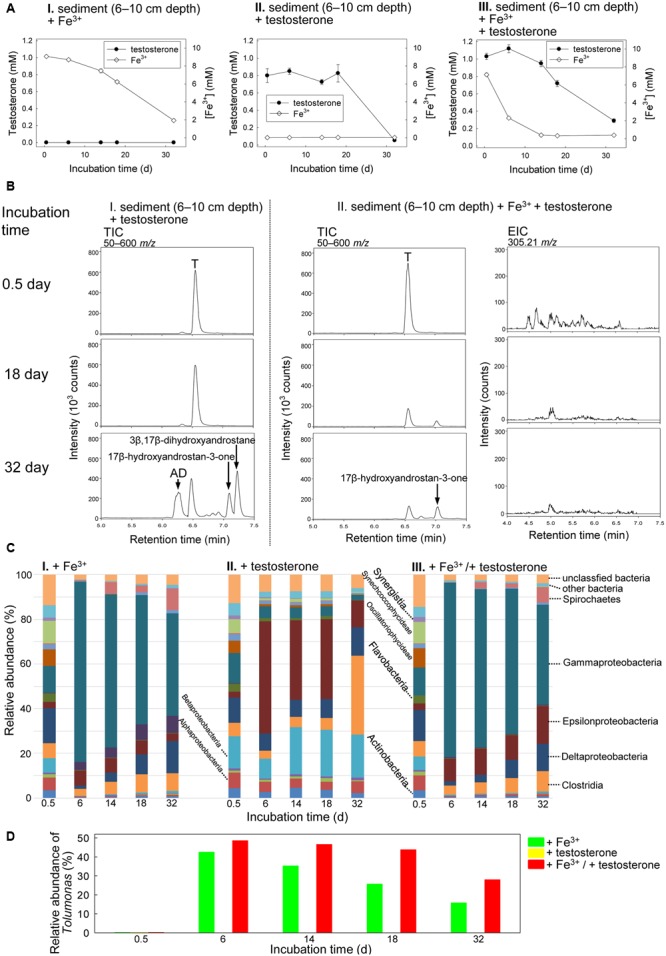
Current knowledge on the biochemical mechanisms underlying microbial steroid metabolism in anaerobic ecosystems is extremely limited. Sulfate, nitrate, and iron [Fe (III)] are common electron acceptors for anaerobes in estuarine sediments. Here, we investigated anaerobic testosterone metabolism in anaerobic sediments collected from the estuary of Tamsui River, Taiwan. The anaerobic sediment samples were spiked with testosterone (1 mM) and individual electron acceptors (10 mM), including nitrate, Fe3+, and sulfate. The analysis of androgen metabolites indicated that testosterone biodegradation under denitrifying conditions proceeds through the 2,3-seco pathway, whereas testosterone biodegradation under iron-reducing conditions may proceed through an unidentified alternative pathway. Metagenomic analysis and PCR-based functional assays suggested that Thauera spp. were the major testosterone degraders in estuarine sediment samples incubated with testosterone and nitrate. Thauera sp. strain GDN1, a testosterone-degrading betaproteobacterium, was isolated from the denitrifying sediment sample. This strain tolerates a broad range of salinity (0-30 ppt). Although testosterone biodegradation did not occur under sulfate-reducing conditions, we observed the anaerobic biotransformation of testosterone to estrogens in some testosterone-spiked sediment samples. This is unprecedented since biotransformation of androgens to estrogens is known to occur only under oxic conditions. Our metagenomic analysis suggested that Clostridium spp. might play a role in this anaerobic biotransformation. These results expand our understanding of microbial metabolism of steroids under strictly anoxic conditions.

Keywords: Illumina MiSeq; Thauera; androgen; biodegradation; estrogen; estuary; sediment; testosterone.


Figures

FIGURE 1
FIGURE 1
Androgen metabolites identified in Steroidobacter denitrificans DSM 18526 grown anaerobically on testosterone. AtcABC, 1-testosterone hydratase/dehydrogenase. The carbon numbering system of steroids is as shown for testosterone.
FIGURE 2
FIGURE 2
Anaerobic testosterone catabolism in subsurface layer sediment (0–5 cm depth)–river water mixture spiked with testosterone or nitrate or both. (A) The consumption of testosterone and nitrate in the sediment treatments. Data are shown as means ± SD of three experimental measurements. (B) UPLC–ESI–MS analysis of the ethyl acetate extracts of the sediment treatments. (Left panel) Total ion chromatograms of subsurface layer sediment–river water mixture spiked with testosterone and nitrate. (Middle panel) Extracted ion chromatograms for 2,3-SAOA (m/z = 305.21; expected retention time = 5.56 min) in subsurface layer sediment–river water mixture spiked with testosterone and nitrate. (Right panel) MS spectra of the authentic standard (top) and 2,3-SAOA extracted from the denitrifying sediment treatment. Abbreviations: ADD, androsta-1,4-diene-3,17-dione; DT, 1-dehydrotestosterone; T, testosterone. (C) Class-level phylogenetic analysis (Illumina MiSeq) revealed the temporal changes in the bacterial community structures in various sediment treatment samples. The non-metric multidimensional scaling (nMDS) analysis of the bacterial community structures in the treatments is shown in Supplementary Figure S7A. The further analyses of the community structures are shown in Supplementary Figure S8 and Tables S3–S5. (D) Thauera spp. (I) and Pseudomonas spp. (II) were enriched in subsurface layer sediment–river water mixture spiked with testosterone and nitrate.
FIGURE 3
FIGURE 3
(A) Agarose gel electrophoresis revealed that atcA-like PCR products were detected only in subsurface layer sediment–river water mixture spiked with testosterone and nitrate. PCR products with the expected size of approximately 1200 bp were amplified from the androgen-degrading denitrifier, Sdo. denitrificans. The full-length gel is presented in Supplementary Figure S3. (B) Maximum Likelihood tree of atcA gene fragments obtained from the subsurface layer sediment–river water mixture incubated with testosterone and nitrate for 4 days. Refer to Supplementary Table S2 for individual atcA sequences. The gene encoding the large subunit (MhyADHL) of 3-hydroxycyclohexanone dehydrogenase from Alicycliphilus denitrificans served as an outgroup sequence. (C) The phylogenetic tree of 16S rRNA gene of strain GDN1. All sequences were aligned by MUSCLE. Both of the evolutionary histories were inferred by using the Maximum Likelihood method. Bootstrap values are based on 1000 replicates. Numbers shown around branches are bootstrap percentages for clades supported above the 50% level. Branch support was determined by bootstrapping 1000 times. The unit for each of the scale bar is nucleotide substitutions per site.
FIGURE 4
FIGURE 4
Salinity tolerance of Thauera sp. strain GDN1. Strain GDN1 was anaerobically grown in the denitrifying medium containing 1 mM testosterone, 10 mM acetate, and 0–50 ppt NaCl. Bacterial growth was measured as the total protein concentration in the cultures. Data are shown as means ± SD of three experimental measurements.
FIGURE 5
FIGURE 5
Anaerobic testosterone metabolism in middle layer sediment (6–10 cm depth)–river water mixture spiked with testosterone or Fe3+ or both. (A) Consumption of testosterone and Fe3+ in the sediment treatments. Data are shown as means ± SD of three experimental measurements. (B) UPLC–ESI–MS analysis of the ethyl acetate extracts of the sediment treatments. (Left panel) Total ion chromatograms of middle layer sediment–river water mixture spiked with testosterone alone. (Middle panel) Total ion chromatograms of middle layer sediment–river water mixture spiked with testosterone and Fe3+. (Right panel) Extracted ion chromatograms for 2,3-SAOA (m/z = 305.21; expected retention time = 5.56 min) in middle layer sediment–river water mixture spiked with testosterone and Fe3+. AD, androst-4-en-3,17-dione; T, testosterone. (C) Class-level phylogenetic analysis (Illumina MiSeq) revealed the temporal changes in the bacterial community structures in various sediment treatments. The nMDS analysis of the bacterial community structures in the treatments is shown in Supplementary Figure S7B. (D) Genus-level phylogenetic analysis revealed the temporal changes in the relative abundance of Tolumonas spp. in the sediment treatments.
FIGURE 6
FIGURE 6
Anaerobic testosterone metabolism in bottom layer sediment (11–15 cm depth)–river water mixture spiked with testosterone or sulfate or both. (A) Consumption of testosterone and sulfate in the sediment treatments. Data are shown as means ± SD of three experimental measurements. (B) HPLC–UV analysis of the ethyl acetate extracts of bottom layer sediment–river water mixture spiked with testosterone and sulfate. During anaerobic incubation, estrogens accumulated in the treatment samples. AD, androst-4-en-3,17-dione; ADD, androsta-1,4-diene-3,17-dione; E1, estrone; E2, 17β-estradiol; T, testosterone. (C) Class-level phylogenetic analysis (Illumina MiSeq) revealed the temporal changes in the bacterial community structures in various sediment treatment samples. The nMDS analysis of the bacterial community structures in the treatments is shown in Supplementary Figure S7C. (D) Genus-level phylogenetic analysis revealed the temporal changes in the relative abundance of Clostridium spp. in the testosterone-spiked sediment treatments.

References

    1. Adams M. A., Teeter J. H., Katz Y., Johnson P. B. (1987). Sex-pheromones of the Sea Lamprey (Petromyzon marinus) –Steroid studies. J. Chem. Ecol. 13 387–395. 10.1007/BF01025898 - DOI - PubMed
    1. Bergstrand L. H., Cardenas E., Holert J., Van Hamme J. D., Mohn W. W. (2016). Delineation of steroid-degrading microorganisms through comparative genomic analysis. mBio 7:e00166 10.1128/mBio.00166-16 - DOI - PMC - PubMed
    1. Bortone S. A., Davis W. P., Bundrick C. M. (1989). Morphological and behavioral characters in mosquitofish as potential bioindication of exposure to kraft mill effluent. Bull. Environ. Contam. Toxicol. 43 370–377. 10.1007/BF01701871 - DOI - PubMed
    1. Boyle E. A., Edmond J. M., Sholkovitz E. R. (1977). The mechanism of iron removal in estuaries. Geochim. Cosmochim. Acta 41 1313–1324. 10.1021/acs.est.5b03419 - DOI
    1. Chang H., Wan Y., Hu J. (2009). Determination and source apportionment of five classes of steroid hormones in urban rivers. Environ. Sci. Technol. 43 7691–7698. 10.1021/es803653j - DOI - PubMed

LinkOut - more resources