

Heart regeneration and the cardiomyocyte cell cycle
- PMID: 28849267
- PMCID: PMC5780532
- DOI: 10.1007/s00424-017-2061-4
Heart regeneration and the cardiomyocyte cell cycle
Abstract
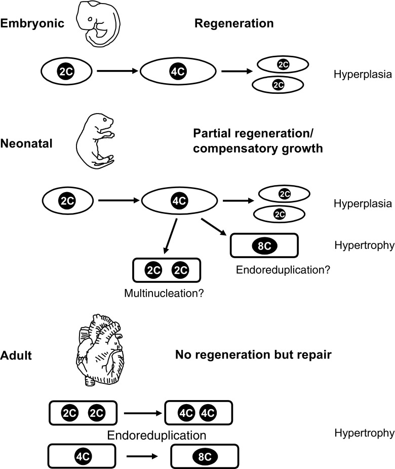
Cardiovascular disease and in particular, heart failure are still main causes of death; therefore, novel therapeutic approaches are urgently needed. Loss of contractile substrate in the heart and limited regenerative capacity of cardiomyocytes are mainly responsible for the poor cardiovascular outcome. This is related to the postmitotic state of differentiated cardiomyocytes, which is partly due to their polyploid nature caused by cell cycle variants. As such, the cardiomyocyte cell cycle is a key player, and its manipulation could be a promising strategy for enhancing the plasticity of the heart by inducing cardiomyocyte proliferation. This review focuses on the cardiac cell cycle and its variants during postnatal growth, the different regenerative responses of the heart in dependance of the developmental stage and on manipulations of the cell cycle. Because a therapeutic goal is to induce authentic cell division in cardiomyocytes, recent experimental approaches following this strategy are also discussed.
Keywords: Cardiomyocyte; Cell growth; Heart; Regeneration; Transgenic animal models.
Figures
References
-
- Andersen DC, Jensen CH, Baun C, Hvidsten S, Zebrowski DC, Engel FB, Sheikh SP. Persistent scarring and dilated cardiomyopathy suggest incomplete regeneration of the apex resected neonatal mouse myocardium—180 days follow up study. J Mol Cell Cardiol. 2016;90:47–52. doi: 10.1016/j.yjmcc.2015.11.031. - DOI - PubMed
Publication types
MeSH terms
LinkOut - more resources
Full Text Sources
Other Literature Sources
Research Materials
