Review
. 2018:415:1-38.
doi: 10.1007/82_2017_44.

The Treponema pallidum Outer Membrane

Affiliations
Review

The Treponema pallidum Outer Membrane

Justin D Radolf et al. Curr Top Microbiol Immunol. 2018.

Abstract

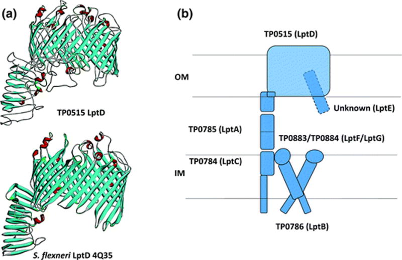
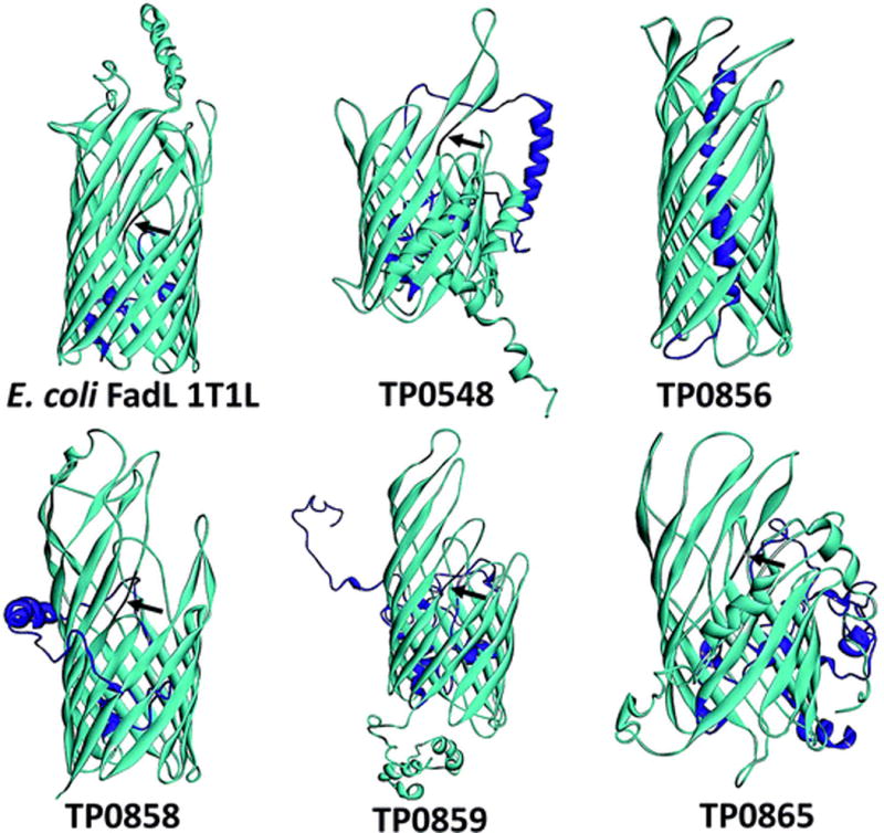
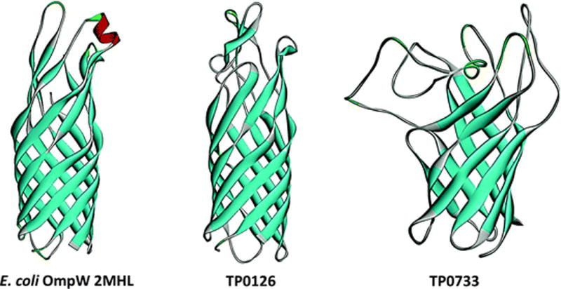
The outer membrane (OM) of Treponema pallidum, the uncultivatable agent of venereal syphilis, has long been the subject of misconceptions and controversy. Decades ago, researchers postulated that T. pallidum's poor surface antigenicity is the basis for its ability to cause persistent infection, but they mistakenly attributed this enigmatic property to the presence of a protective outer coat of serum proteins and mucopolysaccharides. Subsequent studies revealed that the OM is the barrier to antibody binding, that it contains a paucity of integral membrane proteins, and that the preponderance of the spirochete's immunogenic lipoproteins is periplasmic. Since the advent of recombinant DNA technology, the fragility of the OM, its low protein content, and the lack of sequence relatedness between T. pallidum and Gram-negative outer membrane proteins (OMPs) have complicated efforts to characterize molecules residing at the host-pathogen interface. We have overcome these hurdles using the genomic sequence in concert with computational tools to identify proteins predicted to form β-barrels, the hallmark conformation of OMPs in double-membrane organisms and evolutionarily related eukaryotic organelles. We also have employed diverse methodologies to confirm that some candidate OMPs do, in fact, form amphiphilic β-barrels and are surface-exposed in T. pallidum. These studies have led to a structural homology model for BamA and established the bipartite topology of the T. pallidum repeat (Tpr) family of proteins. Recent bioinformatics has identified several structural orthologs for well-characterized Gram-negative OMPs, suggesting that the T. pallidum OMP repertoire is more Gram-negative-like than previously supposed. Lipoprotein adhesins and proteases on the spirochete surface also may contribute to disease pathogenesis and protective immunity.


Figures

Fig. 1
Fig. 1
The T. pallidum cell envelope. a T. pallidum’s major immunogens are associated with the protoplasmic cylinder, not the outer membrane. Reactivity with human syphilitic serum of proteins extracted with Triton X-114 from whole T. pallidum cells (lane 1), protoplasmic cylinders (lane 2), and solubilized outer membranes (lane 3); reproduced from reference (Radolf et al. 1988). b Freeze-fracture EM reveals scarce intramembranous particles (IMPs) within the T. pallidum OM. Convex and concave leaflets of the OM are indicated. Bar, 0.5 μM. Reproduced from reference (Radolf et al. 1994). c Deep etching reveals that OM intramembranous particles are surface-exposed. Arrowheads indicate the boundaries separating the bacterial surface from the convex fracture face. Particles on the convex fracture face and the treponemal surface are indicated by thin and medium-thickness arrows, respectively. Bar, 0.5 μM. Reproduced from reference (Bourell et al. 1994). d TX-114 phase partitioning reveals that the syphilis spirochete’s major immunogens (based on reactivity with human syphilitic serum) possess hydrophobic character. Lanes: 1. Percoll-purified T. pallidum. 2. TX-114-insoluble material. 3. TX114 detergent-enriched phase proteins. 4. aqueous phase proteins. Reproduced from Reference (Radolf et al. 1988). e Scanning probe microscopy reveals rare particles on the T. pallidum surface; reproduced with permission from reference (Liu et al. 2010). f Cryoelectron microscopy (longitudinal slice) showing, from the inside out, cytoplasmic filaments (red line), cytoplasmic membrane (green line), lipoprotein layer (purple circles), peptidoglycan layer (tan line), flagellar filament (thick blue line), and outer membrane (green line). Bar, 50 nM. Reproduced with permission from reference (Liu et al. 2010). g [3H]palmitate-labeled lipids were extracted from isolated T. pallidum outer membranes and separated by two-dimensional thin layer chromatography. GL glycolipids; CL cardiolipin; PC phosphatidylcholine; PS phosphatidylserine; PG phosphatidylglycerol; O origin. Reproduced from reference (Radolf et al. 1995b)
Fig. 2
Fig. 2
Bipartite topology of Tpr C/D and I (Nichols strain). a Domain architectures of T. denticola major outer surface protein (MOSP) and TprC/D/I/F subfamily members. The signal sequences of all three proteins are shown in blue. The portions of TprC and TprI colored in black and yellow, respectively, denote the TprC- and TprI-specific regions of each protein (TprCSp and TprISp). The green regions in TprI and TprF denote regions present in TprI and TprF but not TprC (TprI/FSp). Reproduced with permission from reference (Anand et al. 2015)). b The MOSPC domains of TrpC and TprI are solely responsible for membrane insertion and pore formation by the full-length proteins. Liposomes were reconstituted with folded, full-length recombinant proteins (TprCFl and TprIFl), TprF, or the MOSPC (TprCC and TprIC) or MOSPN (TprCN and TprIN) domains of TprC and TprI followed by sucrose density gradient ultracentrifugation and immunoblot analysis. The top fractions (TF) contain liposome-incorporated material, whereas the middle and bottom fractions (MF and BF, respectively) contain unincorporated material. The bar graphs show pore formation by the same proteins, along with E. coli OmpF (positive) and OmpA (negative) controls, measured by efflux of Tb(DPA)33 encapsulated into liposomes (100% efflux = the degree of quenching obtained by detergent lysis). Statistical significance compared with E. coli OmpF was assigned according to the following scheme: * P < 0.05; ** P < 0.0001. Reproduced from Reference (Anand et al. 2015). c Bipartite topology of native TprC and TprI in live treponemes. Motile T. pallidum were encapsulated in gel microdroplets and probed with 1:100 dilutions of rat antisera against TprCN, TprCC, or FlaA without (intact I) or with the removal of OMs (Permeabilization P) by pre-incubation with 0.10% Triton X-100. Antibody binding was detected with goat anti-rat Alexa Fluor 488 (green) conjugate. Given that TprCC antibodies are highly cross-reactive with TprIC, both TprC and TprI are being labeled. Reproduced from reference (Anand et al. 2015)
Fig. 3
Fig. 3
Native TprC and TprI are amphiphilic but tethered to the peptidoglycan sacculus, whereas TprF is tightly bound to the peptidoglycan sacculus. a Triton X-114 phase partitioning of T. pallidum lysates without (−) or with (+) pre-solubilization with 2% DDM. Whole cells (WC), Triton X-114-insoluble material (Ins), and aqueous and Triton X-114-enriched phases (Aq and Det, respectively) were separated by SDS-PAGE followed by immunoblotting using antisera specific for TprC (top), TprI (middle), or TprI and F (bottom). Arrowheads in bottom panel indicate TprF; TprI is the larger protein. Reproduced from reference (Anand et al. 2015). b Extensively washed Triton X-114-insoluble material visualized in negatively stained whole mounts by transmission electron microscopy. Previous studies have shown that this material contains the peptidoglycan sacculus (Radolf et al. 1989a). Reproduced from reference (Anand et al. 2015). c Bipartite model for Tpr C/D and TprI. d Structural model of TprC (Nichols) generated using TMBpro (Randall et al. 2008) predicts a 10-stranded β-barrel
Fig. 4
Fig. 4
TprC and TprI, but not TprF, expressed in E. coli with PelB signal sequences display bipartite topology. IFA of intact (I) or permeabilized (P) E. coli C41 (DE3) expressing TprC, TprI, or TprF with a PelB signal sequence were probed with rat antisera against TprCN, TprCC, and Skp (periplasmic control). Antibody binding was detected with goat anti-rat Alexa Fluor 488 conjugate. Reproduced from reference (Anand et al. 2015)
Fig. 5
Fig. 5
Variation in TprK is attributed to gene conversion wherein variant DNA segments adjacent to tprD recombine with variable regions (V1–V7) of tprK to generate new TprK mosaics. Reproduced with permission from reference (Ho and Lukehart 2011)
Fig. 6
Fig. 6
TP0326/BamA, the central component of the T. pallidum β-barrel assembly machine (BAM) complex. a Schematic of BamA bipartite topology showing five N-terminal periplasmic POTRA domains and a C-terminal β-barrel. b A homology model based upon the solved structure of Neisseria gonorrhoeae BamA (Noinaj et al. 2013) predicts that TP0326 contains a 16-stranded β-barrel with characteristic BamA features, including three extracellular loops (L4, L6, and L7) that occlude the barrel’s extracellular opening. Reproduced from reference (Luthra et al. 2015a). c L4 is an immunodominant surface feature of BamA. Multiple-sequence alignment of the L4 regions of BamAs from geographically diverse T. pallidum strains, sequences amplified from skin biopsy specimens from two secondary syphilis patients (Cali-77 and Cali-84) enrolled at our Cali, Colombia, study site, and the Gauthier strain of the T. pallidum subsp. pertenue. All strains of T. pallidum subsp. pertenue in the database have same the L4 sequence. Immunoblot relativities of pooled IRS and pooled sera from U.S. (HSSU) and Colombian (HSSC) HIV-negative patients with secondary syphilis against L4, L6, and L7 loop peptides and a control peptide L3β6 derived from a portion of the barrel not surface-exposed. NRS and NHS, normal rabbit and normal human serum, respectively. Reproduced from reference (Luthra et al. 2015a). d Identification of the BAM complex in T. pallidum. Lysates solubilized with graded concentrations of the detergent DDM were separated by Blue-Native PAGE followed by immunoblot analysis with antisera directed against the POTRA arm of TP0326/BamA. Reproduced from reference (Desrosiers et al. 2011)
Fig. 7
Fig. 7
T. pallidum’s LptD ortholog. a Homology model for TP0515 based upon the solved structure of Shigella flexneri LptD (PDB 4Q35). The model and figure were generated using I-TASSER (Yang and Zhang 2015) and Discovery Studio, respectively. b T. pallidum contains orthologs for all of the components of the E. coli LptD complex except LptE
Fig. 8
Fig. 8
T. pallidum contains five FadL orthologs. Structural models and figures for TP0548, TP0856, TP0858, TP0859, and TP0865 were generated using I-TASSER (Yang and Zhang 2015) and Discovery Studio (BIOVIA 2015), respectively. The N-terminal hatch domains are shown in magenta, while the kinks in the third transmembrane strands of TP0548, TP0858, TP0859, and TP0865 are shown in black and indicated by arrowheads. E. coli FadL (PDB 1T1L) is shown for comparison
Fig. 9
Fig. 9
T. pallidum TolC/OprJ/OprN orthologs and putative TolC complex. a Structural models for T. pallidum’s OprJ (TP0966), OprN (TP0967), and TolC (TP0969) orthologs. The models for TP0966 and TP0967 are based upon Pseudomonas aeruginosa OprJ (PDB 5AZS) and OprN (PDB 5AZO), while that for TP0969 is based upon E. coli TolC (PDB 2VDE). Structural models and figures were generated using SWISS-MODEL (Biasini et al. 2014) and Discovery Studio (BIOVIA 2015), respectively. b Schematic for T. pallidum’s putative AcrAB–TolC complex
Fig. 10
Fig. 10
OmpW orthologs in T. pallidum. Structural models for TP0126 and TP0733 are based upon Pseudomonas aeruginosa OprG (2×27) and E. coli OmpW (2MHL). Structural models and figures were generated using SWISS-MODEL (Biasini et al. 2014) and Discovery Studio (BIOVIA 2015), respectively

References

    1. Akins DR, Purcell BK, Mitra MM, Norgard MV, Radolf JD. Lipid modification of the 17-kilodalton membrane immunogen of Treponema pallidum determines macrophage activation as well as amphiphilicity. Infect Immun. 1993;61:1202–1210. - PMC - PubMed
    1. Alderete JF, Baseman JB. Surface-associated host proteins on virulent Treponema pallidum. Infect Immun. 1979;26:1048–1056. - PMC - PubMed
    1. Anand A, Luthra A, Dunham-Ems S, Caimano MJ, Karanian C, LeDoyt M, Cruz AR, Salazar JC, Radolf JD. TprC/D (Tp0117/131), a trimeric, pore-forming rare outer membrane protein of Treponema pallidum, has a bipartite domain structure. J Bacteriol. 2012;194:2321–2333. - PMC - PubMed
    1. Anand A, Luthra A, Edmond ME, Ledoyt M, Caimano MJ, Radolf JD. The major outer sheath protein (Msp) of Treponema denticola has a bipartite domain architecture and exists as periplasmic and outer membrane-spanning conformers. J Bacteriol. 2013;195:2060–2071. - PMC - PubMed
    1. Anand A, LeDoyt M, Karanian C, Luthra A, Koszelak-Rosenblum M, Malkowski MG, Puthenveetil R, Vinogradova O, Radolf JD. Bipartite topology of Treponema pallidum repeat proteins C/D and I: outer membrane insertion, trimerization, and porin function require a C-terminal β-barrel domain. J Biol Chem. 2015;290:12313–12331. - PMC - PubMed

MeSH terms

Substances

LinkOut - more resources