. 2017 Sep 12;56(36):4757-4761.
doi: 10.1021/acs.biochem.7b00551. Epub 2017 Sep 1.

Cancer-Associated Mutations Mapped on High-Resolution Structures of the U2AF2 RNA Recognition Motifs

Affiliations

Cancer-Associated Mutations Mapped on High-Resolution Structures of the U2AF2 RNA Recognition Motifs

Eliezra Glasser et al. Biochemistry. .

Abstract

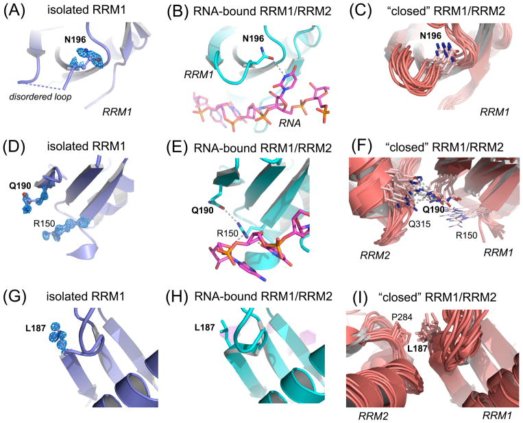
Acquired point mutations of pre-mRNA splicing factors recur among cancers, leukemias, and related neoplasms. Several studies have established that somatic mutations of a U2AF1 subunit, which normally recognizes 3' splice site junctions, recur among myelodysplastic syndromes. The U2AF2 splicing factor recognizes polypyrimidine signals that precede most 3' splice sites as a heterodimer with U2AF1. In contrast with those of the well-studied U2AF1 subunit, descriptions of cancer-relevant U2AF2 mutations and their structural relationships are lacking. Here, we survey databases of cancer-associated mutations and identify recurring missense mutations in the U2AF2 gene. We determine ultra-high-resolution structures of the U2AF2 RNA recognition motifs (RRM1 and RRM2) at 1.1 Å resolution and map the structural locations of the mutated U2AF2 residues. Comparison with prior, lower-resolution structures of the tandem U2AF2 RRMs in the RNA-bound and apo states reveals clusters of cancer-associated mutations at the U2AF2 RRM-RNA or apo-RRM1-RRM2 interfaces. Although the role of U2AF2 mutations in malignant transformation remains uncertain, our results show that cancer-associated mutations correlate with functionally important surfaces of the U2AF2 splicing factor.


Figures

Figure 1
Figure 1
U2AF2 missense mutations in neoplasms mapped on RRM structures. (A) Non-redundant U2AF2 missense mutations in cancer genomes mapped on the protein domains. Line heights are proportional to the number of independent observations for each mutation summed among independent patient samples. Amino acids that are changed in three or more samples are labeled. RNA recognition motifs (RRM1 and RRM2) are blue and other domains black. Residue numbers of domain boundaries are given below the schematic diagram. RS, arginine-serine-rich; ULM, U2AF ligand motif; UHM, U2AF homology motif. (B–C) Crystal structures of U2AF2 RRM1 (B) and RRM2 (C) at 1.07 and 1.11 Å resolutions. Views are rotated 180° about the y-axis. An affected residue (D231) located beyond the C-terminus of the crystallized construct (P229) is indicated in italicized parentheses. (D) The RNA-bound, “open” U2AF2 conformation (PDB ID 5EV4). (E) A typical “closed” U2AF2 conformation in the absence of RNA (PDB ID 2YH0). For clarity, a single representative of the NMR/PRE-fitted ensemble is shown in (E). Affected residues in (B–E) are space-filling atoms colored by the number of non-redundant mutations: 5, firebrick; 4, red; 3, orange; 2, yellow-orange.
Figure 2
Figure 2
U2AF2 hotspot residues show local structural changes between the isolated RRM1 (slate) and the linked, tandem U2AF2 RRMs, either a crystal structure bound to a Py tract oligonucleotide (cyan protein and magenta oligonucleotide, PDB ID 5EV4) or an NMR/PRE-based model in the apo-state (salmon, PDB ID 2YH0). Structures were superimposed by matching Cα-atoms and are shown from identical viewpoints. Feature enhanced maps (FEM) contoured at 1σ level are shown for new RRM1 structure. (A) N196 is poorly ordered and precedes a disordered loop of the isolated RRM1 structure (dashed blue line). (B) This loop folds for RNA binding; N196 forms a well-defined hydrogen bond (dashed gray line) with the rU6 base. (C) N196 is qualitatively similar in the “closed” NMR model. (D) Q190 is well-defined in the FEM map of high resolution apo-RRM1 despite lack of contacts. (E) In the oligonucleotide-bound structure, Q190 switches conformation to accept a hydrogen bond from the RNA-bound R150. (F) RRM1 Q190 mediates a hydrogen bond with RRM2 Q315 in the NMR model of the tandem U2AF2 RRMs in the “closed” conformation. (G) The FEM electron density for L187 of the isolated RRM1 structure is well-defined and remains similar in the RNA-bound state (H). The L187 position differs in the “closed” U2AF2 model (I), in which it makes a hydrophobic contact with P284 in RRM2.

References

    1. Dvinge H, Kim E, Abdel-Wahab O, Bradley RK. Nat Rev Cancer. 2016;16:413–430. - PMC - PubMed
    1. Zamore PD, Green MR. EMBO J. 1991;10:207–214. - PMC - PubMed
    1. Yoshida K, Sanada M, Shiraishi Y, Nowak D, Nagata Y, Yamamoto R, Sato Y, Sato-Otsubo A, Kon A, Nagasaki M, Chalkidis G, Suzuki Y, Shiosaka M, Kawahata R, Yamaguchi T, Otsu M, Obara N, Sakata-Yanagimoto M, Ishiyama K, Mori H, Nolte F, Hofmann WK, Miyawaki S, Sugano S, Haferlach C, Koeffler HP, Shih LY, Haferlach T, Chiba S, Nakauchi H, Miyano S, Ogawa S. Nature. 2011;478:64–69. - PubMed
    1. Gao J, Aksoy BA, Dogrusoz U, Dresdner G, Gross B, Sumer SO, Sun Y, Jacobsen A, Sinha R, Larsson E, Cerami E, Sander C, Schultz N. Sci Signal. 2013;6:pl1. - PMC - PubMed
    1. Cerami E, Gao J, Dogrusoz U, Gross BE, Sumer SO, Aksoy BA, Jacobsen A, Byrne CJ, Heuer ML, Larsson E, Antipin Y, Reva B, Goldberg AP, Sander C, Schultz N. Cancer Discov. 2012;2:401–404. - PMC - PubMed

Publication types

LinkOut - more resources