Review
. 2017 Oct;591(20):3190-3210.
doi: 10.1002/1873-3468.12827. Epub 2017 Sep 15.

RNA-virus proteases counteracting host innate immunity

Affiliations
Review

RNA-virus proteases counteracting host innate immunity

Jian Lei et al. FEBS Lett. 2017 Oct.

Abstract

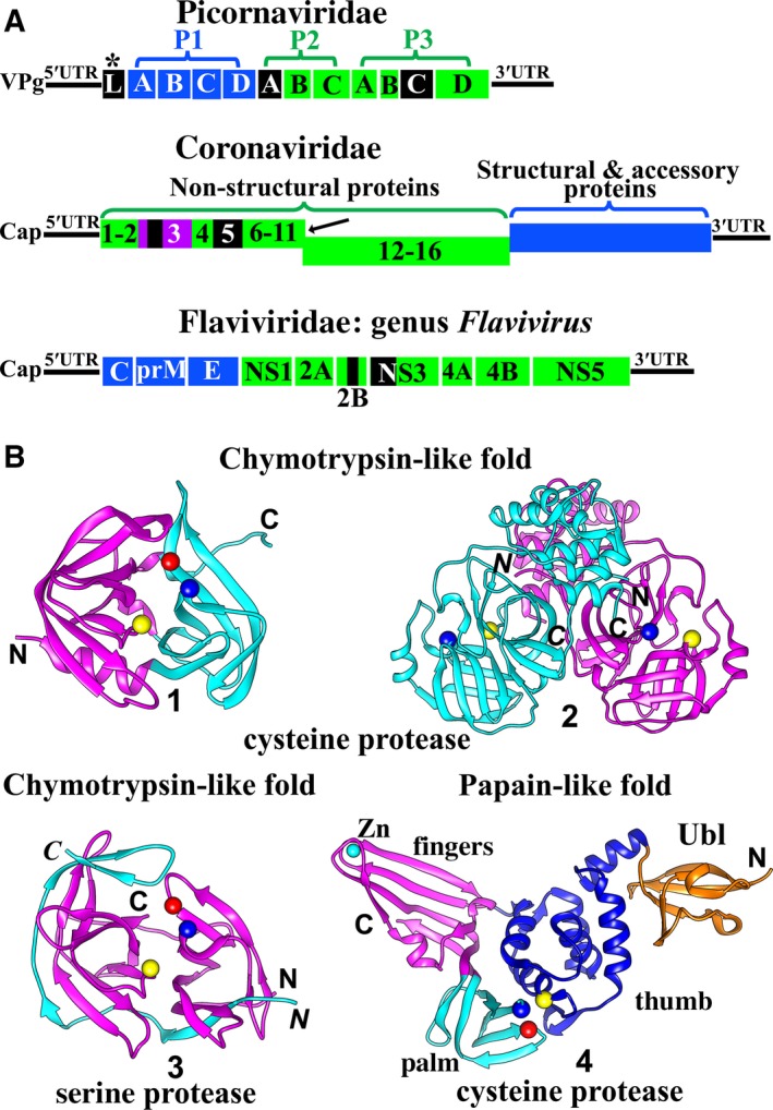
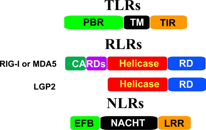
Virus invasion triggers host immune responses, in particular, innate immune responses. Pathogen-associated molecular patterns of viruses (such as dsRNA, ssRNA, or viral proteins) released during virus replication are detected by the corresponding pattern-recognition receptors of the host, and innate immune responses are induced. Through production of type-I and type-III interferons as well as various other cytokines, the host innate immune system forms the frontline to protect host cells and inhibit virus infection. Not surprisingly, viruses have evolved diverse strategies to counter this antiviral system. In this review, we discuss the multiple strategies used by proteases of positive-sense single-stranded RNA viruses of the families Picornaviridae, Coronaviridae, and Flaviviridae, when counteracting host innate immune responses.

Keywords: cleavage of host proteins; innate immunity; viral protease.


Figures

Figure 1
Figure 1
(A) Genome organization of picornaviruses, coronaviruses, and flaviviruses. All structural and accessory proteins are shown in blue. Nonstructural proteins are shown in green, with the exception of proteases, which are shown in black. Picornaviridae : The 5′ end of the picornavirus genomic RNA is covalently bound to VPg (viral protein genome‐linked). The genome encodes a polyprotein comprising the three regions P1 (structural proteins), P2, and P3 (nonstructural proteins). Generally, two viral proteases, 2Apro and 3Cpro, cleave the polyprotein into mature proteins. P1 is processed to yield 1A (VP4), 1B (VP2), 1C (VP3), and 1D (VP1); P2 is processed to 2A, 2B, and 2C; while P3 cleavage products are 3A, 3B, 3C, and 3D. *In some picornavirus genera (e. g. Aphthovirus, Cardiovirus), a third viral protease exists, the leader protease (Lpro). It auto‐cleaves itself from the polyprotein. Coronaviridae:CoVs possess the largest genome of all known RNA viruses. The 5′ genomic RNA carries a methylated cap. Two open‐reading frames (ORFs), 1a and 1b, occupy the 5′‐terminal two thirds of the CoV genome. ORF1a encodes the polyprotein 1a (Nsp1‐11), while ORF1a plus ORF1b are translated into the polyprotein 1ab (Nsp1‐16); this involves a (‐1) ribosomal frameshift overreading the stop codon of ORF1a (indicated by the black arrow). The 3′‐proximal third encodes the structural and accessory proteins. The polyproteins pp1a and pp1ab are processed by the viral proteases PL pro (within Nsp3; Nsp3 is purple) and Mpro (3CL pro, Nsp5). The genomes of members of the Flaviviridae differ between genera. Here, a genome of a member of the genus Flavivirus is shown as an example. The 5′‐capped genome encodes a polyprotein, which is cleaved into three structural proteins as well as seven nonstructural proteins by host and viral proteases. Flaviviruses have only one protease, the NS2B/NS3pro. NS2B is a cofactor for the NS3 serine protease. (B) Structures of proteases of +ssRNA viruses. The fold of most RNA‐virus proteases belongs to either the chymotrypsin‐like class or the papain‐like class. The chymotrypsin fold consists of two β‐barrel domains, while the typical papain‐like fold contains an α‐helical domain and a predominantly β‐sheet domain. The catalytic residues are located in the cleft between the two domains in both chymotrypsin‐like and papain‐like proteases. Picornavirus 2Apro, 3Cpro, coronavirus 3CL pro (Mpro), HCV and pestivirus NS3/NS4Apros, as well as flavivirus NS2B/NS3pro, adopt the chymotrypsin‐like fold, whereas picornavirus Lpro and coronavirus PL pro feature the papain‐like fold. 1) The structure of enterovirus D68 3Cpro 19 (PDB entry: 3ZV8). The Cα atoms of the catalytic triad Cys–His–Glu are shown as yellow, blue, and red spheres, respectively. 2) The structure of transmissible gastroenteritis virus (TGEV, a CoV) 3CL pro (Mpro) 24 (PDB entry: 1LVO). Dimerization of the 3CL pro (Mpro) is a prerequisite for its activity. The two protomers are displayed in cyan and purple. The catalytic dyad Cys–His (Cα atoms shown as yellow and blue spheres) is located within the chymotrypsin‐like subdomain of each monomer. An additional α‐helical domain also exists in each protomer. 3) The structure of Zika virus NS2B/NS3pro 22 (PDB entry 5LC0). The NS3 protease is shown in purple and the NS2B cofactor is in cyan. The Cα atoms of the catalytic triad Ser–His–Asp are shown as yellow, blue, and red spheres, resp. 4, The structure of MERS‐CoV PL pro 26 (PDB entry 4P16). In the coronavirus PL pro, the β‐sheet domain is larger than in the canonical papain‐like fold and divided into two subdomains, fingers (purple) and palm (cyan); together with the thumb subdomain (α‐helical domain; blue), an extended right‐hand fold is the result. A ubiquitin‐like (Ubl) domain (orange) is located in the N‐terminal region of the PL pro. The Cα atoms of the catalytic triad residues Cys–His–Asp are shown as yellow, blue, and red spheres, resp. All figures in (B) have been prepared by using UCSF Chimera 183.
Figure 2
Figure 2
Schematic overview of host innate immune pathways and their disruption by proteases of RNA viruses. The actions of viral proteases of three (+)ssRNA virus families, namely Picornaviridae, Coronaviridae, and Flaviviridae, are illustrated by triangle and square symbols. Triangles indicate cleavage of the target protein, while squares symbolize interaction with this protein in the absence of cleavage. Blue spheres indicate phosphorylation, and (Ub)x means polyubiquitination. (A) The TLR (Toll‐like receptor), RLR (retinoic acid‐inducible gene‐I (RIG‐I)‐like receptor), and NLR (nucleotide‐binding oligomerization domain (NOD)‐like receptor) signaling pathways. TLR7 detects ssRNA and triggers downstream signaling via the adaptor, MyD88 (myeloid differentiation primary response gene 88). Subsequently, MyD88 recruits IRAK4 (interleukin‐1 receptor‐associated kinase 4) to activate IRAK1/2, then IRAKs dissociate from MyD88 and bind to TNF receptor‐associated factor 6 (TRAF6). TRAF6 activates TAK1 (TGF‐β‐activated kinase 1). TAK1 further recruits TAB1/2/3 (TAK1‐binding protein 1/2/3), to activate IKKα/β/γ (IκB kinase alpha, beta, and gamma; IKKγ is also named NEMO). Then, the IKKs mediate the phosphorylation of IκB, the NF‐κB inhibitor. Phosphorylated IκB is degraded and releases NF‐κB to induce production of TNFs (tumor necrosis factors) and other cytokines. This pathway thus comprises TLR7→MyD88→IRAK4/1/2→TRAF 6TAK1/TAB1–3→IKKα/β/γ→IκB→NF‐κB. Also, TRAF6, IRAK4, TRAF3, IKKα, and IRAK1 form a complex. In this complex, both IKKα and IRAK1 activate the IRF7 (interferon regulatory factor 7) but not the IRF3 pathway (see red arrows). TLR3 detects dsRNA and triggers TRAF3 and TRAF6 by the mediator, TRIF (TIR domain‐containing adaptor protein‐inducing IFNβ). TRAF3 activates the TBK1/IKKε (TANK‐binding kinase 1/IκB kinase epsilon)‐mediated IRF3/7 pathway. TANK (TRAF family member‐associated NF‐κB activator) and IKKγ can activate TBK1/IKKε. TBK1/IKKε further stimulate the IRF3/7 pathway. In addition, STING (stimulator of interferon genes) can upregulate IRF3 signaling. The main cascade of this pathway thus comprises TLR3→TRIFTRAF 3TBK1/IKKε→IRF3/7. TRAF6 and TRAF3 are typed in bold to indicate that they are located at central positions of pathways. The downstream cascade of TRAF6 activating the NF‐κB pathway is the same as for the TLR7 pathway. RLRs detect ssRNA/dsRNA and trigger the activation of TRAF3 and TRAF6 by the mediator, MAVS (mitochondrial antiviral‐signaling protein; also known as IPS‐1, Cardif, VISA). The downstream signaling pathway is the same as for the TLR3 pathway. NLRs include NLRP3 and NLRC2 (also named NOD2). NLRP3 does not directly bind the viral RNA. The viral ssRNA or dsRNA causes many intracellular changes (such as reactive oxygen species (ROS) formation and lysosomal maturation), NLRP3 is sensitive to these changes and forms oligomers interacting with ASC (apoptosis‐associated speck‐like protein) and procaspase‐1, collectively called ‘inflammasome complex’. Subsequently, procaspase‐1 is activated, thus leading to the maturation of pro‐IL‐1β and pro‐IL‐18. NLRC2 directly interacts with ssRNA, then it recruits MAVS to activate the IRF3 pathway. Also, it can activate TRAF6 to stimulate the NF‐κB pathway. (B) The JAKSTAT signaling pathway. IFNα or IFNβ are produced via the IRF3/7 pathway. They bind the IFNAR1/2 (interferon alpha/beta receptor 1/2), leading to the activation of TYK2 (tyrosine kinase 2) and JAK1 (Janus kinase 1). These kinases phosphorylate STAT1 (signal transducer and activator of transcription 1) and STAT2. Subsequently, the phosphorylated STAT1/2 interact with IRF9 to form ISGF3 (IFN‐stimulated gene factor 3). This ternary complex enters the nucleus and promotes the expression of ISGs (interferon‐stimulated genes), such as ISG15, to establish the antiviral status. ISG15 covalently binds target proteins (ISGylation). Coronavirus PL pro can remove ISG15 from ISGylated proteins. Other proteins and abbreviations in this figure: Riplet: an E3 ubiquitin ligase and upstream regulator of RIG‐I; MFN 1/2: mitofusins 1/2. MFN1 and MFN2 regulate mitochondrial fusion; MFN1 is required for the RLR signaling pathway; IMPβ1: importin β1, a nucleocytoplasmic transport receptor, plays roles in the nucleocytoplasmic trafficking of IRF3 as well as NF‐κB p65; MDM2: a p53 degradation stimulator blocks the p53–IRF7–IFNβ signaling pathway; ADNP: activity‐dependent neuroprotective protein, a transcription factor, can bind to IFNα promoter sites (IPS) upon induction by Lpro; MC: mitochondrion; ER: endoplasmic‐reticulum.
Figure 3
Figure 3
Schematic presentation of individual TLR, RLR, and NLR domains. TLRs contain three domains: the N‐terminal PAMP‐binding region (PBR), the transmembrane region (TM), and the C‐terminal intracellular Toll/IL‐R homology (TIR) domain. RIG‐I and MDA5 comprise an N‐terminal two‐CARDs (caspase‐recruiting domains) domain, the central helicase domain, and the C‐terminal repressor domain (RD). The CARDs domain is absent in LGP2. NLRs have various domain architectures. They mainly contain three domains: the variable N‐terminal effector‐binding domain (EFB), the middle NACHT (domain existing in NAIP,CIITA,HET‐E and TP‐1) domain, and the C‐terminal leucine‐rich repeat (LRR) domain.

References

    1. Adams MJ, Lefkowitz EJ, King AMQ, Harrach B, Harrison RL, Knowles NJ, Kropinski AM, Krupovic M, Kuhn JH, Mushegian AR et al (2017) Changes to taxonomy and the International Code of Virus Classification and Nomenclature ratified by the International Committee on Taxonomy of Viruses. Arch Virol 162, 2505–2538. - PubMed
    1. Jiang P, Liu Y, Ma HC, Paul AV and Wimmer E (2014) Picornavirus morphogenesis. Microbiol Mol Biol Rev 78, 418–437. - PMC - PubMed
    1. Norder H, De Palma AM, Selisko B, Costenaro L, Papageorgiou N, Arnan C, Coutard B, Lantez V, De Lamballerie X, Baronti C et al (2011) Picornavirus non‐structural proteins as targets for new anti‐virals with broad activity. Antiviral Res 89, 204–218. - PubMed
    1. Seipelt J, Guarné A, Bergmann E, James M, Sommergruber W, Fita I and Skern T (1999) The structures of picornaviral proteinases. Virus Res 62, 159–168. - PubMed
    1. Eckerle I, Müller MA, Kallies S, Gotthardt DN and Drosten C (2013) In‐vitro renal epithelial cell infection reveals a viral kidney tropism as a potential mechanism for acute renal failure during Middle East Respiratory Syndrome (MERS) Coronavirus infection. Virol J 10, 359. - PMC - PubMed

MeSH terms