. 2017 Nov;108(11):2195-2203.
doi: 10.1111/cas.13388. Epub 2017 Sep 17.

Tumor endothelial cells with high aldehyde dehydrogenase activity show drug resistance

Affiliations

Tumor endothelial cells with high aldehyde dehydrogenase activity show drug resistance

Kyoko Hida et al. Cancer Sci. 2017 Nov.

Abstract

Tumor blood vessels play an important role in tumor progression and metastasis. We previously reported that tumor endothelial cells (TEC) exhibit several altered phenotypes compared with normal endothelial cells (NEC). For example, TEC have chromosomal abnormalities and are resistant to several anticancer drugs. Furthermore, TEC contain stem cell-like populations with high aldehyde dehydrogenase (ALDH) activity (ALDHhigh TEC). ALDHhigh TEC have proangiogenic properties compared with ALDHlow TEC. However, the association between ALDHhigh TEC and drug resistance remains unclear. In the present study, we found that ALDH mRNA expression and activity were higher in both human and mouse TEC than in NEC. Human NEC:human microvascular endothelial cells (HMVEC) were treated with tumor-conditioned medium (tumor CM). The ALDHhigh population increased along with upregulation of stem-related genes such as multidrug resistance 1, CD90, ALP, and Oct-4. Tumor CM also induced sphere-forming ability in HMVEC. Platelet-derived growth factor (PDGF)-A in tumor CM was shown to induce ALDH expression in HMVEC. Finally, ALDHhigh TEC were resistant to fluorouracil (5-FU) in vitro and in vivo. ALDHhigh TEC showed a higher grade of aneuploidy compared with that in ALDHlow TEC. These results suggested that tumor-secreting factor increases ALDHhigh TEC populations that are resistant to 5-FU. Therefore, ALDHhigh TEC in tumor blood vessels might be an important target to overcome or prevent drug resistance.

Keywords: Aldehyde dehydrogenase (ALDH); angiogenesis; endothelial cell; resistance; tumor.


Figures

Figure 1
Figure 1
Expression of aldehyde dehydrogenase (ALDH) in mouse tumor endothelial cells (TEC). (a) ALDH mRNA was analyzed in mouse TEC (mTEC) and mouse normal EC (mNEC) using real‐time RTPCR (*P < 0.01). (b) ALDH activity was measured in mTEC and mNEC using flow cytometry and the ALDEFLUOR kit (StemCell Technologies, Durham, NC, USA). DEAB is a specific ALDH inhibitor. Percentage of tumor endothelial cell containing stem‐like populations with high ALDH activity (ALDH high TEC) is shown. (c). Double immunofluorescence staining for endothelial marker CD31 and ALDH in normal mouse tissue (dermis) and super‐metastatic human melanoma (A375SM) xenografts. Merged image (DAPI) shows colocalization of ALDH (green) and CD31. Triangles show ALDH‐negative endothelial cells, whereas arrowheads point to ALDH‐positive endothelial cells. Scale bar, 50 μm.
Figure 2
Figure 2
Expression of aldehyde dehydrogenase (ALDH) in human tumor endothelial cells (TEC). (a) Double immunofluorescence staining for endothelial marker CD31 (red) and ALDH (green) in human normal kidney tissues and human renal cell carcinoma (RCC). Triangles show ALDH‐negative endothelial cells, whereas arrowheads point to ALDH‐positive endothelial cells. Scale bar, 50 μm. (b) ALDH mRNA expression was compared between human normal EC (hNEC) and human TEC (hTEC) isolated from non‐cancerous tissue and RCC tissue, respectively (eight patients) using real‐time RTPCR (*P < 0.01). (c) ALDH activity was analyzed in hNEC and hTEC using flow cytometry and the ALDEFLUOR kit (StemCell Technologies, Durham, NC, USA).
Figure 3
Figure 3
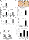
Induction of stem‐like phenotype by tumor‐conditioned medium (CM). (a) Expression of aldehyde dehydrogenase (ALDH), MDR1, CD90, ALP, and Oct‐4, normalized to GAPDH, was measured in human microvascular endothelial cells (HMVEC) after tumor CM treatment using real‐time RTPCR (*P < 0.01). (b) ALDH activity was analyzed using flow cytometry after treatment with tumor CM for 24 h. Proportion of high ALDH activity tumor endothelial cells (ALDH high TEC) was measured and counted. (c) HMVEC were photographed at 72 h after seeding into microwells that contained culture medium. Of note, HMVEC treated with tumor CM showed spheroid morphology with a smooth surface and high circularity. Scale bars, 50 μm. (d). Number of sphere‐forming cells in HMVEC treated with control CM or tumor CM (*< 0.05). (e) Platelet‐derived growth factor (PDGF)‐A concentration was determined in control CM and tumor CM (*P < 0.01). (f) HMVEC were treated by tumor CM or PDGF‐A. ALDH expression was determined by real‐time RTPCR (*P < 0.01).
Figure 4
Figure 4
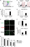
High aldehyde dehydrogenase activity tumor endothelial cells (ALDH high TEC) show resistance to fluorouracil (5‐FU). (a) ALDH high TEC and low ALDH activity TEC (ALDH low TEC) in the indicated gates were sorted using FACS Aria II (BD Biosciences, San Jose, CA, USA). (b) ALDH mRNA levels were analyzed using real‐time RTPCR in the sorted ALDH high/low TEC (*< 0.01). (c) The dead cell population treated with 5‐FU was analyzed using flow cytometry as detected for propidium iodide (PI)‐ or annexin V‐positive cells (red box). (d) Percentages of dead cells were compared between ALDH high TEC and ALDH low TEC (*< 0.01). (e) Double immunofluorescence staining for endothelial marker CD31 (green) and ALDH (red) in super‐metastatic human melanoma (A375SM) tumor tissues following injection of vehicle (control) or 5‐FU. White arrowheads show ALDH and CD31‐double‐positive blood EC. Scale bar, 200 μm. (f) Microvessel density (MVD) was calculated from the CD31‐positive area as a percentage of the total area by Image J in control‐ and 5‐FU‐treated tumors. n = 5 (*P < 0.01). (g) Percentage of ALDH‐positive cells in the total CD31‐positive cells was calculated using Image J in control‐ and 5‐FU‐treated tumors. n = 5 (*P < 0.01). (h) Sensitivity to 5‐FU in ALDH siRNA‐transfected TEC was compared with that of control siRNA‐transfected TEC and non‐treated TEC by the MTS assay. N.S., not significant.
Figure 5
Figure 5
High aldehyde dehydrogenase activity tumor endothelial cells (ALDH high TEC) show a higher aneuploidy grade. (a) Cell cycle distribution was analyzed using propidium iodide (PI). Red line, cell population in G2/M phase. (b) FISH analysis carried out using a spectrum red‐conjugated mouse chromosome‐17 locus‐specific probe (red spot). Nuclei are stained with DAPI (blue). (c) Quantification of chromosome 17 FISH signals in cells. Red rectangles show cells with five or more obtained signals.

Similar articles

Cited by

References

    1. Folkman J. Angiogenesis: an organizing principle for drug discovery? Nat Rev Drug Discov 2007; 6: 273–86. - PubMed
    1. Auerbach R, Akhtar N, Lewis RL, Shinners BL. Angiogenesis assays: problems and pitfalls. Cancer Metastasis Rev 2000; 19: 167–72. - PubMed
    1. Kerbel RS, Yu J, Tran J et al Possible mechanisms of acquired resistance to anti‐angiogenic drugs: implications for the use of combination therapy approaches. Cancer Metastasis Rev 2001; 20: 79–86. - PubMed
    1. Kesari S, Schiff D, Doherty L et al Phase II study of metronomic chemotherapy for recurrent malignant gliomas in adults. Neuro Oncol 2007; 9: 354–63. - PMC - PubMed
    1. Krzyzanowska MK, Tannock IF, Lockwood G, Knox J, Moore M, Bjarnason GA. A phase II trial of continuous low‐dose oral cyclophosphamide and celecoxib in patients with renal cell carcinoma. Cancer Chemother Pharmacol 2007; 60: 135–41. - PubMed

MeSH terms

LinkOut - more resources