

Dynamic imaging demonstrates that pulsed electromagnetic fields (PEMF) suppress IL-6 transcription in bovine nucleus pulposus cells
- PMID: 28851112
- PMCID: PMC5873378
- DOI: 10.1002/jor.23713
Dynamic imaging demonstrates that pulsed electromagnetic fields (PEMF) suppress IL-6 transcription in bovine nucleus pulposus cells
Abstract
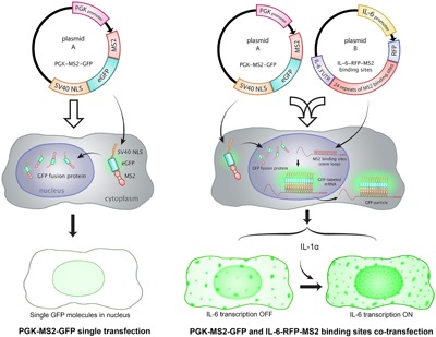
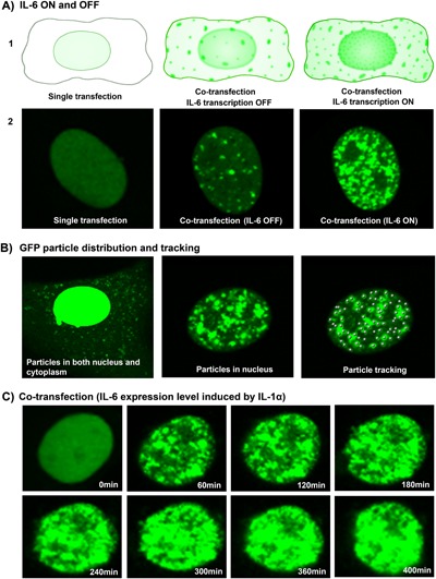
Inflammatory cytokines play a dominant role in the pathogenesis of disc degeneration. Pulsed electromagnetic fields (PEMF) are noninvasive biophysical stimulus that has been used extensively in the orthopaedic field for many years. However, the specific cellular responses and mechanisms involved are still unclear. The objective of this study was to assess the time-dependent PEMF effects on pro-inflammatory factor IL-6 expression in disc nucleus pulposus cells using a novel green fluorescence protein (GFP) reporter system. An MS2-tagged GFP reporter system driven by IL-6 promoter was constructed to visualize PEMF treatment effect on IL-6 transcription in single living cells. IL-6-MS2 reporter-labeled cells were treated with IL-1α to mimic the in situ inflammatory environment of degenerative disc while simultaneously exposed to PEMF continuously for 4 h. Time-lapse imaging was recorded using a confocal microscope to track dynamic IL-6 transcription activity that was demonstrated by GFP. Finally, real-time RT-PCR was performed to confirm the imaging data. Live cell imaging demonstrated that pro-inflammatory factor IL-1α significantly promoted IL-6 transcription over time as compared with DMEM basal medium condition. Imaging and PCR data demonstrated that the inductive effect of IL-1α on IL-6 expression could be significantly inhibited by PEMF treatment in a time-dependent manner (early as 2 h of stimulus initiation). Our data suggest that PEMF may have a role in the clinical management of patients with chronic low back pain. Furthermore, this study shows that the MS2-tagged GFP reporter system is a useful tool for visualizing the dynamic events of mechanobiology in musculoskeletal research. © 2017 The Authors. Journal of Orthopaedic Research® Published by Wiley Periodicals, Inc. on behalf of Orthopaedic Research Society. J Orthop Res 35:778-787, 2018.
Keywords: IL-6 mRNA expression; MS2-GFP reporter; dynamic imaging; pulsed electromagnetic fields; spine/disc biology.
© 2017 The Authors. Journal of Orthopaedic Research® Published by Wiley Periodicals, Inc. on behalf of Orthopaedic Research Society.
Figures


Similar articles
-
Pulsed Electromagnetic Fields Reduce Interleukin-6 Expression in Intervertebral Disc Cells Via Nuclear Factor-κβ and Mitogen-Activated Protein Kinase p38 Pathways.Spine (Phila Pa 1976). 2019 Nov 15;44(22):E1290-E1297. doi: 10.1097/BRS.0000000000003136. Spine (Phila Pa 1976). 2019. PMID: 31689248
-
Pulsed electromagnetic field (PEMF) treatment reduces expression of genes associated with disc degeneration in human intervertebral disc cells.Spine J. 2016 Jun;16(6):770-6. doi: 10.1016/j.spinee.2016.01.003. Epub 2016 Jan 15. Spine J. 2016. PMID: 26780754
-
Hypotonicity differentially affects inflammatory marker production by nucleus pulposus tissue in simulated disc degeneration versus herniation.J Orthop Res. 2019 May;37(5):1110-1116. doi: 10.1002/jor.24268. Epub 2019 Apr 1. J Orthop Res. 2019. PMID: 30835843 Free PMC article.
-
Pulsed Electromagnetic Fields (PEMF)-Physiological Response and Its Potential in Trauma Treatment.Int J Mol Sci. 2023 Jul 8;24(14):11239. doi: 10.3390/ijms241411239. Int J Mol Sci. 2023. PMID: 37510998 Free PMC article. Review.
-
Veterinary applications of pulsed electromagnetic field therapy.Res Vet Sci. 2018 Aug;119:1-8. doi: 10.1016/j.rvsc.2018.05.005. Epub 2018 May 7. Res Vet Sci. 2018. PMID: 29775839 Review.
Cited by
-
Pulsed electromagnetic field applications: A corporate perspective.J Orthop Translat. 2017 Mar 31;9:60-68. doi: 10.1016/j.jot.2017.02.006. eCollection 2017 Apr. J Orthop Translat. 2017. PMID: 29662800 Free PMC article. Review.
-
In Vitro Validation of Pulsed Electromagnetic Field (PEMF) as an Effective Countermeasure Against Inflammatory-Mediated Intervertebral Disc Degeneration.JOR Spine. 2025 May 19;8(2):e70077. doi: 10.1002/jsp2.70077. eCollection 2025 Jun. JOR Spine. 2025. PMID: 40391205 Free PMC article.
-
Pulsed Electromagnetic Fields for Cervical Spine Fusion in Patients with Risk Factors for Pseudarthrosis.Orthop Rev (Pavia). 2024 Oct 3;16:122534. doi: 10.52965/001c.122534. eCollection 2024. Orthop Rev (Pavia). 2024. PMID: 39698480 Free PMC article.
-
Gathering Evidence to Leverage Musculoskeletal Magnetic Stimulation Towards Clinical Applicability.Small Sci. 2024 Feb 26;4(5):2300303. doi: 10.1002/smsc.202300303. eCollection 2024 May. Small Sci. 2024. PMID: 40213569 Free PMC article.
-
Coupling of pulsed electromagnetic fields (PEMF) therapy to molecular grounds of the cell.Am J Transl Res. 2018 May 15;10(5):1260-1272. eCollection 2018. Am J Transl Res. 2018. PMID: 29887943 Free PMC article. Review.
References
-
- Katz JN. 2006. Lumbar disc disorders and low‐back pain: socioeconomic factors and consequences. J Bone Joint Surg Am 88:21–24. - PubMed
-
- Hoy D, March L, Brooks P, et al. 2010. The global burden of low back pain: estimates from the Global Burden of Disease study. Ann Rheum Dis 73:968–974. - PubMed
-
- Martin BI, Deyo RA, Mirza SK, et al. 2008. Expenditures and health status among adults with back and neck problems. JAMA 299:656–664. - PubMed
-
- Kao TH, Peng YJ, Tsou HK, et al. 2014. Nerve growth factor promotes expression of novel genes in intervertebral disc cells that regulate tissue degradation: laboratory investigation. J Neurosurg Spine 21:653–661. - PubMed
Publication types
MeSH terms
Substances
Grants and funding
LinkOut - more resources
Full Text Sources
Other Literature Sources
