. 2017 Aug 29;16(1):147.
doi: 10.1186/s12943-017-0717-5.

Phospholipid Phosphatase 4 promotes proliferation and tumorigenesis, and activates Ca2+-permeable Cationic Channel in lung carcinoma cells

Affiliations

Phospholipid Phosphatase 4 promotes proliferation and tumorigenesis, and activates Ca2+-permeable Cationic Channel in lung carcinoma cells

Xin Zhang et al. Mol Cancer. .

Abstract

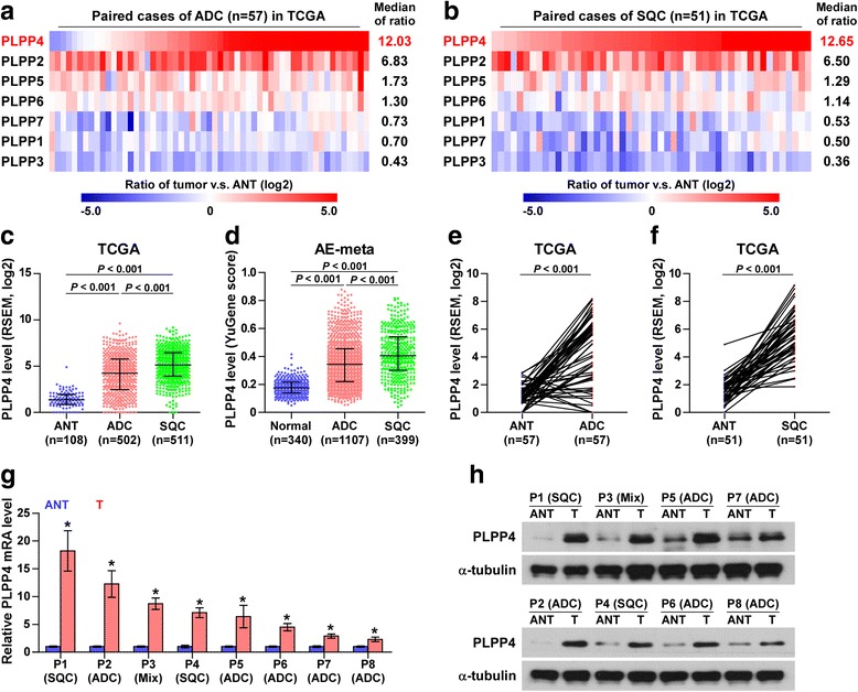
Background: Phospholipid phosphatase 4 (PPAPDC1A or PLPP4) has been demonstrated to be involved in the malignant process of many cancers. The purpose of this study was to investigate the clinical significance and biological roles of PLPP4 in lung carcinoma.

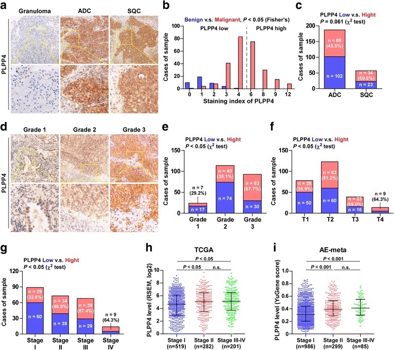
Methods: PLPP4 expression was examined in 8 paired lung carcinoma tissues by real-time PCR and in 265 lung carcinoma tissues by immunohistochemistry (IHC). Statistical analysis was performed to evaluate the clinical correlation between PLPP4 expression and clinicopathological features and survival in lung carcinoma patients. In vitro and in vivo assays were performed to assess the biological roles of PLPP4 in lung carcinoma. Fluorescence-activated cell sorting, Western blotting and luciferase assays were used to identify the underlying pathway through which PLPP4 silencing mediates biological roles in lung carcinoma.

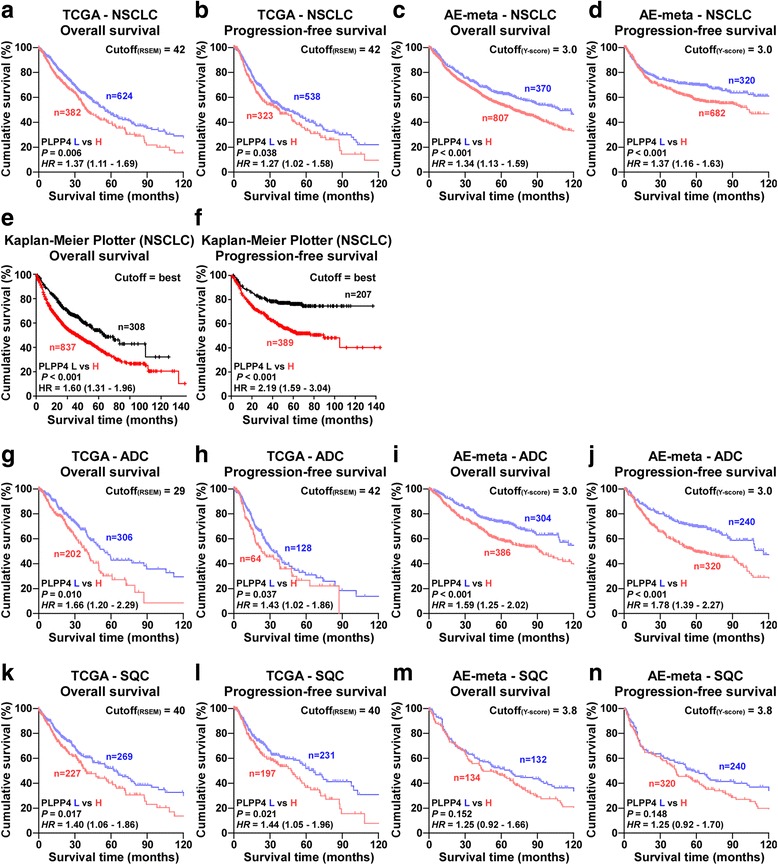
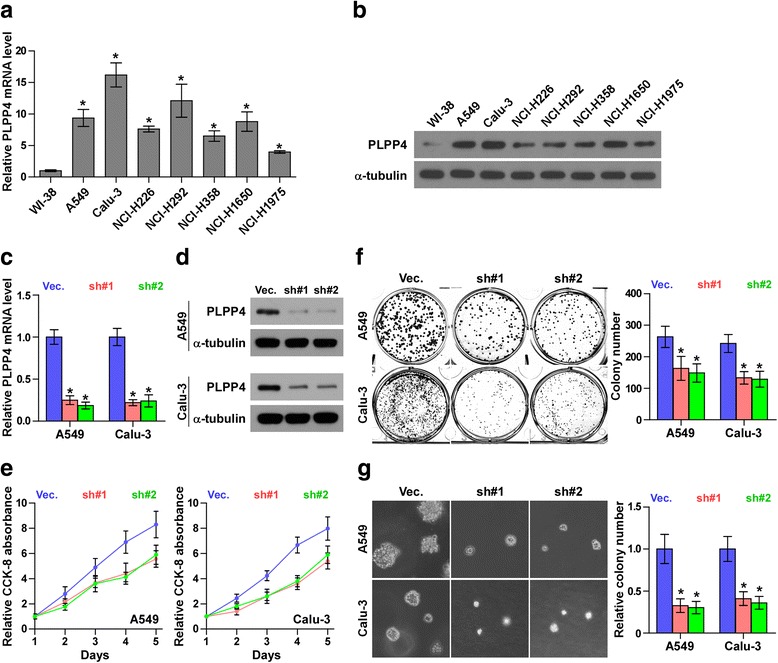
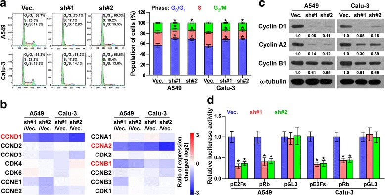
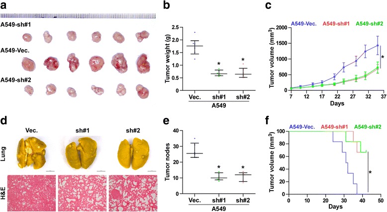
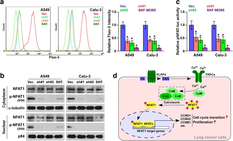
Results: PLPP4 is differentially elevated in lung adenocarcinoma (ADC) and lung squamous cell carcinoma (SQC) tissues. Statistical analysis demonstrated that high expression of PLPP4 significantly and positively correlated with clinicopathological features, including pathological grade, T category and stage, and poor overall and progression-free survival in lung carcinoma patients. Silencing PLPP4 inhibits proliferation and cell cycle progression in vitro and tumorigenesis in vivo in lung carcinoma cells. Our results further reveal that PLPP4 silencing inhibits Ca2+-permeable cationic channel, suggesting that downregulation of PLPP4 inhibits proliferation and tumorigenesis in lung carcinoma cells via reducing the influx of intracellular Ca2+.

Conclusion: Our results indicate that PLPP4 may hold promise as a novel marker for the diagnosis of lung carcinoma and as a potential therapeutic target to facilitate the development of novel treatment for lung carcinoma.

Keywords: Ca2+-permeable cationic channel; Cell cycle; Lung carcinoma; PLPP4; Proliferation; Therapeutic target; Tumorigenesis.


Conflict of interest statement

Ethics approval and consent to participate

This study was conducted in compliance with the declaration of Helsinki regarding ethical principles for medical research involving human subjects. The research protocols were approved by the Ethnic Committee of the Jiangmen Central Hospital, Affiliated Jiangmen Hospital of Sun Yat-sen University. The ethics approval numbers of patient related experiments was 20170718A. The ethics approval statements for animal work were provided by Comments of laboratory animal ethical Committee of Guangdong Medical University. The ethics approval numbers of animal work was GDY1701033.

Consent for publication

Not applicable.

Competing interests

The authors declare that they have no competing interests.

Publisher’s Note

Springer Nature remains neutral with regard to jurisdictional claims in published maps and institutional affiliations.

Figures

Fig. 1
Fig. 1
PLPP4 is upregulated in lung carcinoma. a PLPP1-7 expression levels in the RNA sequencing profiles of 57 paired lung adenocarcinoma (ADC) tissues from TCGA dataset. b PLPP1-7 expression levels in the RNA sequencing profile of 51 paired lung squamous cell carcinoma (SQC) tissues from TCGA dataset. c Expression of PLPP4 in the adjacent normal tissues (ANT), ADC tissues and SQC tissues in TCGA lung carcinoma profiles. d Expression of PLPP4 in the adjacent normal tissues (ANT), ADC tissues and SQC tissues in the lung carcinoma profiles from ArrayExpress. e PLPP4 expression was upregulated in the RNA sequencing profile of 57 ADC tissues from TCGA dataset compared with that in ANT. f PLPP4 expression was upregulated in the RNA sequencing profile of 51 SQC tissues from TCGA dataset compared with that in ANT. g and h mRNA and protein expression levels of PLPP4 in eight paired lung tissues, including 5 paired ADC tissues, 2 paired SQC tissues and 1 lung adenosquamous carcinoma tissues (Mix). α-Tubulin was used as a loading control. The average PLPP4 mRNA expression level was normalized to the expression of GAPDH. Each bar represents the mean values ± SD of three independent experiments. *P < 0.05
Fig. 2
Fig. 2
High PLPP4 expression correlates with advanced clinicopathological features in lung carcinoma patients. a Representative images of PLPP4 expression in lung granuloma tissues, ADC tissues and SQC tissues. b The number of lung granuloma tissues and lung carcinoma tissues stratified by IHC staining index. c Percentages and number of samples showing high or low PLPP4 expression in ADC and SQC tissues. d Representative images of PLPP4 expression in lung carcinoma tissues with different clinical grade. e Percentages and number of samples showing high or low PLPP4 expression in lung carcinoma tissues with different grades. f Percentages and number of samples showing high or low PLPP4 expression in lung carcinoma tissues with tumor volume. g Percentages and number of samples showing high or low PLPP4 expression in lung carcinoma tissues with different stages. h and i Expression levels of PLPP4 in lung carcinoma tissues with different stages from lung carcinoma datasets from TCGA and ArrayExpress
Fig. 3
Fig. 3
High PLPP4 expression correlates with poor survival in lung carcinoma patients. a-f Overall survival and progression-free survival curves from TCGA, ArrayExpress and Kaplan-Meier Plotter profiles for non-small-cell lung carcinoma (NSCLC) patients stratified by high and low expression of PLPP4. g-j Overall survival and progression-free survival curves from TCGA and ArrayExpress profiles for ADC patients stratified by high and low expression of PLPP4. k-n Overall survival and progression-free survival curves from TCGA and ArrayExpress profiles for SQC patients stratified by high and low expression of PLPP4
Fig. 4
Fig. 4
Silencing PLPP4 inhibits proliferation in lung carcinoma cells. a and b Real-time PCR and Western blotting analysis of PLPP4 expression in WI-38 and lung carcinoma cell lines. GAPDH was used as the endogenous control for RT-PCR and α-Tubulin was used as the loading control in the Western blot. Each bar represents the mean values ± SD of three independent experiments. *P < 0.05. c and d Real-time PCR and Western blot of the indicated lung carcinoma cells transfected with PLPP4-RNAi-vector, PLPP4-RNAi#1 and PLPP4-RNAi#2. GAPDH was used as the endogenous control for RT-PCR and α-Tubulin was used as the loading control in the Western blot. Each bar represents the mean values ± SD of three independent experiments. *P < 0.05. e CCK8 assays revealed that silencing PLPP4 reduced cell viability in lung carcinoma cells. Each bar represents the mean values ± SD of three independent experiments. *P < 0.05. f Silencing PLPP4 reduced the mean colony number according to the colony formation assay. Each bar represents the mean values ± SD of three independent experiments. *P < 0.05. g Representative micrographs and colony numbers from the anchorage-independent growth assay. Each bar represents the mean values ± SD of three independent experiments. *P < 0.05
Fig. 5
Fig. 5
Silencing PLPP4 inhibited cell cycle progression in lung carcinoma cells. a Flow cytometric analysis of the indicated lungcarcinoma cells. Each bar represents the mean values ± SD of three independent experiments. *P < 0.05. b Real-time PCR analysis of multiple cell cycle regulators in the indicated cells. Transcript levels were normalized by GAPDH expression. c Western blot analysis of multiple cell cycle regulators expression in the indicated cells. α-Tubulin was used as the loading control. Protein expression levels of cyclin D1, A2 and B1 were quantified by densitometry using Image J Software, and normalized to the corresponding expression levels of α-tubulin. Sample 1 was used as the standard. d Relative E2F and Rb reporter activity in the indicated cells. Each bar represents the mean values ± SD of three independent experiments. *P < 0.05
Fig. 6
Fig. 6
PLPP4 silencing inhibits the tumorigenesis and lung colonization of lung carcinoma cells in vivo. a Images of excised tumors from six BALB/c mice at 30 days after injection with the indicated cells. b Average weight of excised tumors from the indicated mice. Each bar represents the median values ± quartile values. *P < 0.05. c Tumor volumes were measured every five days. Each bar represents the median values ± quartile values. d and e In vivo lung metastasis assays of A549 cells with PLPP4 knockdown. Lung metastases in mice were confirmed by H&E staining. The number of lung tumor nests in each group was counted under a low power field (LPF) and is presented as the median values ± quartile values (right panel). *P < 0.05. f Kaplan-Meier survival curves of the indicated mice
Fig. 7
Fig. 7
Silencing PLPP4 inhibits Ca2+-permeable cationic channel in lung carcinoma cells. a FACS analysis showed that silencing PLPP4 decreased intracellular Ca2+ in the indicated cells. Each bar represents the mean values ± SD of three independent experiments. *P < 0.05. b Western blot analysis of cytoplasmic and nuclear expression of the S54 phosphorylated NFAT1 in the indicated cells. α-Tubulin was used as the loading control and the nuclear protein p84 was used as the nuclear protein marker. c Relative NFAT reporter activity in the indicated cells. Each bar represents the mean values ± SD of three independent experiments. *P < 0.05. d Hypothetical model illustrating that activation of Ca2+-permeable cationic channel by PLPP4 contributes to proliferation and cell cycle progression in lung cancer cells

References

    1. Herbst RS, Heymach JV, Lippman SM. Lung cancer. N Engl J Med. 2008;359:1367–1380. doi: 10.1056/NEJMra0802714. - DOI - PMC - PubMed
    1. Gilligan D, Nicolson M, Smith I, Groen H, Dalesio O, Goldstraw P, Hatton M, Hopwood P, Manegold C, Schramel F, et al. Preoperative chemotherapy in patients with resectable non-small cell lung cancer: results of the MRC LU22/NVALT 2/EORTC 08012 multicentre randomised trial and update of systematic review. Lancet. 2007;369:1929–1937. doi: 10.1016/S0140-6736(07)60714-4. - DOI - PubMed
    1. Waggoner DW, Xu J, Singh I, Jasinska R, Zhang QX, Brindley DN. Structural organization of mammalian lipid phosphate phosphatases: implications for signal transduction. Biochim Biophys Acta. 1999;1439:299–316. doi: 10.1016/S1388-1981(99)00102-X. - DOI - PubMed
    1. Sciorra VA, Morris AJ. Roles for lipid phosphate phosphatases in regulation of cellular signaling. Biochim Biophys Acta. 2002;1582:45–51. doi: 10.1016/S1388-1981(02)00136-1. - DOI - PubMed
    1. Sydorenko V, Shuba Y, Thebault S, Roudbaraki M, Lepage G, Prevarskaya N, Skryma R. Receptor-coupled, DAG-gated Ca2+−permeable cationic channels in LNCaP human prostate cancer epithelial cells. J Physiol. 2003;548:823–836. doi: 10.1113/jphysiol.2002.036772. - DOI - PMC - PubMed