Review
. 2017 Aug 29;16(1):145.
doi: 10.1186/s12943-017-0706-8.

Role of exosomal proteins in cancer diagnosis

Affiliations
Review

Role of exosomal proteins in cancer diagnosis

Weihua Li et al. Mol Cancer. .

Abstract

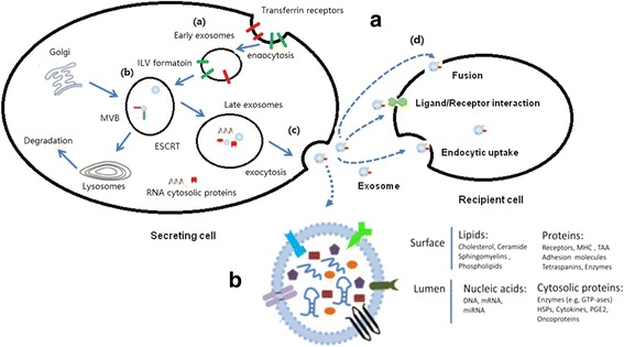
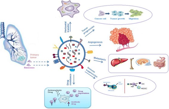
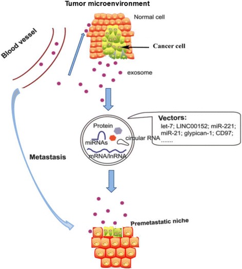
Exosomes are emerging as a new type of cancer biomarkers. Exosome is a bilayered nano-sized vesicle secreted by various living cells in all body fluids. Based on the expanding albeit incomplete knowledge of their biogenesis, secretion by cells and cancer cell-specific molecular and genetic contents, exosomes are viewed as promising, clinically-relevant surrogates of cancer progression and response to therapy. Preliminary proteomic, genetic and functional profiling of cancer cell-derived or cancer plasma-derived exosomes confirms their unique characteristics. Alterations in protein or nucleic acid profiles of exosomes in plasma correlate with pathological processes of many diseases including cancer. However, previous studies on exosome application in cancer diagnosis and treatment mainly focussed on miRNAs. With the development of rapid large-scale production, purification, extraction and screening of exosomal contents, exosomal protein application can be explored for early stage cancer diagnosis, monitoring and prognosis evaluation. Here, we summarized the recent developments in application of exosomal proteins for cancer diagnosis.

Keywords: Biomarker; Cancer; Diagnose; Exosome.


Conflict of interest statement

Competing interests

The authors declare that they have no competing interests.

Publisher’s Note

Springer Nature remains neutral with regard to jurisdictional claims in published maps and institutional affiliations.

Figures

Fig. 1
Fig. 1
Biogenesis, release, structure, and uptake of exosomes. In A, exosomes are repressed by small vesicles of different sizes that are formed as the ILV by budding into early endosomes and multivesicular body and are released by fusion of multivesicular body fuse with lysosomes. (a). By endocytosis of membrane segments, the initial endosome arises, containing receptors and transmembrane proteins of the plasma membrane. (b). Instead of lysosomal degradation, the matured late endosome transforms by inward budding of tiny vesicles into a multivesicular body. Furthermore, the exosomal cargo as proteins and miRNA, is selectively loaded into the vesicles. (c). Exosomes are released into the extracellular space by fusion of the multivesicular body with the plasma membrane. (d). Cell-secreted exosomes can travel through biological fluids (e.g. serum, lymph) and be internalized by recipient cells. Exosomes transfer information to the target cells through three main ways: (1) direct fusion with plasma membrane; (2) receptor-ligand interaction; (3) endocytosis by phagocytosis. In B, The membrane of the MVBs bulges inward to form exosomes. During this process, proteins (e.g., receptor, cytoplasmic proteins, tetraspanin), nucleic acids (e.g., DNA, mRNA, miRNA), and lipids (e.g., cholesterol, ceramide) are packed into exosomes in a cell type-dependent manner
Fig. 2
Fig. 2
Role of exosomes in sustaining cancer resistance networks. Exosome mediated export of biological material can induce a microenvironment favorable for resistance. Exosome released factor can promote a EMT cell morphology, resulting in stemness; b promote fibroblast like cell formation that causes desmoplatic reaction (stromal reaction); c promote immune escape mechanisms and d promote angiogenesis and metastasis. The miRNAs expelled by exosomes can regulate multiple signaling pathways that cumulatively promote resistant phenotype of most tumors
Fig. 3
Fig. 3
Role of exosomes in NSCLC. Exosomes have a key role in: 1 horizontal transfer of mRNAs and miRNAs from cancer cells to cells of microenvironment; 2 tumor progression, inducing cells motility; 3 angiogenesis; 4: metastatization; 5: immunosuppression; 6: drug resistance
Fig. 4
Fig. 4
The schematic representation of the role that exosomes play in gastric cancer carcinogenesis and metastasis is summarized in the figure

References

    1. Mignot G, Roux S, Thery C, Segura E, Zitvogel L. Prospects for exosomes in immunotherapy of cancer. J Cell Mol Med. 2006;10:376–388. doi: 10.1111/j.1582-4934.2006.tb00406.x. - DOI - PMC - PubMed
    1. Johnstone RM, Bianchini A, Teng K. Reticulocyte maturation and exosome release: transferrin receptor containing exosomes shows multiple plasma membrane functions. Blood. 1989;74:1844–1851. - PubMed
    1. Fader CM, Savina A, Sanchez D, Colombo MI. Exosome secretion and red cell maturation: exploring molecular components involved in the docking and fusion of multivesicular bodies in K562 cells. Blood Cells Mol Dis. 2005;35:153–157. doi: 10.1016/j.bcmd.2005.07.002. - DOI - PubMed
    1. Raposo G, Nijman HW, Stoorvogel W, Liejendekker R, Harding CV, Melief CJ, Geuze HJ. B lymphocytes secrete antigen-presenting vesicles. J Exp Med. 1996;183:1161–1172. doi: 10.1084/jem.183.3.1161. - DOI - PMC - PubMed
    1. Clayton A, Turkes A, Navabi H, Mason MD, Tabi Z. Induction of heat shock proteins in B-cell exosomes. J Cell Sci. 2005;118:3631–3638. doi: 10.1242/jcs.02494. - DOI - PubMed

Publication types