Clinical Trial
. 2017 Nov 9;130(19):2131-2145.
doi: 10.1182/blood-2017-05-782466. Epub 2017 Aug 29.

An aberrant NOTCH2-BCR signaling axis in B cells from patients with chronic GVHD

Affiliations
Clinical Trial

An aberrant NOTCH2-BCR signaling axis in B cells from patients with chronic GVHD

Jonathan C Poe et al. Blood. .

Abstract

B-cell receptor (BCR)-activated B cells contribute to pathogenesis in chronic graft-versus-host disease (cGVHD), a condition manifested by both B-cell autoreactivity and immune deficiency. We hypothesized that constitutive BCR activation precluded functional B-cell maturation in cGVHD. To address this, we examined BCR-NOTCH2 synergy because NOTCH has been shown to increase BCR responsiveness in normal mouse B cells. We conducted ex vivo activation and signaling assays of 30 primary samples from hematopoietic stem cell transplantation patients with and without cGVHD. Consistent with a molecular link between pathways, we found that BCR-NOTCH activation significantly increased the proximal BCR adapter protein BLNK. BCR-NOTCH activation also enabled persistent NOTCH2 surface expression, suggesting a positive feedback loop. Specific NOTCH2 blockade eliminated NOTCH-BCR activation and significantly altered NOTCH downstream targets and B-cell maturation/effector molecules. Examination of the molecular underpinnings of this "NOTCH2-BCR axis" in cGVHD revealed imbalanced expression of the transcription factors IRF4 and IRF8, each critical to B-cell differentiation and fate. All-trans retinoic acid (ATRA) increased IRF4 expression, restored the IRF4-to-IRF8 ratio, abrogated BCR-NOTCH hyperactivation, and reduced NOTCH2 expression in cGVHD B cells without compromising viability. ATRA-treated cGVHD B cells had elevated TLR9 and PAX5, but not BLIMP1 (a gene-expression pattern associated with mature follicular B cells) and also attained increased cytosine guanine dinucleotide responsiveness. Together, we reveal a mechanistic link between NOTCH2 activation and robust BCR responses to otherwise suboptimal amounts of surrogate antigen. Our findings suggest that peripheral B cells in cGVHD patients can be pharmacologically directed from hyperactivation toward maturity.


Figures

Figure 1.
Figure 1.
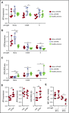
NOTCH2 ligation heightens ex vivo active cGVHD B-cell responses to minimal BCR engagement by surrogate antigen. B cells were magnetically purified to >95% from viably frozen PBMCs from HCT patients who at the time of sample collection had active cGVHD (Active; n = 9) or no cGVHD (No; n = 6). After plating B cells onto OP9 stromal cell monolayers or OP9-DL1 cells that express the NOTCH ligand DLL1, cultures were either treated with the γ-secretase inhibitor DAPT (10 μM in DMSO) to block Notch activation, or with DMSO alone. After 30 minutes, agonistic anti-IgM Ab was added to the appropriate wells at a concentration of 0.625 μg/mL. The cells were cultured for 72 hours, harvested, and flow cytometry analysis performed to assess Ki-67 expression. (A) Representative flow cytometry histograms for Ki-67 expression are shown for healthy donor (Healthy), no cGVHD (No), or active cGVHD (Active) donor B cells cultured in the presence of 0.625 μg/mL anti-IgM and plated on OP9 parental cells (top panels) or OP9-DL1 cells (bottom panels). The effect of DAPT treatment on active cGVHD B cells is also shown (right panels). (B-C) Frequency of Ki-67+ B cells for all patients assessed in each group where the B cells were stimulated with 0.625 μg/mL anti-IgM (B) or were not stimulated through the BCR (C). P values were determined using a nonpaired Student t test for intergroup comparisons, and paired Student t test for same group comparisons. (D) Representative flow cytometry histograms showing BLNK expression as assessed by intracellular flow cytometry in B cells from active cGVHD patients stimulated as described for panel A, with plating on OP9 cells or OP9-DL1 cells. (E) Median fluorescence intensity (MFI) expression for BLNK in B cells from n = 4 active cGVHD patients stimulated as described for panel A and cultured on OP9-DL1 cells. In some cultures, DAPT (10 μM) was added to inhibit NOTCH activation, or R406 (0.1 μM) was added to inhibit SYK. DMSO alone used as the vehicle control in parallel. P values were determined using a paired Student t test.
Figure 2.
Figure 2.
Hyperactivation of active cGVHD patient B cells to NOTCH ligand and anti-IgM is NOTCH2-dependent. (A-B) Anti-N2 mAb (N2) or ragweed isotype control mAb (Iso) were diluted in medium and added to 96-well plates containing OP9-DL1 feeder cell monolayers to achieve a final concentration of 1 μg/mL. B cells purified from viably frozen PBMCs of active cGVHD patients (n = 6) were then added to the plates. Following an initial culture period of 30 minutes, agonistic anti-IgM Ab was added to the appropriate wells at a final concentration of 0.625 μg/mL. The cells were cultured for 72 hours, harvested, and flow cytometry analysis performed to assess Ki-67 expression (A), and relative cell size by forward scatter (FSC) (B). P values were determined using a paired Student t test (*P < .05; **P < .01). (C) NOTCH-BCR stimulation with or without specific NOTCH2 blockade with anti-N2, as described in panels A and B. The cells were cultured for 24 hours, the B cells purified away from the feeder cells using magnetic microbeads, and RNA isolated. B-cell RNA was then subjected in NanoString gene-expression profiling to assess differences in NOTCH pathway-regulated genes between groups. Data points represent NanoString nCounter target count number of individual samples for each gene, graphed on a relative scale. Statistical analysis comparing the anti-N2– and isotype control–treated groups was performed using a paired, negative binomial test (*P < .05).
Figure 3.
Figure 3.
NOTCH2 expression is maintained on active cGVHD B cells in the presence of NOTCH ligand and BCR stimulation. (A-B) B cells were purified from viably frozen PBMCs of HCT patients with active cGVHD (Active, n = 4) or no cGVHD (No, n = 4) and plated in medium onto OP9-DL1 feeder cell monolayers. In some wells, the γ-secretase inhibitor DAPT was added for a final concentration of 10 μM, with DMSO alone used as the vehicle control in parallel. Following an initial culture period of 30 minutes, agonistic anti-IgM Ab was then added to the appropriate wells at a concentration of 0.625 μg/mL. The cells were cultured for 72 hours, harvested, and flow cytometry analysis performed to assess NOTCH2 surface expression on CD19+ B cells. (A) Representative histogram overlay showing relative NOTCH2 expression between patient groups and activation conditions (as indicated). (B) MFI values for all patients assessed under conditions of anti-IgM stimulation, either without or with DAPT. (C) B cells were purified from viably frozen PBMCs from healthy donors and then plated onto monolayers of parental OP9 cells or OP9-DL1 cells, with various concentrations of anti-IgM Ab (micrograms per milliliter, indicated by numbers in the OP9 histogram overlay). Following a culture period of 72 hours, the cells were harvested and analysis of NOTCH2 expression on CD19+ cells was performed by flow cytometry. In panel B, P values were determined using a nonpaired Student t test for Active vs No comparison, and a paired Student t test for Active (no DAPT) vs Active (DAPT) comparison. *P < .05.
Figure 4.
Figure 4.
Altered IRF4 and IRF8 gene expression in B cells from active cGVHD patients, with normalization by ATRA. B cells from HCT patients with active cGVHD vs no cGVHD (n = 6), or from healthy donors (n = 6) were cultured in medium alone, or with low-dose (0.625 μg/mL) or high-dose (5 μg/mL) concentrations of anti-IgM for 24 hours. The B cells were then harvested, total RNA isolated, and real-time qPCR analysis performed to assess the abundance of IRF4 and IRF8 transcripts. The relative expression level shown for each gene is normalized to the median value for healthy donors with no anti-IgM stimulation, represented as a value of 1. (A) IRF4 and (B) IRF8 mRNA levels in each of the 3 sample groups with and without BCR stimulation. (C) Normalized IRF4 and IRF8 mRNA expressed as the ratio of IRF4 to that of IRF8 transcripts (IRF4-to-IRF8). P values were determined using a nonpaired Student t test (*P < .05; **P < .01). (D) B cells from HCT patients with active cGVHD (n = 6) were cultured with ATRA (0.1 μM) or vehicle alone, followed 30 minutes later by the addition of 0.625 μg/mL anti-IgM. Following a 24-hour culture period, the B cells were then harvested, total RNA isolated, and real-time qPCR analysis performed to determine relative IRF4 and IRF8 expression, and the IRF4-to-IRF8 ratio, as described for panels B and C. (E) B cells from HCT patients with active cGVHD (n = 4-6) were plated onto OP9-DL1 monolayers in the presence of ATRA (0.1 μM) or vehicle alone. Following an initial culture period of 30 minutes, agonistic anti-IgM Ab was then added to the appropriate wells at a final concentration of 0.625 μg/mL. The cells were cultured for 24 hours, harvested, and intracellular staining with flow cytometry analysis performed to assess IRF4 and IRF8 levels. For each protein, data represent the ratio of ATRA-treated B cells over vehicle-treated B cells, with the dashed line included for reference to a value of 1.0.
Figure 5.
Figure 5.
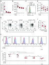
ATRA treatment of B cells from active cGVHD patients enhances genes involved in B-cell maturation. (A) B cells from active cGVHD patients (n = 4) were treated with ATRA (0.1 μM) or vehicle alone for 24 hours. The B cells were harvested, total RNA isolated, and real-time qPCR analysis performed to assess the expression levels of genes of interest. P values were determined using a paired Student t test (*P < .05; **P < .01). (B-C) Purified B cells from active cGVHD patients were plated in medium in the presence of ATRA (0.1 μM) or DMSO vehicle control. Following an initial culture period of 30 minutes, CpG (1 μg/mL) or anti-IgM Ab (0.625 μg/mL) were added alone or in combination. The cells were cultured for 72 hours, harvested, and flow cytometry analysis performed to assess Ki-67 and CD19 expression (B), as well as relative cell size by FSC (C). In panel B, the large gates with numbers outside indicate the frequency of Ki-67+ B cells, and the small gates (arrows) with numbers inside represent the frequency of proliferating, CD19low B cells.
Figure 6.
Figure 6.
ATRA suppresses NOTCH2-BCR hyperresponsiveness of B cells from active cGVHD patients. (A-D) B cells from HCT patients with active cGVHD (n = 6) were plated in medium on monolayers of OP9-DL1 feeder cells and exposed to ATRA (0.1 μM) or vehicle alone. Following an initial culture period of 30 minutes, agonistic anti-IgM Ab was then added to the appropriate wells at a final concentration of 0.625 μg/mL. The cells were cultured for 72 hours, harvested, and flow cytometry analysis performed to assess B-cell Ki-67 expression (A), FSC as a measure of relative cell size (B), cell-surface NOTCH2 expression (C), and number of live cells (D). (E-F) B cells from HCT patients with active cGVHD (n = 4) were plated in medium on monolayers of OP9-DL1 feeder cells and exposed to ATRA (0.1 μM), vehicle alone, anti-N2 mAb, or isotype control mAb. Following an initial culture period of 30 minutes, agonistic anti-IgM Ab was then added to the appropriate wells at a final concentration of 0.625 μg/mL. The cells were cultured for 72 hours, harvested, and flow cytometry analysis performed to assess B-cell surface proteins (E) and intracellular ERK1/2 protein phosphorylation (F). (E) Panel i shows representative dot plots from 1 patient for the treatments indicated; panel ii shows the frequency of CD38+IgD B cells from all 4 patients under the indicated culture conditions. (F) Panel i shows representative histograms from 1 patient for the treatments indicated; panel ii shows phospho-ERK MFI levels in B cells from all 4 patients under the indicated culture conditions. P values were determined using a paired Student t test. n.s., not significant.
Figure 7.
Figure 7.
Model for a NOTCH2-BCR axis in the generation of pathogenic B cells in cGVHD. (A) Reduced activation threshold response to antigen and to NOTCH ligand. High BAFF levels in the cGVHD setting leads to the preferential survival of B cells with MZ-like properties. These B cells are driven by an abnormally low ratio of IRF4 to IRF8, and are dependent on NOTCH2 activation of target genes. BCR-NOTCH2 synergy in these B cells leads to pathogenic Allo-Ab production. (B) ATRA exposure normalizes the phenotype of cGVHD B cells by increasing the ratio of IRF4 to IRF8, eliminating NOTCH2 dependence. This in turn leads to enhanced expression of PAX5 and TLR9, a genetic profile associated with mature follicular B cells capable of producing protective Ab against bacteria and viruses, as well as to vaccines. Importantly, these B cells attain functional memory essential for long-term humoral immunity.

Comment in

Similar articles

Cited by

References

    1. Cutler CS, Koreth J, Ritz J. Mechanistic approaches for the prevention and treatment of chronic GVHD. Blood. 2017;129(1):22-29. - PMC - PubMed
    1. MacDonald KP, Hill GR, Blazar BR. Chronic graft-versus-host disease: biological insights from preclinical and clinical studies. Blood. 2017;129(1):13-21. - PMC - PubMed
    1. Gea-Banacloche J, Komanduri KV, Carpenter P, et al. . National Institutes of Health Hematopoietic Cell Transplantation Late Effects Initiative: The Immune Dysregulation and Pathobiology Working Group Report. Biol Blood Marrow Transplant. 2017;23(6):870-881. - PMC - PubMed
    1. Tivol E, Komorowski R, Drobyski WR. Emergent autoimmunity in graft-versus-host disease. Blood. 2005;105(12):4885-4891. - PMC - PubMed
    1. Allen JL, Fore MS, Wooten J, et al. . B cells from patients with chronic GVHD are activated and primed for survival via BAFF-mediated pathways. Blood. 2012;120(12):2529-2536. - PMC - PubMed

Publication types

MeSH terms