Review
. 2017 Sep 1;144(17):3012-3021.
doi: 10.1242/dev.153171.

Microtubule organization, dynamics and functions in differentiated cells

Affiliations
Review

Microtubule organization, dynamics and functions in differentiated cells

Andrew Muroyama et al. Development. .

Abstract

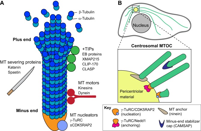
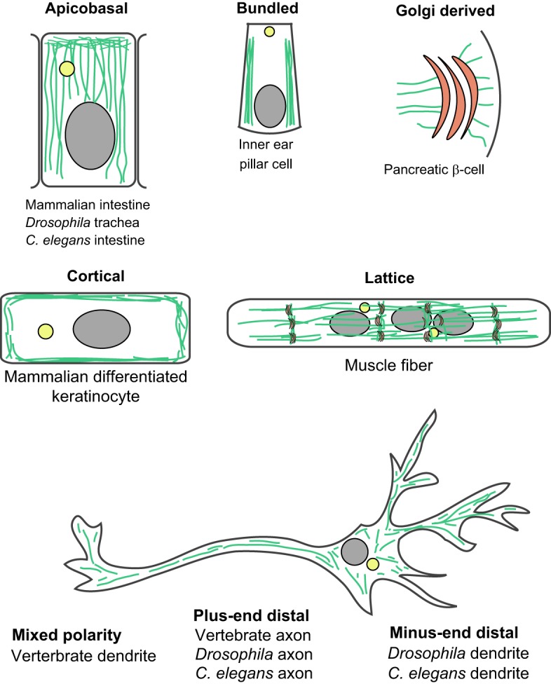
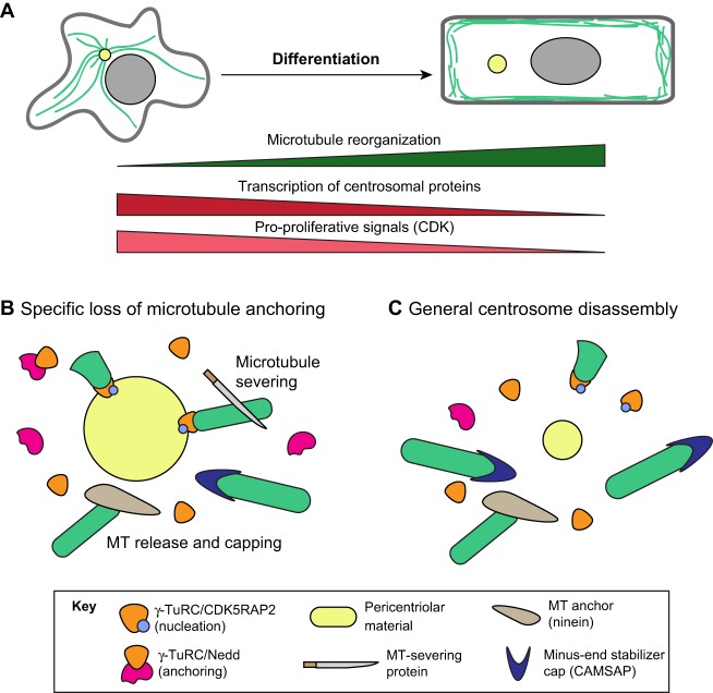
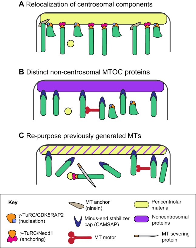
Over the past several decades, numerous studies have greatly expanded our knowledge about how microtubule organization and dynamics are controlled in cultured cells in vitro However, our understanding of microtubule dynamics and functions in vivo, in differentiated cells and tissues, remains under-explored. Recent advances in generating genetic tools and imaging technologies to probe microtubules in situ, coupled with an increased interest in the functions of this cytoskeletal network in differentiated cells, are resulting in a renaissance. Here, we discuss the lessons learned from such approaches, which have revealed that, although some differentiated cells utilize conserved strategies to remodel microtubules, there is considerable diversity in the underlying molecular mechanisms of microtubule reorganization. This highlights a continued need to explore how differentiated cells regulate microtubule geometry in vivo.

Keywords: Centrosome; Differentiation; Microtubule; Nucleation.


Conflict of interest statement

Competing interestsThe authors declare no competing or financial interests.

Figures

Fig. 1.
Fig. 1.
Regulators of microtubule dynamics and organization. (A) Numerous microtubule-associated proteins (MAPs) influence microtubule behavior. Many of these, such as EB proteins, XMAP215, CLIP-170 and CLASP proteins, regulate plus-tip dynamics and are collectively known as microtubule plus-end tracking proteins (+TIPs). Only a few proteins are known to bind specifically to the minus end. One of these, the γ-tubulin ring complex (γ-TuRC), is the primary microtubule nucleator in the cell. Nucleation by γ-TuRCs can be modulated by activators such as CDK5RAP2. Microtubule motors can intrinsically influence microtubule dynamics and also regulate microtubule organization by guiding microtubules along existing filaments. Microtubule-severing proteins induce breaks along the length of the filament to impact microtubule organization within the cell. (B) The centrosome is the primary microtubule organizing center (MTOC) in many proliferative cells. However, note that non-centrosomal microtubules and centrosomal microtubules can co-exist within the same cell. MTOC activity is conferred through both microtubule nucleation and anchoring abilities. CDK5RAP2 and Nedd1, acting via γ-TuRC, can promote these activities, respectively, but both basal activity and other activators are also likely to be involved. Ninein colocalizes with microtubule minus ends and may play a role in anchoring. CAMSAP proteins also preferentially localize to microtubule minus ends and serve to stabilize and potentially cap minus ends.
Fig. 2.
Fig. 2.
Differentiated animal cells form a variety of non-centrosomal MT arrays. Array geometry is highly cell type specific, but similar cells across species form analogous arrays. Selected representative cell types for different array geometries are illustrated.
Fig. 3.
Fig. 3.
Models for differentiation-induced centrosome inactivation. (A) Both transcriptional downregulation of centrosomal components and decreased signaling through cell-cycle regulators accompany microtubule reorganization. (B) Specific loss of microtubule anchoring can be the first step in centrosome inactivation; this can be mediated through increased microtubule severing or delocalization/degradation of specific anchoring factors. (C) Centrosome inactivation can also be caused by a general dispersal of pericentriolar material. Note that the mechanisms illustrated in B and C are not mutually exclusive.
Fig. 4.
Fig. 4.
Potential models of non-centrosomal MTOC activation. (A) Centrosomal proteins can be relocalized to a novel cellular site to re-specify that site for microtubule nucleation and/or anchoring. (B) A distinct set of non-centrosomal proteins can be utilized to generate non-centrosomal MTOCs. (C) Non-centrosomal arrays can be generated through microtubule severing and subsequent reorganization independently of new nucleation. These models are not mutually exclusive.

References

    1. Abal M., Piel M., Bouckson-Castaing V., Mogensen M., Sibarita J. B. and Bornens M. (2002). Microtubule release from the centrosome in migrating cells. J. Cell Biol. 159, 731-737. 10.1083/jcb.200207076 - DOI - PMC - PubMed
    1. Achler C., Filmer D., Merte C. and Drenckhahn D. (1989). Role of microtubules in polarized delivery of apical membrane proteins to the brush border of the intestinal epithelium. J. Cell Biol. 109, 179-189. 10.1083/jcb.109.1.179 - DOI - PMC - PubMed
    1. Ahmad F. J., Yu W., McNally F. J. and Baas P. W. (1999). An essential role for katanin in severing microtubules in the neuron. J. Cell Biol. 145, 305-315. 10.1083/jcb.145.2.305 - DOI - PMC - PubMed
    1. Akhmanova A. and Hoogenraad C. C. (2015). Microtubule minus-end-targeting proteins. Curr. Biol. 25, R162-R171. 10.1016/j.cub.2014.12.027 - DOI - PubMed
    1. Akhmanova A. and Steinmetz M. O. (2008). Tracking the ends: a dynamic protein network controls the fate of microtubule tips. Nat. Rev. Mol. Cell Biol. 9, 309-322. 10.1038/nrm2369 - DOI - PubMed

Publication types