. 2017 Oct 2;214(10):2859-2873.
doi: 10.1084/jem.20171320. Epub 2017 Aug 29.

Gpr158 mediates osteocalcin's regulation of cognition

Affiliations

Gpr158 mediates osteocalcin's regulation of cognition

Lori Khrimian et al. J Exp Med. .

Abstract

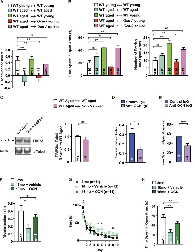
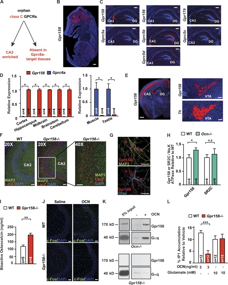
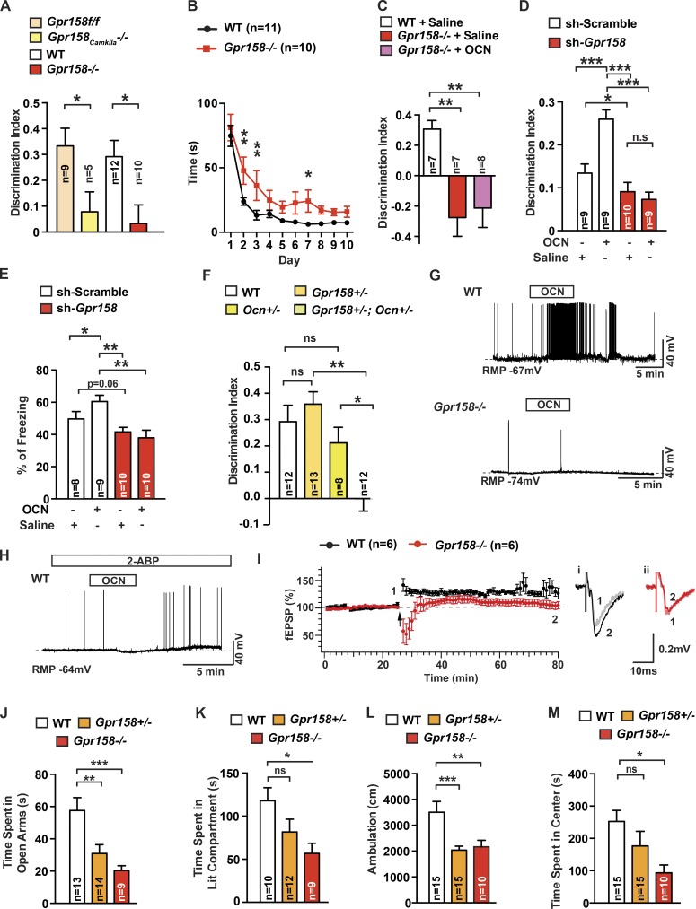
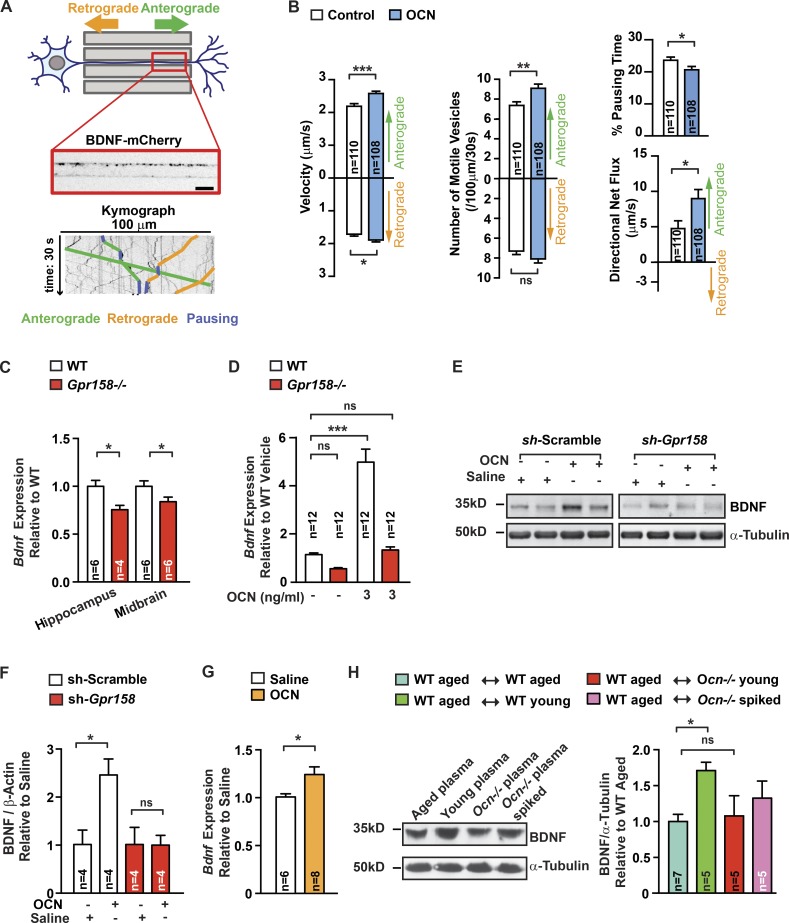
That osteocalcin (OCN) is necessary for hippocampal-dependent memory and to prevent anxiety-like behaviors raises novel questions. One question is to determine whether OCN is also sufficient to improve these behaviors in wild-type mice, when circulating levels of OCN decline as they do with age. Here we show that the presence of OCN is necessary for the beneficial influence of plasma from young mice when injected into older mice on memory and that peripheral delivery of OCN is sufficient to improve memory and decrease anxiety-like behaviors in 16-mo-old mice. A second question is to identify a receptor transducing OCN signal in neurons. Genetic, electrophysiological, molecular, and behavioral assays identify Gpr158, an orphan G protein-coupled receptor expressed in neurons of the CA3 region of the hippocampus, as transducing OCN's regulation of hippocampal-dependent memory in part through inositol 1,4,5-trisphosphate and brain-derived neurotrophic factor. These results indicate that exogenous OCN can improve hippocampal-dependent memory in mice and identify molecular tools to harness this pathway for therapeutic purposes.


Figures

Figure 1.
Figure 1.
OCN is sufficient to improve cognitive function and anxiety-like behaviors. (A and B) NOR (A) and EPMT (B) performed in 3-mo-old (young) WT mice treated with plasma from young WT mice (n = 8–10) or 16-mo-old (aged) WT mice treated with plasma from WT mice, either aged (n = 11–12) or young (n = 11–16), or from young Ocn−/− mice (n = 11–16), or plasma from young Ocn−/− mice supplemented with 90 ng/g BW of OCN (spiked; n = 12–13). For NOR, discrimination index was measured; for EPMT, number of entries and time spent in the open arms were scored (one-way ANOVA followed by a Bonferroni post hoc test compared with WT young). (C) TIMP2 accumulation (representative Western blot, left) and quantification of band intensities (right) in hippocampi of older WT mice receiving plasma from Ocn−/− mice supplemented with 90 ng/g BW of OCN (Student’s t test, n = 4 mice per group). α-Tubulin is used as a loading control. (D and E) NOR (D) and EPMT (E) performed in 18-mo-old mice treated with IgG (n = 9–10) or anti-Ocn immunodepleted plasma (n = 8–9). For NOR, discrimination index was measured. For EPMT, number of entries and time spent in the open arms were scored (Student’s t test). (F–H) NOR (F), Morris water maze test (MWMT; G), and EPMT (H) performed in 3-mo-old (n = 10–15) and older WT mice treated with saline (n = 12–14) or OCN (n = 13–14; 90 ng/h) for 2 mo. For NOR, discrimination index was measured (one-way ANOVA followed by a Bonferroni post hoc test compared with WT young). For MWMT, the graph shows the time to localize a submerged platform in the swimming area (two-way repeated-measures ANOVA followed by a Fisher’s least significantly different post hoc test compared with 3 mo old). For EPMT, time spent in the open arms was measured (one-way ANOVA followed by a Bonferroni post hoc test compared with WT young). Results are given as mean ± SEM. *, P ≤ 0.05; **, P ≤ 0.01; ***, P ≤ 0.001.
Figure 2.
Figure 2.
Identification of a receptor for OCN in the brain. (A) Strategy used to identify an OCN brain receptor. (B) In situ hybridization of Gpr158 in E14.5 WT mouse embryos. Bar, 0.5 mm. (C) In situ hybridization of Gpr158, Gpr156, Gpr179, Gprc5a, Gprc5b, Gprc5c, and Gprc5d in the brain of 10-d-old WT mice. Bar, 200 µm. (D) Expression of Gpr158 and Gprc6a in various tissues of 3-mo-old WT mice (Student’s t test, n = 4 mice per group). In the brain-derived tissues, expression is relative to Gpr158 expression, and in peripheral tissues, expression is relative to Gprc6a. (E) In situ hybridization of Gpr158 in the brain of 3-mo-old WT mice. For the ventral tegmental area (VTA), Th was used as a positive control. Bar, 200 µm. (F) Immunofluorescence of Lacz and Map2 in brain slices of 3-mo-old Gpr158−/− mice. The right-most panel is a 40× magnification of the region indicated in the middle panel. Bar, 50 µm. (G) Immunofluorescence of Gpr158, Map2, and Gfap in primary hippocampal neuronal preparation. Bar, 50 µm. (H) Gpr158 and serotonin receptor 2C (SR2C) accumulation in Ocn−/− and WT hippocampi. Na,K ATPase channel was used as a loading control (Student’s t test, n = 4 mice per group). (I) Bioactive OCN content in the serum of 3-mo-old Gpr158−/− (n = 4) and WT (n = 5 mice; Student’s t test). (J) Immunofluorescence of c-Fos 1 h after stereotaxic injection of either saline or OCN (10 ng) into the anterior hippocampus of 3-mo-old Gpr158−/− and WT mice. Bar, 50 µm. (K) Pull-down assay using biotinylated-OCN on solubilized Ocn−/− or Gpr158−/− hippocampal membranes. Purified proteins were subjected to Western blot analysis using anti-Gpr158 and anti-Gαq antibodies. (L) IP1 accumulation in Gpr158−/− and WT hippocampal neurons treated with saline, OCN, or glutamate as a positive control for 1 h (Student’s t test, n = 12). Results are given as mean ± SEM. *, P ≤ 0.05; **, P ≤ 0.01; ***, P ≤ 0.001.
Figure 3.
Figure 3.
Functional evidence that OCN signals through Gpr158. (A) NOR performed in 3-mo-old Gpr158f/f (n = 9), Gpr158CamkIIa−/− (n = 5), Gpr158−/− (n = 10), and WT (n = 12) littermates. Discrimination index was measured (Student’s t test). (B) MWMT performed in 3-mo-old Gpr158−/− (n = 10) and WT (n = 9) littermates. The graph shows the time needed to localize a submerged platform in the swimming area (two-way repeated-measures ANOVA followed by Fisher’s least significantly different [LSD] post hoc test). (C) NOR performed in 4-mo-old WT (n = 7) or Gpr158−/− mice treated with saline (n = 7) or OCN (n = 8; 90 ng/hr) for 1 mo. Discrimination index was measured (one-way ANOVA compared with WT followed by Bonferroni’s post hoc test). (D and E) NOR (D) and contextual fear conditioning (CFC; E) performed in 3-mo-old shRNA scramble- or shRNA Gpr158-injected mice. After a 10-d recovery, mice were injected with saline (n = 8–10) or OCN (10 ng; n = 9 or 10). For NOR, discrimination index was measured; for CFC, percentage freezing 24 h after training was measured (two-way repeated-measures ANOVA followed by Fisher’s LSD post hoc test). (F) NOR performed in 3-mo-old Gpr158+/−;Ocn+/− (n = 12), Ocn+/− (n = 8), and Gpr158+/− (n = 13) littermates. Discrimination index was measured for each group (one-way ANOVA followed by Tukey’s post hoc test). (G) OCN’s influence on spontaneous action potential (AP) frequency in WT or Gpr158−/− CA3 pyramidal neurons (n = 6). The bars above recording traces indicate the application of OCN (10 ng/ml). RMP, resting membrane potential. (H) OCN’s effect on spontaneous AP frequency in WT CA3 pyramidal neurons pretreated with 2-aminoethoxydiphenyl borate (2-ABP; n = 4). The bars above the recording traces indicate the application of OCN (10 ng/ml) and 2-ABP (50 µM). (I) LTP in the MF-CA3 synapse from WT and Gpr158−/− (Student’s t test, n = 6) hippocampal slices. Left: the time course of field excitatory postsynaptic currents (fEPSCs) recorded extracellularly in the CA3 area with a bipolar electrode placed in the mossy fiber (MF) from WT or Gpr158−/− slices. Two trains of tetanic stimulation (arrow) were applied to the MF-CA3 synapse (100 Hz, 1-s duration, and 10-s interval) once a stable baseline of fEPSC was recorded. Representative traces of fEPSPs recorded from WT (i) and Gpr158−/− slices (ii). Traces recorded before (1) and 50 min after (2) two trains of tetanic stimulations. (J–M) EPMT, dark-to-light transition test (DLT), and OFT performed in 3-mo-old Gpr158−/− (n = 9–10), Gpr158+/− (n = 12–15), and WT (n = 13–15) littermates. For EPMT, time spent in the open arms was scored; for DLT, time spent in the lit compartment was measured; for OFT, total ambulation and time spent in the center of the arena were measured (one-way ANOVA compared with WT followed by Bonferroni’s post hoc test). Results are given as mean ± SEM. *, P ≤ 0.05; **, P ≤ 0.01; ***, P ≤ 0.001.
Figure 4.
Figure 4.
BDNF as a mediator of OCN function in the brain. (A) Representation of the microfluidic device (top) composed of somatic and synaptic chambers connected by 500-µm-long microchannels. BDNF-mCherry-positive vesicles were imaged in a 100-µm-long distal region of axons for 30 s. Kymographs (bottom) extracted from the movies. Examples of traces of anterograde (green), retrograde (orange), and pausing (blue) vesicles are depicted on the kymograph. Bar, 10 µm. (B) Quantification of BDNF-mCherry transport in axons of hippocampal neurons cultured in microchambers treated with OCN (n = 108) or vehicle (n = 110) for 4 h (Mann-Whitney U test) compared with control condition. Anterograde and retrograde mean velocities of vesicles per axon, the number of anterograde and retrograde vesicles that are motile in a 100-µm length of axon during 30 s, the percentage of time spent by vesicles in pause, and the directional flux of vesicles within axons were measured. (C and D) Bdnf expression (quantitative PCR [qPCR]) in midbrain and hippocampi of 6-mo-old Gpr158−/− (n = 4–6) and WT (n = 6) littermates (Student’s t test; C) and in Gpr158−/− and WT hippocampal neurons (one-way ANOVA compared with vehicle-treated cells followed by Bonferroni’s post hoc test, n = 12) treated with either saline or OCN for 4 h (D). (E and F) BDNF accumulation (representative Western blot, E) and quantification of band intensities in hippocampi of 3-mo-old shRNA scramble- or shRNA Gpr158-injected (F). After a 10 d-long recovery, mice were injected with saline or OCN (10 ng) 16 h before collections (two-way repeated-measures ANOVA followed by Fisher’s least significantly different post hoc test, n = 4 per group). (G) Bdnf expression (qPCR) in hippocampi of 16-mo-old WT mice injected i.p. with either saline (n = 6) or OCN (n = 8; 500 ng/g BW) 2 h before collection (Student’s t test). (H) BDNF accumulation (representative Western blot, left) and quantification of band intensities (right) in hippocampi of older WT mice receiving plasma from older (n = 7) or young WT (n = 5) mice, from young Ocn−/− (n = 5) mice, or from young Ocn−/− mice supplemented with OCN (n = 5; 90 ng/g BW; one-way ANOVA compared with older WT mice receiving plasma from older WT mice, followed by Bonferroni’s post hoc test). α-Tubulin used as a loading control. Results are given as mean ± SEM. *, P ≤ 0.05; **, P ≤ 0.01; ***, P ≤ 0.001.

References

    1. Broadbent N.J., Gaskin S., Squire L.R., and Clark R.E.. 2010. Object recognition memory and the rodent hippocampus. Learn. Mem. 17:5–11. 10.1101/lm.1650110 - DOI - PMC - PubMed
    1. Castellano J.M., Mosher K.I., Abbey R.J., McBride A.A., James M.L., Berdnik D., Shen J.C., Zou B., Xie X.S., Tingle M., et al. . 2017. Human umbilical cord plasma proteins revitalize hippocampal function in aged mice. Nature. 544:488–492. 10.1038/nature22067 - DOI - PMC - PubMed
    1. Chiappe A., Gonzalez G., Fradinger E., Iorio G., Ferretti J.L., and Zanchetta J.. 1999. Influence of age and sex in serum osteocalcin levels in thoroughbred horses. Arch. Physiol. Biochem. 107:50–54. 10.1076/apab.107.1.50.4357 - DOI - PubMed
    1. Crawley J.N. 1985. Exploratory behavior models of anxiety in mice. Neurosci. Biobehav. Rev. 9:37–44. 10.1016/0149-7634(85)90030-2 - DOI - PubMed
    1. David D.J., Samuels B.A., Rainer Q., Wang J.W., Marsteller D., Mendez I., Drew M., Craig D.A., Guiard B.P., Guilloux J.P., et al. . 2009. Neurogenesis-dependent and -independent effects of fluoxetine in an animal model of anxiety/depression. Neuron. 62:479–493. 10.1016/j.neuron.2009.04.017 - DOI - PMC - PubMed