. 2017 Sep 29;121(8):930-940.
doi: 10.1161/CIRCRESAHA.117.311327. Epub 2017 Aug 29.

MerTK Cleavage on Resident Cardiac Macrophages Compromises Repair After Myocardial Ischemia Reperfusion Injury

Affiliations

MerTK Cleavage on Resident Cardiac Macrophages Compromises Repair After Myocardial Ischemia Reperfusion Injury

Matthew DeBerge et al. Circ Res. .

Abstract

Rationale: Clinical benefits of reperfusion after myocardial infarction are offset by maladaptive innate immune cell function, and therapeutic interventions are lacking.

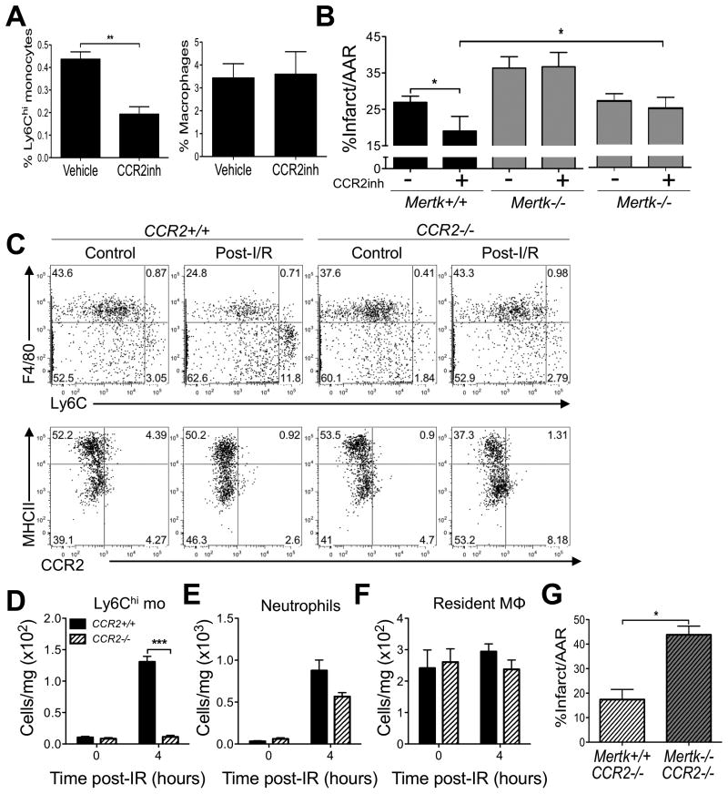
Objective: We sought to test the significance of phagocytic clearance by resident and recruited phagocytes after myocardial ischemia reperfusion.

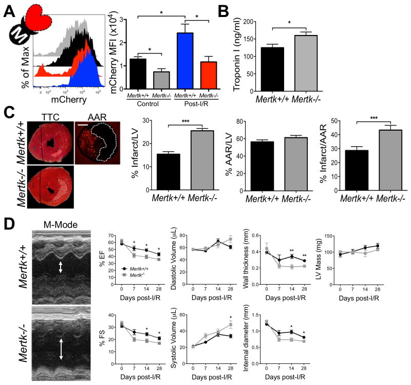
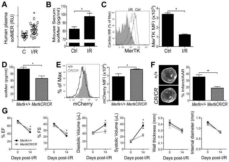
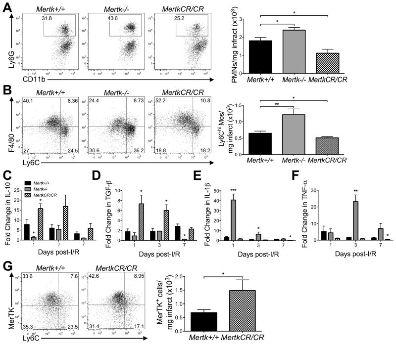
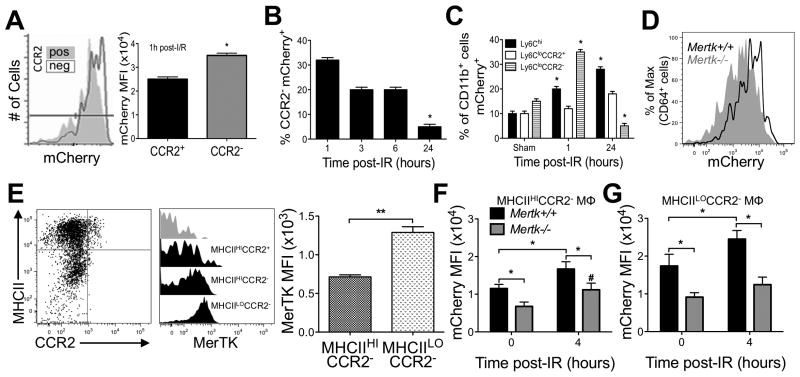
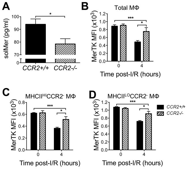
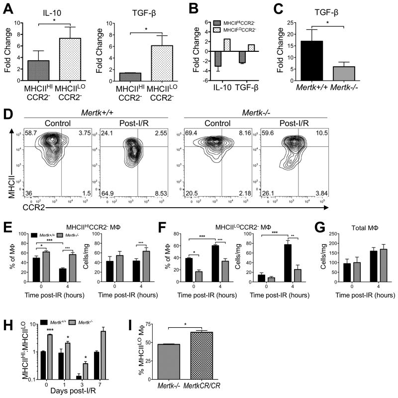
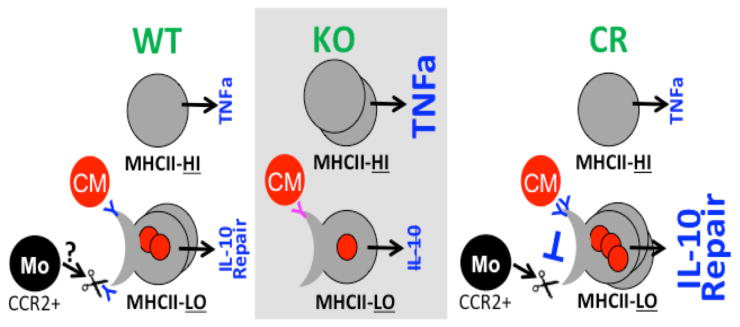
Methods and results: In humans, we discovered that clinical reperfusion after myocardial infarction led to significant elevation of the soluble form of MerTK (myeloid-epithelial-reproductive tyrosine kinase; ie, soluble MER), a critical biomarker of compromised phagocytosis by innate macrophages. In reperfused mice, macrophage Mertk deficiency led to decreased cardiac wound debridement, increased infarct size, and depressed cardiac function, newly implicating MerTK in cardiac repair after myocardial ischemia reperfusion. More notably, Mertk(CR) mice, which are resistant to cleavage, showed significantly reduced infarct sizes and improved systolic function. In contrast to other cardiac phagocyte subsets, resident cardiac MHCIILOCCR2- (major histocompatibility complex II/C-C motif chemokine receptor type 2) macrophages expressed higher levels of MerTK and, when exposed to apoptotic cells, secreted proreparative cytokines, including transforming growth factor-β. Mertk deficiency compromised the accumulation of MHCIILO phagocytes, and this was rescued in Mertk(CR) mice. Interestingly, blockade of CCR2-dependent monocyte infiltration into the heart reduced soluble MER levels post-ischemia reperfusion.

Conclusions: Our data implicate monocyte-induced MerTK cleavage on proreparative MHCIILO cardiac macrophages as a novel contributor and therapeutic target of reperfusion injury.

Keywords: efferocytosis; inflammation; ischemia reperfusion injury; macrophage; phagocytosis.


Figures

Figure 1
Figure 1. Post-reperfusion phagocytosis via MerTK occurs within hours and is cardio-protective after infarction
Myocardial ischemia reperfusion (I/R) injury was induced as described in methods. A Hearts were harvested at 4 hours after I/R injury from MHC-mCherry mice, which express mCherry in cardiomyocytes. mCherry levels were analyzed in cardiac CD11b+Ly6CloLy6G-F4/80+ macrophages in Mertk+/+ and Mertk-/- mice. B Serum troponin levels on day 3 post-I/R. C Representative TTC heart stained sections after I/R injury and fluorescent bead injection to delineate AAR. Bar graphs are quantification of % Infarct/LV, % AAR/LV, and % Infarct/AAR on day 7 post-I/R. D Echocardiographic analysis of LV function demonstrating impaired LV function in Mertk-/- hearts. *p<0.05, ***p<0.001.
Figure 2
Figure 2. Post-reperfusion phagocytosis via MerTK is impaired in humans and mice by MerTK cleavage
A solMER (ELISA) from human plasma in patients with stable cardiovascular disease (CVD) vs. STE-MI patients at day 1 post-reperfusion revealing increased MerTK cleavage in STEMI patients. B solMER (ELISA) from mice pre and post-I/R revealing increased MerTK cleavage after I/R injury. C Flow cytometry analysis of MerTK on cardiac macrophages showing a reduction in cell-surface MerTK after I/R (Gray represents Mertk-/- control). D Characterization of MertkCR/CR mice for serum solMER levels post-I/R. E Flow cytometry analysis of mCherry expression in F4/80+ phagocytes 4h post-I/R revealing enhanced phagocytosis of cardiomyocytes by MertkCR/CR macrophages. F Quantification of % Infarct/AAR on day 7 post-I/R showing reduced infarct size in MertkCR/CR hearts. G Echocardiographic analysis of LV function demonstrating improved LV function by MertkCR/CR hearts. *p<0.05.
Figure 3
Figure 3. Inhibition of MerTK cleavage promotes inflammation resolution after I/R
Flow cytometry analysis of A neutrophils and B Ly6Chi monocytes in the infarcted myocardium on day 3 post-I/R revealing increased inflammatory cells in Mertk-/- hearts and reduced inflammatory cells in MertkCR/CR hearts compared to Mertk+/+. Gene expression analysis of C IL-10, D TGF-β, E IL-1β, and F TNF-α in the infarcted heart demonstrating enhanced inflammation in Mertk-/- hearts and enhanced resolution in MertkCR/CR hearts compared to Mertk+/+ hearts. G Flow cytometry of phagocytes on day 3 post-I/R revealing increased pro-reparative, MerTK+ phagocytes in the hearts of MertkCR/CR mice. *p<0.05, **p<0.01, ***p<0.001.
Figure 4
Figure 4. Resident CCR2- cardiac macrophages participate in early phagocytosis and suppression of infarct size through MerTK
A Cardiac CD11b+mCherry+ phagocytes in CCR2+ and CCR2- phagocytes post-I/R. B Time course of CD11b+CCR2-mCherry+ cardiac phagocytes post-I/R. C Distribution % of CD11b+mCherry+ cells in heart for Ly6Chi monocytes, Ly6CloCCR2+, and Ly6CloCCR2- phagocytes. *p<0.05 vs. sham. #p<0.05 vs. sham. D mCherry expression in Mertk+/+ and Mertk-/- cardiac CD64+ macrophages 6h post-I/R. E Flow cytometry of MerTK expression on MHCIIHICCR2-, MHCIILOCCR2-, and MHCIIHICCR2+ cardiac resident macrophages showing variable MerTK expression on the different subsets. Flow cytometry analysis of mCherry expression by F MHCIIHICCR2- and G MHCIILOCCR2- resident macrophages revealing impaired cardiomyocyte phagocytosis in Mertk-/- resident macrophages compared to Mertk+/+ resident macrophages. *p<0.05, **p<0.01.
Figure 5
Figure 5. MerTK on non-recruited cardiac macrophages promotes cardio-protection after I/R
A Percent of Ly6Chi monocytes and cardiac macrophages in the hearts of Mertk+/+ on day 3 post-I/R after CCR2-antagonism (RS50493). B Infarct measurements on day 3 post-I/R in Mertk+/+ and Mertk-/- mice following treatment with CCR2 antagonist. Third pair of bars = normalized infarct conditions. C Flow cytometry analysis showing the changes in MHCIIHICCR2- and MHCIILOCCR2- cardiac resident macrophages after I/R in CCR2+/+ and CCR2-/- hearts. Quantification of D Ly6Chi monocytes, E neutrophils, and F resident macrophages showing reduced cellular recruitment in CCR2-/- hearts compared to CCR2+/+ hearts. G Infarct sizes in Mertk+/+CCR2-/- and Mertk-/-CCR2-/- mice. *p<0.05, ***p<0.001.
Figure 6
Figure 6. Resident macrophages are susceptible to MERTK cleavage by CCR2+ recruited cells
A Serum solMER 3 days post-I/R in Ccr2+/+ and Ccr2-/- mice. Expression of MerTK on B total cardiac resident macrophages and C MHCIIHICCR2- or D MHCIILOCCR2- resident macrophage subsets revealing reduced MerTK cleavage degradation in CCR2-/- hearts after I/R. *p<0.05, ***p<0.001.
Figure 7
Figure 7. Relative to MHCIIHI cardiac macrophages (MΦs), MHCIILO cardiac MΦs have elevated MerTK, are more phagocytic, and require MerTK for anti-inflammatory cytokine production and a balanced MHCIIHI to MHCIILO cell ratio
A mCherry+ MHCIILO and MHCIIHI macrophages were sorted from heart post-I/R and assessed for gene expression of IL-10 and TGF-β. B MHCIILO and MHCIIHI macrophages were sorted from naive hearts and assessed for IL-10 and TGF-β after ex vivo challenge with apoptotic cells (AC). C AC-induced TGF-β production by macrophages requires MerTK. D MHCII skewing of cardiac CD64+ macrophages at steady-state and after I/R. Quantification of E MHCIIHICCR2-, F MHCIILOCCR2-, and G total macrophages demonstrating a reduction in MHCIILOCCR2- macrophages in Mertk-/- hearts compared to Mertk+/+ hearts.. H-I MHCIIHI to MHCIILO ratios of cardiac CD64+ macrophages post-I/R. I Cardiac CD64+ MHCIILO macrophages are rescued in MertkCR/CR mice post-I/R. *p<0.05, **p<0.01, ***p<0.001.
Figure 8
Figure 8. Working model of causes and consequences of phagocytosis, MerTK, and MerTK cleavage post-myocardial ischemia/reperfusion (I/R)
Under wild type (WT) conditions, MerTK levels are highest on MHCIILO cardiac phagocytes, which are required to promote dead cardiomyocyte (CM) phagocytic removal and pro-reparative cytokines after I/R. I/R leads to CCR2-dependent monocyte recruitment and MerTK cleavage, which compromises MerTK function. Without MerTK (knockout/KO), MHCIILO macrophages are reduced in favor of skewing toward MHCIIHI macrophages. Alternative receptors (pink) promote reduced levels of phagocytosis. Under cleavage-resistant/CR or potential therapeutic conditions, this increases MHCIILO cardiac macrophages and promotes MHCIILO macrophage-mediated repair after phagocytosis.

Similar articles

Cited by

References

    1. Whelan RS, Kaplinskiy V, Kitsis RN. Cell death in the pathogenesis of heart disease: Mechanisms and significance. Annu Rev Physiol. 2010;72:19–44. - PubMed
    1. Bulluck H, Yellon DM, Hausenloy DJ. Reducing myocardial infarct size: Challenges and future opportunities. Heart (British Cardiac Society) 2016;102:341–348. - PMC - PubMed
    1. Howangyin KY, Zlatanova I, Pinto C, Ngkelo A, Cochain C, Rouanet M, Vilar J, Lemitre M, Stockmann C, Fleischmann BK, Mallat Z, Silvestre JS. Myeloid-epithelial-reproductive receptor tyrosine kinase and milk fat globule epidermal growth factor 8 coordinately improve remodeling after myocardial infarction via local delivery of vascular endothelial growth factor. Circulation. 2016;133:826–839. - PMC - PubMed
    1. Wan E, Yeap XY, Dehn S, Terry R, Novak M, Zhang S, Iwata S, Han X, Homma S, Drosatos K, Lomasney J, Engman DM, Miller SD, Vaughan DE, Morrow JP, Kishore R, Thorp EB. Enhanced efferocytosis of apoptotic cardiomyocytes through myeloid-epithelial-reproductive tyrosine kinase links acute inflammation resolution to cardiac repair after infarction. Circulation research. 2013;113:1004–1012. - PMC - PubMed
    1. Lemke G, Rothlin CV. Immunobiology of the tam receptors. Nature reviews Immunology. 2008;8:327–336. - PMC - PubMed

MeSH terms

LinkOut - more resources