

EPAC-RAP1 Axis-Mediated Switch in the Response of Primary and Metastatic Melanoma to Cyclic AMP
- PMID: 28851815
- PMCID: PMC6309370
- DOI: 10.1158/1541-7786.MCR-17-0067
EPAC-RAP1 Axis-Mediated Switch in the Response of Primary and Metastatic Melanoma to Cyclic AMP
Abstract
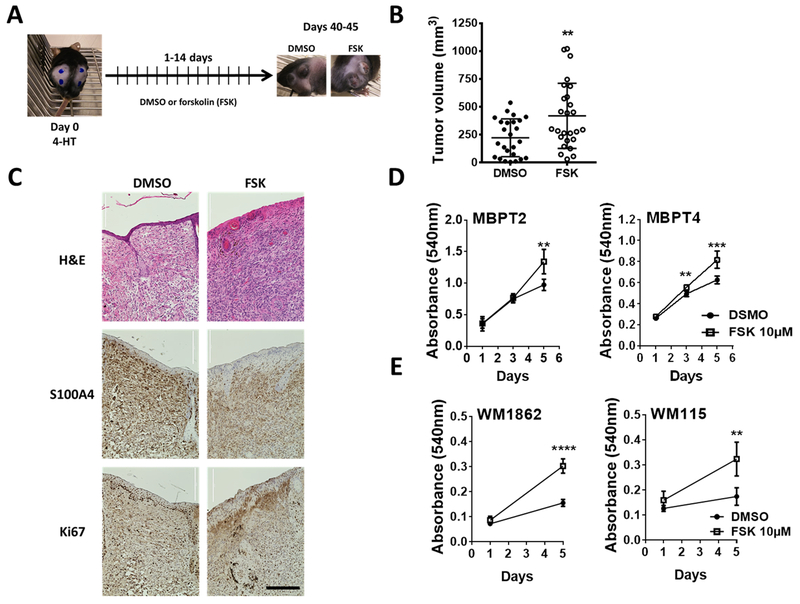
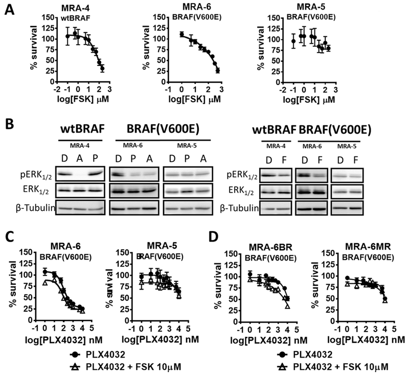
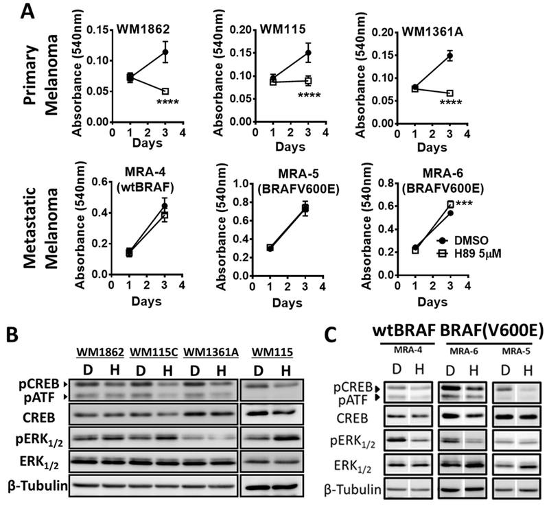
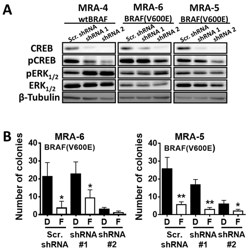
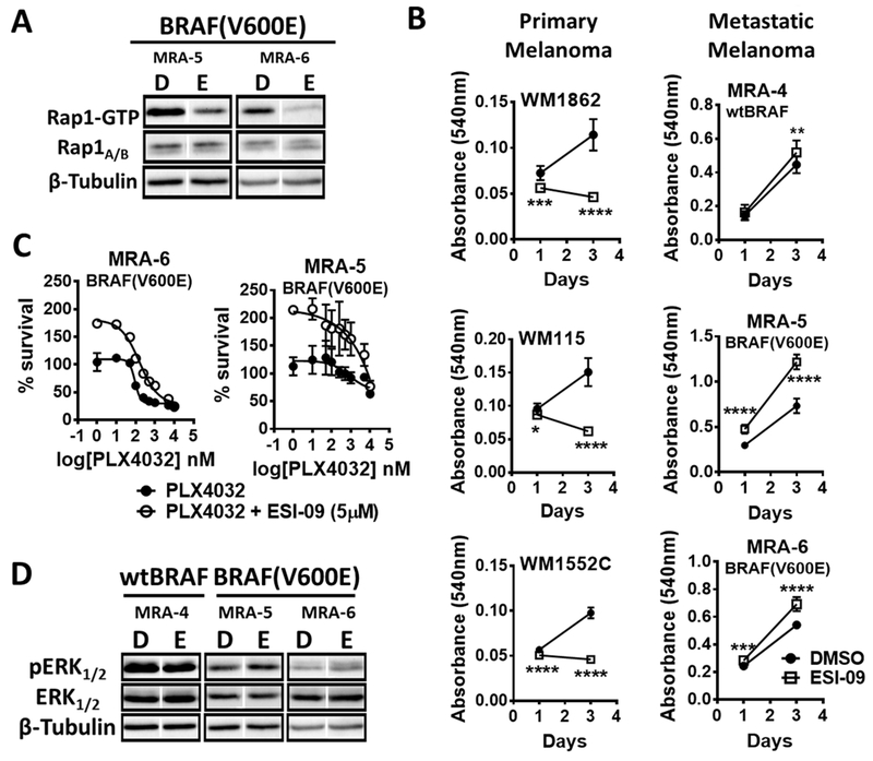
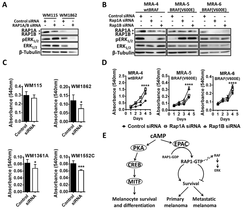
Cyclic AMP (cAMP) is an important second messenger that regulates a wide range of physiologic processes. In mammalian cutaneous melanocytes, cAMP-mediated signaling pathways activated by G-protein-coupled receptors (GPCR), like melanocortin 1 receptor (MC1R), play critical roles in melanocyte homeostasis including cell survival, proliferation, and pigment synthesis. Impaired cAMP signaling is associated with increased risk of cutaneous melanoma. Although mutations in MAPK pathway components are the most frequent oncogenic drivers of melanoma, the role of cAMP in melanoma is not well understood. Here, using the Braf(V600E)/Pten-null mouse model of melanoma, topical application of an adenylate cyclase agonist, forskolin (a cAMP inducer), accelerated melanoma tumor development in vivo and stimulated the proliferation of mouse and human primary melanoma cells, but not human metastatic melanoma cells in vitro The differential response of primary and metastatic melanoma cells was also evident upon pharmacologic inhibition of the cAMP effector protein kinase A. Pharmacologic inhibition and siRNA-mediated knockdown of other cAMP signaling pathway components showed that EPAC-RAP1 axis, an alternative cAMP signaling pathway, mediates the switch in response of primary and metastatic melanoma cells to cAMP. Evaluation of pERK levels revealed that this phenotypic switch was not correlated with changes in MAPK pathway activity. Although cAMP elevation did not alter the sensitivity of metastatic melanoma cells to BRAF(V600E) and MEK inhibitors, the EPAC-RAP1 axis appears to contribute to resistance to MAPK pathway inhibition. These data reveal a MAPK pathway-independent switch in response to cAMP signaling during melanoma progression.Implications: The prosurvival mechanism involving the cAMP-EPAC-RAP1 signaling pathway suggest the potential for new targeted therapies in melanoma. Mol Cancer Res; 15(12); 1792-802. ©2017 AACR.
©2017 American Association for Cancer Research.
Figures
Similar articles
-
Elevated cyclic AMP levels promote BRAFCA/Pten-/- mouse melanoma growth but pCREB is negatively correlated with human melanoma progression.Cancer Lett. 2018 Feb 1;414:268-277. doi: 10.1016/j.canlet.2017.11.027. Epub 2017 Nov 24. Cancer Lett. 2018. PMID: 29179997 Free PMC article.
-
Cyclic AMP signaling as a mediator of vasculogenic mimicry in aggressive human melanoma cells in vitro.Cancer Res. 2009 Feb 1;69(3):802-9. doi: 10.1158/0008-5472.CAN-08-2391. Epub 2009 Jan 27. Cancer Res. 2009. PMID: 19176384
-
Functional status and relationships of melanocortin 1 receptor signaling to the cAMP and extracellular signal-regulated protein kinases 1 and 2 pathways in human melanoma cells.Int J Biochem Cell Biol. 2012 Dec;44(12):2244-52. doi: 10.1016/j.biocel.2012.09.008. Epub 2012 Sep 19. Int J Biochem Cell Biol. 2012. PMID: 23000456
-
Cyclic AMP (cAMP) signaling in melanocytes and melanoma.Arch Biochem Biophys. 2014 Dec 1;563:22-7. doi: 10.1016/j.abb.2014.07.003. Epub 2014 Jul 10. Arch Biochem Biophys. 2014. PMID: 25017568 Review.
-
Regulatory actions of 3',5'-cyclic adenosine monophosphate on osteoclast function: possible roles of Epac-mediated signaling.Ann N Y Acad Sci. 2018 Dec;1433(1):18-28. doi: 10.1111/nyas.13861. Epub 2018 May 30. Ann N Y Acad Sci. 2018. PMID: 29846007 Review.
Cited by
-
Notch signaling activation induces cell death in MAPKi-resistant melanoma cells.Pigment Cell Melanoma Res. 2019 Jul;32(4):528-539. doi: 10.1111/pcmr.12764. Epub 2019 Feb 3. Pigment Cell Melanoma Res. 2019. PMID: 30614626 Free PMC article.
-
RBM10 inhibits cell proliferation of lung adenocarcinoma via RAP1/AKT/CREB signalling pathway.J Cell Mol Med. 2019 Jun;23(6):3897-3904. doi: 10.1111/jcmm.14263. Epub 2019 Apr 6. J Cell Mol Med. 2019. PMID: 30955253 Free PMC article.
-
Elevated cyclic AMP levels promote BRAFCA/Pten-/- mouse melanoma growth but pCREB is negatively correlated with human melanoma progression.Cancer Lett. 2018 Feb 1;414:268-277. doi: 10.1016/j.canlet.2017.11.027. Epub 2017 Nov 24. Cancer Lett. 2018. PMID: 29179997 Free PMC article.
-
Molecular Mechanism of Perfluorooctane Sulfonate-Induced Lung Injury Mediated by the Ras/Rap Signaling Pathway in Mice.Toxics. 2025 Apr 20;13(4):320. doi: 10.3390/toxics13040320. Toxics. 2025. PMID: 40278636 Free PMC article.
-
Melanoma Progression Inhibits Pluripotency and Differentiation of Melanoma-Derived iPSCs Produces Cells with Neural-like Mixed Dysplastic Phenotype.Stem Cell Reports. 2019 Jul 9;13(1):177-192. doi: 10.1016/j.stemcr.2019.05.018. Epub 2019 Jun 20. Stem Cell Reports. 2019. PMID: 31231022 Free PMC article.
References
-
- Rodríguez CI, Setaluri V. Cyclic AMP (cAMP) signaling in melanocytes and melanoma. Arch Biochem Biophys 2014 - PubMed
-
- Schiöth HB, Phillips SR, Rudzish R, Birch-Machin MA, Wikberg JE, Rees JL. Loss of function mutations of the human melanocortin 1 receptor are common and are associated with red hair. Biochem Biophys Res Commun 1999;260:488–91 - PubMed
-
- Kennedy C, ter Huurne J, Berkhout M, Gruis N, Bastiaens M, Bergman W , et al. Melanocortin 1 receptor (MC1R) gene variants are associated with an increased risk for cutaneous melanoma which is largely independent of skin type and hair color. J Invest Dermatol 2001;117:294–300 - PubMed
-
- Kadekaro AL, Chen J, Yang J, Chen S, Jameson J, Swope VB , et al. Alpha-melanocyte-stimulating hormone suppresses oxidative stress through a p53-mediated signaling pathway in human melanocytes. Mol Cancer Res 2012;10:778–86 - PubMed
MeSH terms
Substances
Grants and funding
LinkOut - more resources
Full Text Sources
Other Literature Sources
Medical
Molecular Biology Databases
Research Materials
