. 2017 Aug 29;7(1):9886.
doi: 10.1038/s41598-017-06907-4.

A unique tolerizing dendritic cell phenotype induced by the synthetic triterpenoid CDDO-DFPA (RTA-408) is protective against EAE

Affiliations

A unique tolerizing dendritic cell phenotype induced by the synthetic triterpenoid CDDO-DFPA (RTA-408) is protective against EAE

Hsi-Ju Wei et al. Sci Rep. .

Abstract

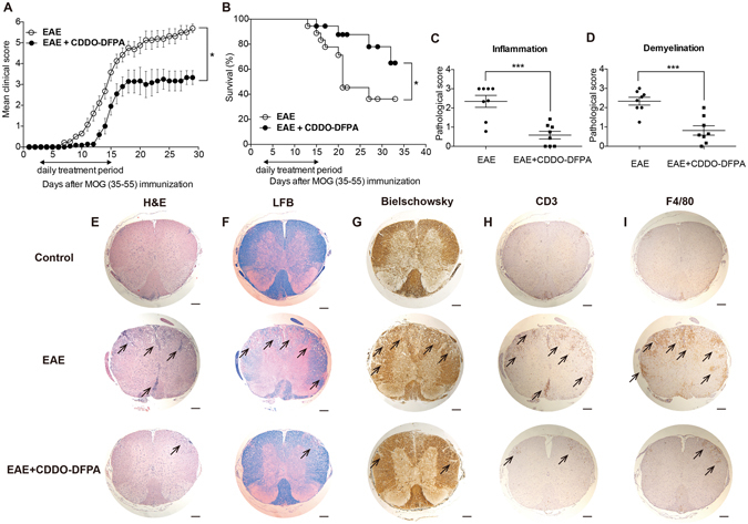
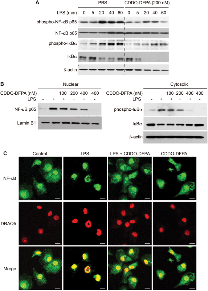
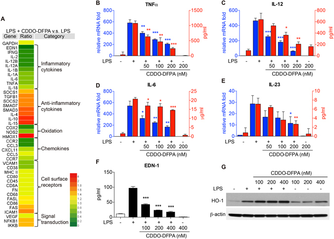
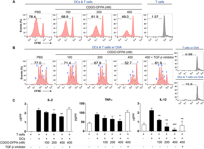
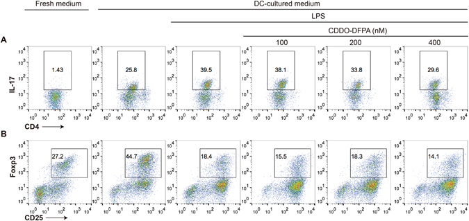
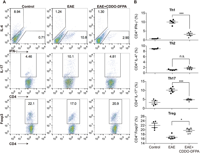
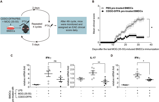
Tolerogenic dendritic cells (DCs) have emerged as relevant clinical targets for the treatment of multiple sclerosis and other autoimmune disorders. However, the pathways essential for conferring the tolerizing DC phenotype and optimal methods for their induction remain an intense area of research. Triterpenoids are a class of small molecules with potent immunomodulatory activity linked to activation of Nrf2 target genes, and can also suppress the manifestations of experimental autoimmune encephalomyelitis (EAE). Here we demonstrate that DCs are a principal target of the immune modulating activity of triterpenoids in the context of EAE. Exposure of DCs to the new class of triterpenoid CDDO-DFPA (RTA-408) results in the induction of HO-1, TGF-β, and IL-10, as well as the repression of NF-κB, EDN-1 and pro-inflammatory cytokines IL-6, IL-12, and TNFα. CDDO-DFPA exposed DCs retained expression of surface ligands and capacity for antigen uptake but were impaired to induce Th1 and Th17 cells. TGF-β was identified as the factor mediating suppression of T cell proliferation by CDDO-DFPA pretreated DCs, which failed to passively induce EAE. These findings demonstrate the potential therapeutic utility of CDDO-DFPA in the treatment and prevention of autoimmune disorders, and its capacity to induce tolerance via modulation of the DC phenotype.


Conflict of interest statement

The authors declare that they have no competing interests.

Figures

Figure 1
Figure 1
CDDO-DFPA is protective against clinical pathology of EAE. EAE was induced in age-matched female C57BL/6 mice (8 to 10 weeks old), by MOG (35–55) immunization. Pertussis toxin (PTX) was also injected immediately and again 2 days later. CDDO-DFPA was administered (i.p. injection) daily from day 3 to day 15. (A) A clinical score was assigned to each mouse daily. All data are presented as the mean ± S.E.M. *P < 0.05. Multiple t-tests with Holm-Sidak analysis. (B) Survival curve for immunized mice (Kaplan-Meier survival curve followed by the Mental-Cox log-rank test within 35 days (n = 11–12 mice in each group). *P < 0.05. Mice were sacrificed at day 21 and representative sections of lumbar spinal cord were prepared from control mice and from mice immunized with MOG and subsequently treated with either CDDO-DFPA or vehicle control. Tissues were stained with hematoxylin and eosin (H&E) to assess inflammation (C,E), with Luxol fast blue (LFB) to assess myelin content (D,F), and with Bielschowsky stain to measure axonal loss (G). CD3 and F4/80 antibodies were used for immunohistochemical localization of T cells (H) and macrophages (I), respectively (each indicated by arrows). A pathologist blinded to subject identity scored sections taken from each animal for H&E inflammation (C) and LFB demyelination (D) on the scale of 0 to 3. Scale bars, 100 μm. Quantification data in panel C and D were presented as the mean ± S.E.M. (n = 8 mice in each group). ***P < 0.001, Unpaired student t-test.
Figure 2
Figure 2
CDDO-DFPA suppresses NF-κB signaling in LPS-activated BMDCs. (A) CDDO-DFPA impairs time-dependent induction of NF-κB in BMDCs expanded from the normal bone marrow of C57BL/6 mice that were pretreated in either the presence or absence of CDDO-DFPA (200 nM) for 1 hour before LPS (100 ng/ml) treatment for the indicated intervals. (B) BMDCs were pretreated in the presence or absence of CDDO-DFPA (100–400 nM) for 1 hour, followed by stimulation with LPS for 10 mins (100 ng/ml). The cytosolic, nuclear, and total cellular lysates were analyzed for phospho- and total NF-κB p65 and IκBα, β-actin, and Lamin B1 expression by Western blotting. All depicted blots are cropped and respective full-length blots are presented in the Supplementary Figure S6. (C) BMDCs were pretreated in the presence or absence of CDDO-DFPA (400 nM) for 1 hour, followed by stimulation with LPS for 10 mins (100 ng/ml). Cells were fixed and stained with NF-κB and DRAQ5 (nucleus), and images were acquired by confocal microscopy. All experiments were repeated a minimum of three times.
Figure 3
Figure 3
A unique CDDO-DFPA-induced transcriptome in LPS-activated BMDCs. (A) Cells were pretreated in the presence or absence of CDDO-DFPA (200 nM) for 1 hour before exposure to LPS (100 ng/ml for 3 hours). Cells were harvested, and RNA was extracted for qRT-PCR array. Expression of each gene was normalized by the control gene (GAPDH) in its own group and then normalized to the average value of each gene within groups. A total of 41 genes known to be related to DC maturation and mediators of DC modulated T cell responses were sorted and classified from 69 genes. The heat map was drawn using the HemI (Heat map illustrator) with the default value. (BE) Cells were pretreated in the presence or absence of CDDO-DFPA (50–200 nM) for 1 hour prior to addition of LPS (100 ng/ml), and either harvested for RNA extraction (4 hours) or allowed to condition culture medium for 24 hours prior to collection for cytokine analyses. The levels of TNFα, IL-12, IL-6, and IL-23 were measured by qRT-PCR and ELISA. (F,G) Conditioned medium and cell protein lysate (12 hours) were collected for analyses. The levels of EDN-1, HO-1, and β-actin expression were analyzed by ELISA and Western blotting. Depicted blots are cropped and respective full-length blots are presented in the supplementary Figure S6. The results are expressed as mean ± S.D. of three experiments. *P < 0.05, **P < 0.01, ***P < 0.001 compared with the LPS-treated groups. Unpaired student t-test.
Figure 4
Figure 4
CDDO-DFPA exposed DCs suppress T cell proliferation. DCs were pretreated with CDDO-DFPA (400 nM) for 1 hour only, then washed and co-cultured with CFSE stained T cells at a 1:10 ratio. (A) In an in vitro model of allogeneic T cell stimulation, splenic T cells and DCs were isolated from C57BL/6 and BALB/c mice respectively and T cell proliferation was determined by flow cytometry at day 6. (B) In a syngeneic model, splenic T cells and DCs were isolated from C57BL/6 OTII transgenic mice and C57BL/6 mice respectively. CDDO-DFPA pretreated DCs were co-cultured with CFSE stained T cells with (w/) or without (w/o) OVA addition during incubation in presence or absence of TGF-β receptor inhibitor EW-7197 (5 μM). T cell proliferation was determined by flow cytometry at day 2. Graphs depict the percentage of dividing T cells relative to numbers T cell division. (C) Medium conditioned by the co-culture DCs and C57BL/6 OTII transgenic T cells was also collected to assay levels of IL-2, TNFα, and IL-12 by ELISA. The data is a representation of 3 independent experiments. The results in panel C are expressed as mean ± S.D. *P < 0.05, **P < 0.01, ***P < 0.001 compared with the co-cultured untreated group. Unpaired student t-test.
Figure 5
Figure 5
CDDO-DFPA-exposed DCs suppress Th17 induction via soluble mediators. BMDCs were pretreated in the presence or absence of CDDO-DFPA (100–400 nM) for 1 hour and washed before incubation with LPS (100 ng/ml) for 24 hours. DC conditioned medium was collected and stored in −80 °C. Splenic CD4+ T cells were isolated from C57BL/6 mice and incubated under the indicated conditions with the addition of DC-cultured medium. T cells were stimulated for 2 days (Th17) or 3 days (Treg) with plate-bound CD3 and CD28 antibodies and in the presence of differentiation factors (TGF-β and IL-6 for Th17, TGF-β only for Treg). Subsets of IL-17+ Th17 (A) and CD25+/Foxp3+ Treg (B) were analyzed by flow cytometry. Similar results were obtained in three independent experiments.
Figure 6
Figure 6
CDDO-DFPA increased Treg and suppressed Th1 and Th17 responses during EAE induction. (A) Mice were immunized with MOG (35–55) and treated with either vehicle control or CDDO-DFPA daily from day 3 to day 15 by i.p injection. Mice were sacrificed at day 17 and splenocytes were harvested and stimulated with PMA/ionomycin/Golgistop for 4 hours and subjected to flow cytometry to determine the frequency of Th1, Th2, Th17, and Treg subsets among CD4+ T cells based on their expression of IFN-γ, IL-4, IL-17, and Foxp3, respectively. (B) Quantification of data was presented as the mean ± S.E.M. (n = 4–5 mice in each group). *P < 0.05, ***P < 0.001, One-way ANOVA with the Bonferroni corrections.
Figure 7
Figure 7
Passively DC-induced EAE is abrogated by DC exposure to CDDO-DFPA. (A) EAE induction by MOG (35–55)-pulsed BMDCs. Mature BMDCs were treated in the presence or absence of CDDO-DFPA (400 nM) and subsequently pulsed with MOG (35–55) for 4 hours. A total of 200 μl of 2 × 106 cells were administered by subcutaneous injected into the flank region of C57BL/6 mice once each week for a total of four injections. Each time, PTX was administered by i.p. injection immediately and again 2 days later (day 0 on 4th cycle). (B) Clinical scores were recorded after all four injections, using standard criteria. All data were presented as the mean ± S.E.M. *P < 0.05. Multiple t-tests with Holm-Sidak analysis (n = 7 mice in each group). (C and D) These mice were sacrificed on day 17 and mRNA expression of IFN-γ and IL-17 in the spleen (C) and IFN-γ mRNA expression in the lumbar spinal cord were measured by qRT-PCR. The results in panel C and D were presented as the mean ± S.E.M. (n = 3–6 mice in each group). *P < 0.05, **P < 0.01, One-way ANOVA with the Bonferroni corrections.

Similar articles

Cited by

References

    1. Coutant F, Miossec P. Altered dendritic cell functions in autoimmune diseases: distinct and overlapping profiles. Nature reviews. Rheumatology. 2016;12:703–715. doi: 10.1038/nrrheum.2016.147. - DOI - PubMed
    1. Devi KS, Anandasabapathy N. The origin of DCs and capacity for immunologic tolerance in central and peripheral tissues. Seminars in immunopathology. 2016 - PMC - PubMed
    1. Wang L, et al. Selective depletion of CD11c+ CD11b+ dendritic cells partially abrogates tolerogenic effects of intravenous MOG in murine EAE. European journal of immunology. 2016;46:2454–2466. doi: 10.1002/eji.201546274. - DOI - PMC - PubMed
    1. Ballestrero A, et al. Immunotherapy with dendritic cells for cancer. Advanced drug delivery reviews. 2008;60:173–183. doi: 10.1016/j.addr.2007.08.026. - DOI - PubMed
    1. Banchereau J, et al. Harnessing human dendritic cell subsets to design novel vaccines. Annals of the New York Academy of Sciences. 2009;1174:24–32. doi: 10.1111/j.1749-6632.2009.04999.x. - DOI - PMC - PubMed

Publication types

MeSH terms