. 2017 Aug 29;7(1):9610.
doi: 10.1038/s41598-017-09040-4.

Human antigen R-regulated CCL20 contributes to osteolytic breast cancer bone metastasis

Affiliations

Human antigen R-regulated CCL20 contributes to osteolytic breast cancer bone metastasis

Sun Kyoung Lee et al. Sci Rep. .

Abstract

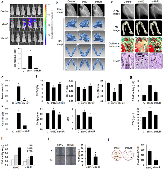
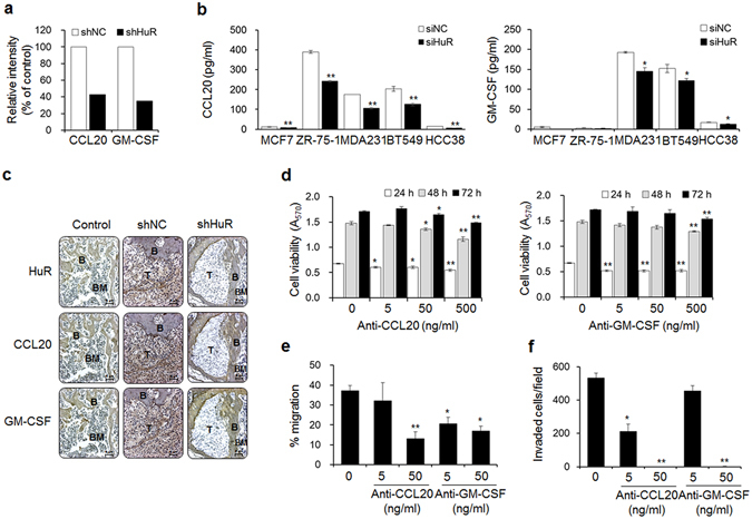
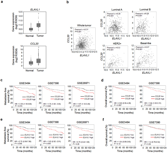
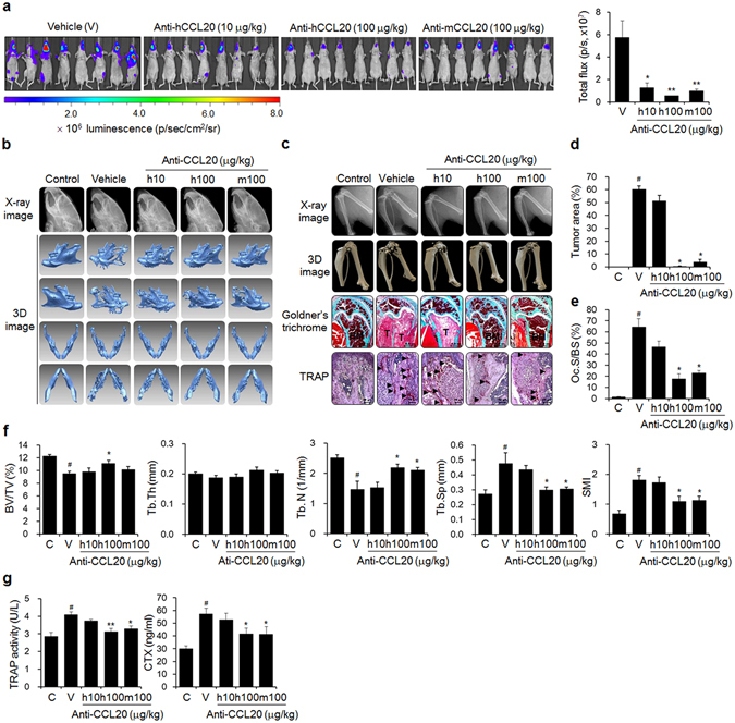
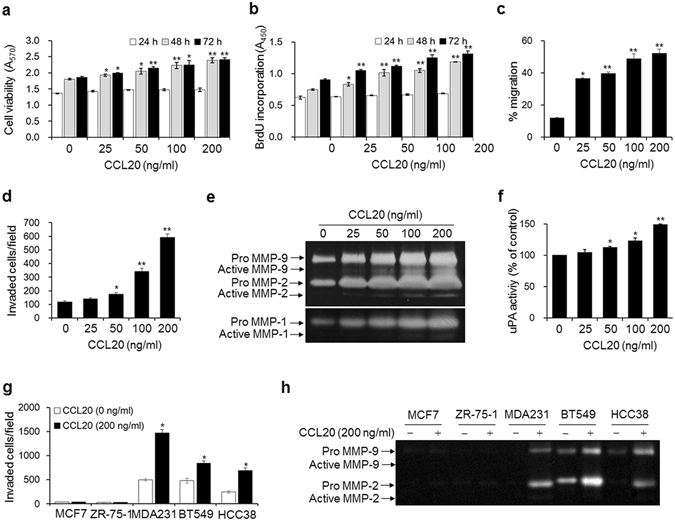
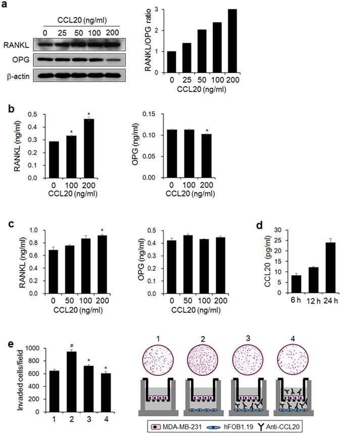
Breast cancer mainly spreads to bone, causing decreased survival of patient. Human antigen R (HuR) and chemokines are important molecules associated with mRNA stability and cell-cell interaction in cancer biology. Here, HuR knockdown inhibited bone metastasis and osteolysis of metastatic breast cancer cells in mice and HuR expression promoted the metastatic ability of cancer cells via CCL20 and GM-CSF. In contrast with the findings for GM-CSF, ELAVL1 and CCL20 expressions were markedly increased in breast tumor tissues and ELAVL1 expression showed a strong positive correlation with CCL20 expression in breast cancer subtypes, particularly the basal-like subtype. Metastasis-free survival and overall survival were decreased in the breast cancer patients with high CCL20 expression. We further confirmed the role of CCL20 in breast cancer bone metastasis. Intraperitoneal administration of anti-CCL20 antibodies inhibited osteolytic breast cancer bone metastasis in mice. Treatment with CCL20 noticeably promoted cell invasion and the secretion of MMP-2/9 in the basal-like triple-negative breast cancer cell lines, not the luminal. Moreover, CCL20 elevated the receptor activator of nuclear factors kappa-B ligand/osteoprotegerin ratio in breast cancer and osteoblastic cells and mediated the crosstalk between these cells. Collectively, HuR-regulated CCL20 may be an attractive therapeutic target for breast cancer bone metastasis.


Conflict of interest statement

The authors declare that they have no competing interests.

Figures

Figure 1
Figure 1
HuR knockdown prevents bone metastasis of breast cancer cells in mice. (a) Bioluminescence images taken 6 weeks after luciferase-transfected HuR-knockdown (shHuR) and control (shNC) breast cancer cells were inoculated into the left cardiac ventricles of nude mice (n = 10). (b,c) X-ray and 3D images of mandibles (b), femora, and tibiae (c) derived from μCT scans performed on week 6. (c) Goldner’s trichrome and TRAP staining of femoral tissues. Arrowheads: TRAP-positive osteoclasts; T: tumor; B: bone; BM: bone marrow. Scale bar: 0.5 mm for Goldner’s trichrome staining and 10 μm for TRAP staining. (d,e) Tumor areas (d) and osteoclast surface per bone surface (Oc.S/BS) (e) values for stained femoral sections. (f) Bone morphometric parameters BV/TV (%), Tb.Th (mm), Tb.N (1/mm), Tb.Sp (mm), and SMI of the mouse femora analyzed by μCT. (g) Serum levels of the bone resorption markers TRAP 5b and CTX quantified using commercial kits. The data are expressed as the mean ± standard error (s.e.m.). # P < 0.05 versus control mice without cancer cells; *P < 0.05, **P < 0.01 versus mice injected with shNC MDA-MB-231 cells. (h) Viabilities of shNC and shHuR MDA-MB-231 cells. The cells were cultured for 24, 48, and 72 h, and cell viability was assessed by MTT assay. (i) Cell migration. Movement of cells in scratched areas was observed under an inverted optical microscope (original magnification, × 40) and measured using ImageJ software at 0 h and 24 h. (j) Cell invasion. Cell invasion was measured by transwell invasion assay. Representative images of cells that invaded the lower surfaces of the filters were captured using an optical microscope (original magnification, × 200). The data are expressed as the mean ± s.e.m. *P < 0.01 versus shNC.
Figure 2
Figure 2
HuR improves the metastatic potential of breast cancer cells via CCL20 and GM-CSF. (a) The relative intensities of CCL20 and GM-CSF spots on membranes of human cytokine Ab arrays treated with conditioned media from shNC and shHuR MDA-MB-231 cells. The relative intensities of the CCL20 and GM-CSF spots were plotted as a % of the control. (b) The levels of secreted CCL20 and GM-CSF in multiple siNC and siHuR breast cancer cell lines. The levels of CCL20 and GM-CSF in the culture medium of each cell line were measured using commercial human CCL20 and GM-CSF ELISA kits, respectively. The data are expressed as the mean ± s.e.m. *P < 0.05, **P < 0.01 versus siNC. (c) Immunohistochemical staining of HuR, CCL20, and GM-CSF in femoral tumor tissues from mice injected with shNC or shHuR breast cancer cells, as shown in Fig. 1. T: tumor; B: bone; BM: bone marrow. Scale bar: 5 μm. (d) Viabilities of breast cancer cells treated with the indicated concentrations of anti-CCL20 or anti-GM-CSF antibody for 24, 48, and 72 h. Cell viability was determined by MTT assay. (e,f) Migration (e) and invasion (f) capabilities of cells treated with anti-CCL20 or anti-GM-CSF antibody. Cell migration and invasion were measured by wound-healing and transwell invasion assays, respectively. The data are expressed as the mean ± s.e.m. *P < 0.05, **P < 0.01 versus cells without anti-CCL20 or anti-GM-CSF.
Figure 3
Figure 3
CCL20 expression is correlated with ELAVL1 expression, distant metastasis-free survival, and overall survival in patients with breast cancer. (a) Expression levels of ELAVL1 and CCL20 in normal and tumor tissues. The data were obtained from the TCGA database. RSEM: RNA-Seq by Expectation Maximization. *P < 0.01 versus normal breast tissues. (b) Scatterplot showing the correlations between ELAVL1 and CCL20 expression in whole breast cancer tissues and in tissues with different tumor subtypes. Pearson’s correlation analysis was performed to assess statistical significance. Normal: normal breast tissues (n = 113); Tumor: whole tumor tissues (n = 1,069); Luminal A subtype (n = 422); Luminal B subtype (n = 194); HER2-enriched subtype (n = 68); Basal-like subtype (n = 142). (c–f) Kaplan-Meier plots derived from clinical datasets (GSE3494: n = 130; GSE7390: n = 198; GSE26971: n = 256) showing the distant metastasis-free survival (c,e) and overall survival (d,f) of breast cancer patients with high or low CCL20 expression (c,d) and with high or low ELAVL1 expression (e,f). P values were determined using the log-rank test. Hazard ratios (HRs) with 95% confidence intervals are shown.
Figure 4
Figure 4
Anti-CCL20 treatment blocks bone metastasis of breast cancer cells in mice. (a) Metastatic progression detected by bioluminescence. Mice (n = 10) were injected with luciferase-transfected MDA-MB-231 cells into the left cardiac ventricles, followed by intraperitoneal administration of anti-human or anti-mouse CCL20 antibody at the indicated doses three times per week for 6 weeks. The control mice were treated with HBSS and PBS instead of the cancer cell suspension and anti-CCL20 antibody, respectively. The vehicle group was administered PBS instead of anti-CCL20. (b,c) X-ray and 3D images of mandibles (b), femora, and tibiae (c) derived from μCT scans. (c) Goldner’s trichrome and TRAP staining of femoral tissue sections. Arrowheads: TRAP-positive osteoclasts; T: tumor; B: bone; BM: bone marrow. Scale bar: 0.5 mm for Goldner’s trichrome staining and 10 μm for TRAP staining. (d,e) Tumor areas and (d) Oc.S/BS (e) values for stained femoral sections. (f) Bone morphometric parameters of the femora, including the BV/TV (%), Tb.Th (mm), Tb.N (1/mm), Tb.Sp (mm), and SMI. (g) Serum levels of the bone resorption markers TRAP 5b and CTX. The data are expressed as the mean ± s.e.m. # P < 0.05 versus control mice (C); *P < 0.05, **P < 0.01 versus vehicle-treated mice (V).
Figure 5
Figure 5
CCL20 enhances the metastatic ability of breast cancer cells. (a) Viabilities of breast cancer cells stimulated with the indicated concentrations of CCL20 for 24, 48, and 72 h. (b–d) Quantity of 5-bromo-2′-deoxyuridine (BrdU) incorporated into newly synthesized DNA (b), migration (c), and invasion (d) in MDA-MB-231 cells stimulated with the indicated concentrations of CCL20 for 24 h. (e,f) MMP-1, MMP-2/9 (e), and uPA (f) activities in conditioned media from MDA-MB-231 cells stimulated with the indicated concentrations of CCL20 for 24 h. MMP-1 and MMP2/9 activities were determined by collagen and gelatin zymography, respectively. The clear bands indicate the activities of the MMPs. uPA activity was measured with a commercial uPA activity assay kit. (g,h) Invasion (g) and MMP-2 and MMP-9 activities in conditioned media (h) of MCF-7, ZR-75-1, BT549, and HCC38 breast cancer cells stimulated with CCL20 (200 ng/ml) for 24 h. The data are expressed as the mean ± s.e.m. *P < 0.05, **P < 0.01 versus cells without CCL20.
Figure 6
Figure 6
CCL20 increases the RANKL/OPG ratio and mediates interactions between human breast cancer cells and osteoblastic cells. (a) RANKL and OPG protein expression in MDA-MB-231 cells stimulated with the indicated concentrations of CCL20 for 24 h. Protein expression was detected by western blotting. β-actin served as a loading control. The graph presents the ratio of the intensity of RANKL to that of OPG after normalization against the intensity of β-actin. (b) Levels of RANKL and OPG in conditioned media from CCL20-stimulated MDA-MB-231 cells. (c) Levels of RANKL and OPG in conditioned media from CCL20-stimulated hFOB1.19 osteoblastic cells. (d) Levels of osteoblastic-derived CCL20 at the indicated time points. (b–d) RANKL, OPG, and CCL20 levels in conditioned media were detected with commercially available ELISA kits. The data are expressed as the mean ± s.e.m. *P < 0.05 versus cells without CCL20. (e) The effect of anti-CCL20 antibody on the invasion of MDA-MB-231 cells exposed to conditioned medium of osteoblastic cells. 1: MDA-MB-231 cells (upper)-media (lower); 2: MDA-MB-231 cells (upper)-hFOB1.19 cells (lower); 3: MDA-MB-231 cells (upper)-hFOB1.19 cells and anti-CCL20 antibody (lower); 4: MDA-MB-231 cells and anti-CCL20 antibody (upper)-hFOB1.19 cells and anti-CCL20 antibody (lower). # P < 0.05 versus 1; *P < 0.01 versus 2.

Similar articles

Cited by

References

    1. Mundy GR. Metastasis to bone: causes, consequences and therapeutic opportunities. Nat. Rev. Cancer. 2002;2:584–593. doi: 10.1038/nrc867. - DOI - PubMed
    1. Roodman GD. Mechanisms of bone metastasis. N. Engl. J. Med. 2004;350:1655–1664. doi: 10.1056/NEJMra030831. - DOI - PubMed
    1. Roodman GD. Bone-breaking cancer treatment. Nat. Med. 2007;13:25–26. doi: 10.1038/nm0107-25. - DOI - PubMed
    1. Bendre, M., Gaddy, D., Nicholas, R. W. & Suva, L. J. Breast cancer metastasis to bone: it is not all about PTHrP. Clin. Orthop. Relat. Res. S39–S45 (2003). - PubMed
    1. Guise TA, et al. Evidence for a causal role of parathyroid hormone-related protein in the pathogenesis of human breast cancer-mediated osteolysis. J. Clin. Invest. 1996;98:1544–1549. doi: 10.1172/JCI118947. - DOI - PMC - PubMed

Publication types

MeSH terms