. 2017 Aug 29;7(1):9622.
doi: 10.1038/s41598-017-08985-w.

The Multi Domain Caldicellulosiruptor bescii CelA Cellulase Excels at the Hydrolysis of Crystalline Cellulose

Affiliations

The Multi Domain Caldicellulosiruptor bescii CelA Cellulase Excels at the Hydrolysis of Crystalline Cellulose

Roman Brunecky et al. Sci Rep. .

Abstract

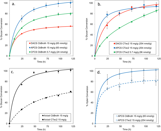
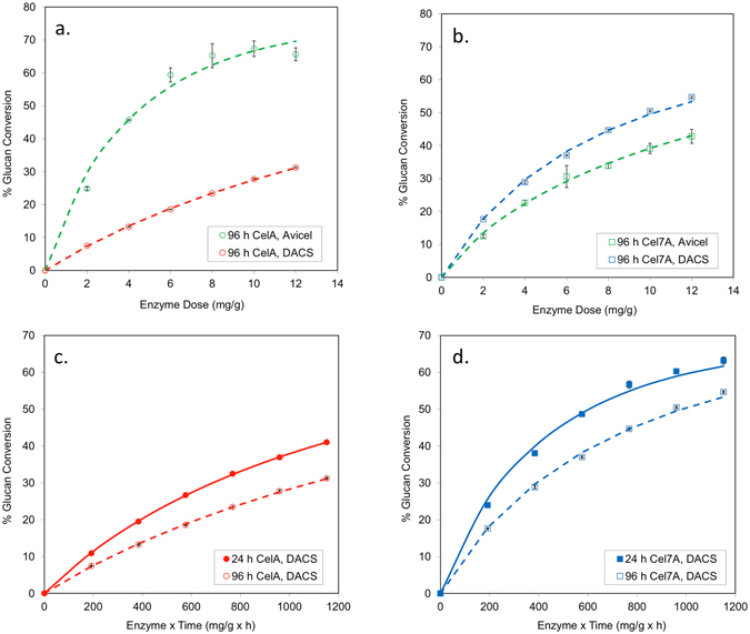
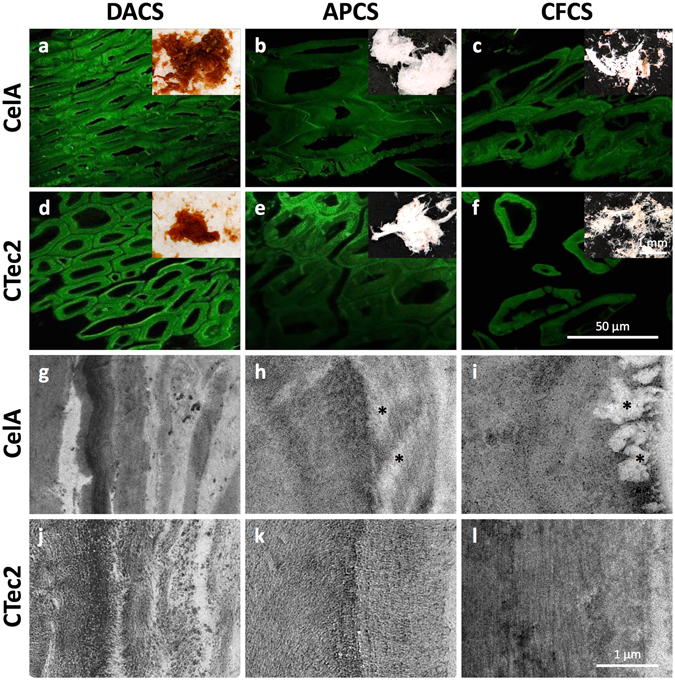
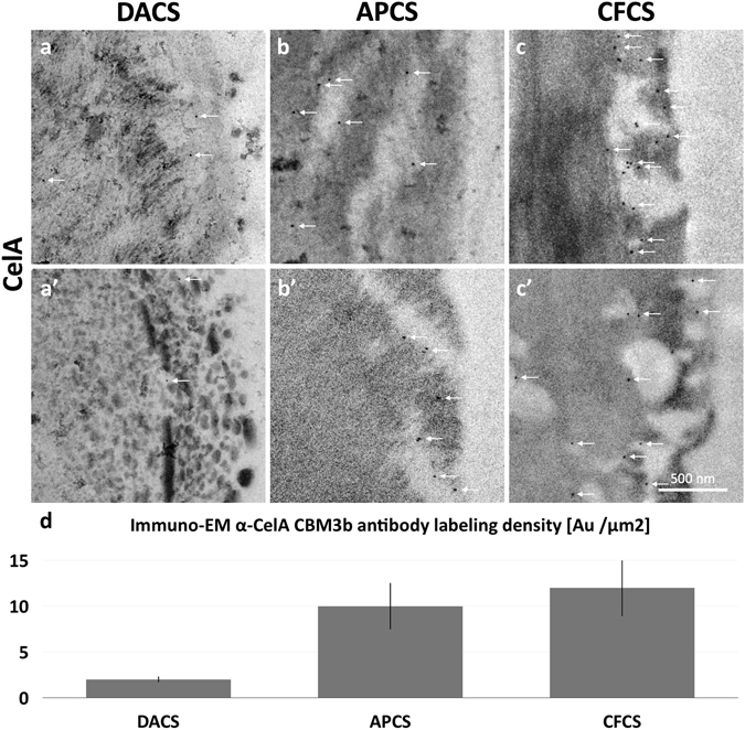
The crystalline nature of cellulose microfibrils is one of the key factors influencing biomass recalcitrance which is a key technical and economic barrier to overcome to make cellulosic biofuels a commercial reality. To date, all known fungal enzymes tested have great difficulty degrading highly crystalline cellulosic substrates. We have demonstrated that the CelA cellulase from Caldicellulosiruptor bescii degrades highly crystalline cellulose as well as low crystallinity substrates making it the only known cellulase to function well on highly crystalline cellulose. Unlike the secretomes of cellulolytic fungi, which typically comprise multiple, single catalytic domain enzymes for biomass degradation, some bacterial systems employ an alternative strategy that utilizes multi-catalytic domain cellulases. Additionally, CelA is extremely thermostable and highly active at elevated temperatures, unlike commercial fungal cellulases. Furthermore we have determined that the factors negatively affecting digestion of lignocellulosic materials by C. bescii enzyme cocktails containing CelA appear to be significantly different from the performance barriers affecting fungal cellulases. Here, we explore the activity and degradation mechanism of CelA on a variety of pretreated substrates to better understand how the different bulk components of biomass, such as xylan and lignin, impact its performance.


Conflict of interest statement

The authors declare that they have no competing interests.

Figures

Figure 1
Figure 1
(a,b) Activity of Cb broth and CTec2, respectively, on Dilute Acid treated Corn Stover, on alkaline peroxide treated corn stover and on Clean fractionated corn stover (c) Conversion of Avicel by Cb broth and CTec2. (d) Xylan conversion of Cb broth and CTec2 of alkaline peroxide treated corn stover.
Figure 2
Figure 2
Digestion of differential crystal index (CI) materials by (a) mix of CelA and Bg and (b) by Cel7A, E1 and Bg indicate that while Cel7A is impacted significantly by CI, CelA is agnostic to CI of materials.
Figure 3
Figure 3
Protein concentration of CelA and Cel7A in the supernatant after incubation with insoluble lignin at 75 °C and 30 °C and at 45 °C and 30 °C, respectively, as measured by densitometry from SDS-Page Gel (Figure S6).
Figure 4
Figure 4
(a) Digestion of dilute acid treated corn stover by CelA is improved in the presence of surfactants (b) The effect of SMWL compounds decreases the performance of CelA on avicel.
Figure 5
Figure 5
Performance of CelA and Cel7A on Avicel and DACS at 50 °C. CelA and Cel7A were incubated with Avicel and DACS at 50 °C for 24 and 96 h. β-glucosidase was added to each of these incubations at 5% total protein. Total enzyme doses ranged from 2 to 48 mg/g and glucose concentrations were measured by HPLC. (a,b) show the relative performance of CelA and Cel7A on these two substrates during 96 h incubations. This demonstrates that CelA is considerably better on Avicel than Cel7A even when tested at the temperature optimum of Cel7A. (c and d) show the 24 h and 96 h incubations for CelA and Cel7A, respectively, on DACS plotted as a function of enzyme x time. Model fits to these data are shown here – see Table 6 for parameter values. This demonstrates that the deviation in Et relationship is similar for CelA and Cel7A on DACS when tested at 50 °C.
Figure 6
Figure 6
(af) CSLM micrographs (with stereoscope micrograph insets) of digested corn stover particles display morphological features typical of materials exposed to DA, AP, and CF pretreatments. Among these, the clearest evidence for cellular dislocation and deconstruction at the tissue scale is seen in the samples exposed to CF pretreatment (c,f). At this scale, there are no clear or consistent differences between the CTec2 and CelA digested samples (gl). TEM micrographs of secondary cell walls from fiber cells show evidence for delamination, wall loosening and enzymatic deconstruction. The DACS micrographs show the coalescence and relocalization of lignin into globules that is typical of dilute acid (g,j) pretreatment. The APCS cell walls (h,k) show lower contrast and reveal cellulose structure due to lignin removal and show evidence for channels or cavities formed in CelA digested material (* k). The digested CFCS samples (i,l) display the clearest evidence for a difference between the deconstruction mechanism of CelA compared to CTec2 revealing completely cleared cavities near the surface of cell wall regions (* i) similar to the cavities previously observed in Avicel particles.
Figure 7
Figure 7
Immuno-EM micrographs reveal the pattern of penetration of CelA enzymes into pretreated corn stover cell walls and confirm that CelA enzymes occupy the cavities generated in APCS and CFCS. (a,a’) The labeled enzymes (white arrows) in DACS samples appear in cleared zones well into the secondary cell wall and do not appear to be associated with surface lignin globules. (b,b’,c,c’) CelA enzyme labeled in APCS and CFCS cell walls were most often found within cavities that connect to the cell wall surface and penetrate into the secondary cell wall. (d) Quantitation of the immune gold α-CelA CBM3 labeling density shows 3–4 times the labeling density in the digested APCS or CFCS compared to DACS.

References

    1. Zverlov, V. et al. Properties and gene structure of a bifunctional cellulolytic enzyme (CelA) from the extreme thermophile ‘Anaerocellum thermophilum’ with separate glycosyl hydrolase family 9 and 48 catalytic domains. Microbiology, 144 (1998). - PubMed
    1. Young J, et al. Deletion of Caldicellulosiruptor bescii CelA reveals its crucial role in the deconstruction of lignocellulosic biomass. Biotechnology for Biofuels. 2014;7(1):1–8. doi: 10.1186/s13068-014-0142-6. - DOI - PMC - PubMed
    1. Brunecky, R. et al. Revealing nature’s cellulase diversity: the digestion mechanism of Caldicellulosiruptor bescii CelA. Science, 342 2013. - PubMed
    1. Bischof RH, Ramoni J, Seiboth B. Cellulases and beyond: the first 70 years of the enzyme producer Trichoderma reesei. Microbial Cell Factories. 2016;15(1) doi: 10.1186/s12934-016-0507-6. - DOI - PMC - PubMed
    1. Kari, J. et al. Kinetics of cellobiohydrolase (Cel7A) variants with lowered substrate affinity. Journal of Biological Chemistry, (2014). - PMC - PubMed

Publication types

LinkOut - more resources