. 2017 Aug 29;7(1):9659.
doi: 10.1038/s41598-017-09260-8.

Aspergillus flavus infection triggered immune responses and host-pathogen cross-talks in groundnut during in-vitro seed colonization

Affiliations

Aspergillus flavus infection triggered immune responses and host-pathogen cross-talks in groundnut during in-vitro seed colonization

Spurthi N Nayak et al. Sci Rep. .

Abstract

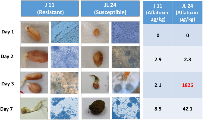
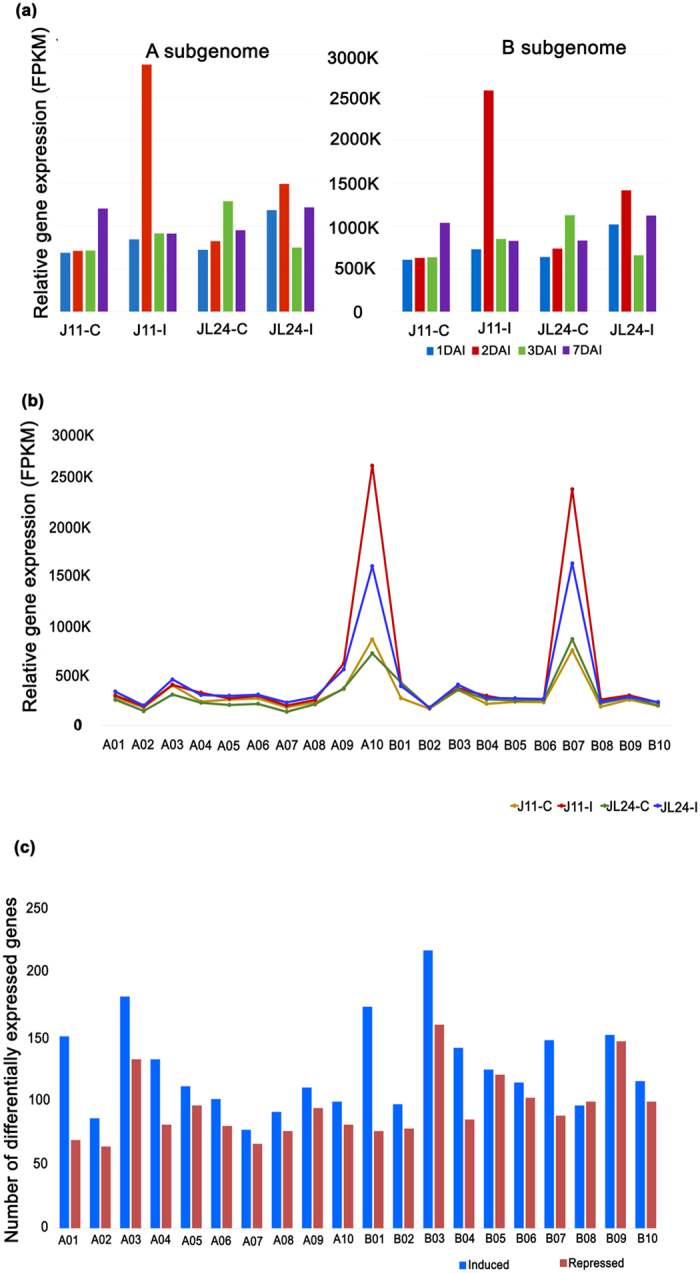
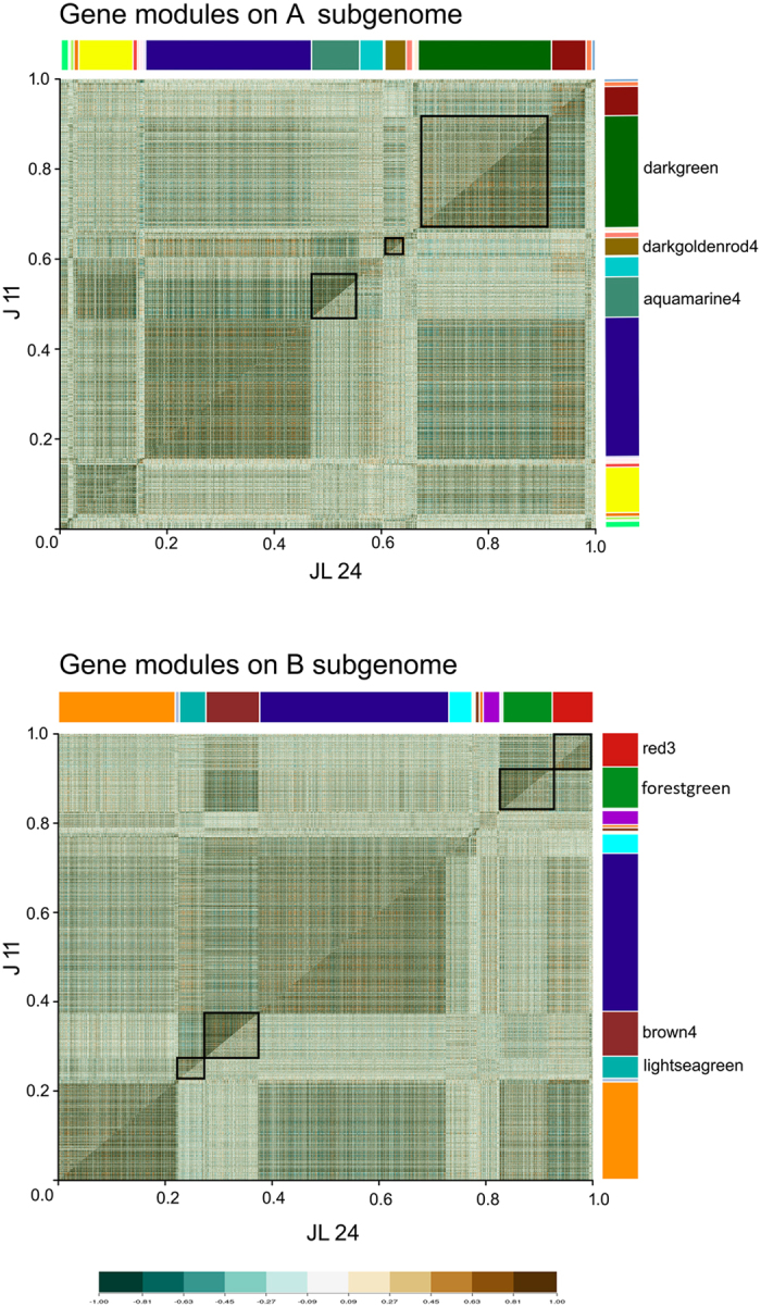
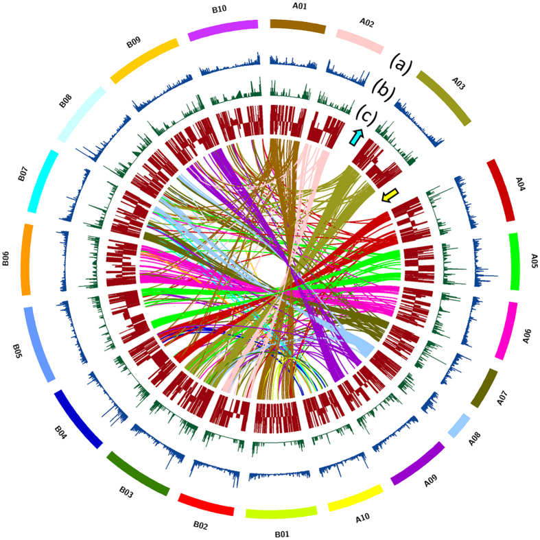
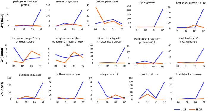
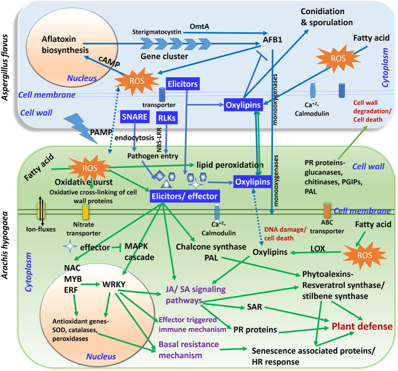
Aflatoxin contamination, caused by fungal pathogen Aspergillus flavus, is a major quality and health problem delimiting the trade and consumption of groundnut (Arachis hypogaea L.) worldwide. RNA-seq approach was deployed to understand the host-pathogen interaction by identifying differentially expressed genes (DEGs) for resistance to in-vitro seed colonization (IVSC) at four critical stages after inoculation in J 11 (resistant) and JL 24 (susceptible) genotypes of groundnut. About 1,344.04 million sequencing reads have been generated from sixteen libraries representing four stages in control and infected conditions. About 64% and 67% of quality filtered reads (1,148.09 million) were mapped onto A (A. duranensis) and B (A. ipaёnsis) subgenomes of groundnut respectively. About 101 million unaligned reads each from J 11 and JL 24 were used to map onto A. flavus genome. As a result, 4,445 DEGs including defense-related genes like senescence-associated proteins, resveratrol synthase, 9s-lipoxygenase, pathogenesis-related proteins were identified. In A. flavus, about 578 DEGs coding for growth and development of fungus, aflatoxin biosynthesis, binding, transport, and signaling were identified in compatible interaction. Besides identifying candidate genes for IVSC resistance in groundnut, the study identified the genes involved in host-pathogen cross-talks and markers that can be used in breeding resistant varieties.


Conflict of interest statement

The authors declare that they have no competing interests.

Figures

Figure 1
Figure 1
Phenotypic observations for seeds of J 11 and JL 24 during in-vitro seed colonization by Aspergillus flavus at different time points along with microscopic observation and aflatoxin estimation. The figure shows the microscopic observations of seed coat after fungal inoculation in J 11 (resistant) and JL 24 (susceptible) that clearly shows the presence of mycelium at Day 3 and sporulation at Day 7 after inoculation.
Figure 2
Figure 2
Gene expression during in-vitro seed colonization of Aspergillus flavus in groundnut. (a) Temporal distribution of relative abundance in expressed genes at different stages of fungal infection. The relative expression is found to be higher in infected samples of J 11 (J11-I) as compared to their control counter parts (J11-C) at 2nd day after inoculation (2DAI). The expression level of infected samples of JL 24 (JL24-I) was higher as compared to JL24-C. (b) Relative abundance of all the genes expressed during in-vitro seed colonization across subgenomes of groundnut progenitors. The relative abundance in terms of FPKM values were mapped onto A10 and B07 of A and B subgenomes, respectively. (c) Number of differentially expressed genes-DEGs (induced and repressed) during in-vitro seed colonization. The highest number of DEGs were mapped onto A03 and B03 of A and B subgenomes, respectively.
Figure 3
Figure 3
Differentially co-expressed modules between J 11 and JL 24 in A and B subgenomes of groundnut. The comparative correlation heat maps with brown color corresponding to positive correlation and blue corresponding to negative correlations are depicted in the figure. The upper diagonal of the main matrix shows a correlation between pairs of genes among J 11 (resistant) transcripts while the lower diagonal of the heat map shows a correlation between the same gene pairs in the JL 24 (susceptible). Modules are identified in the heat map by different built-in color bars on the right side of the heat map. Distinguished gene modules are demarcated by black squares.
Figure 4
Figure 4
Distribution of single nucleotide polymorphisms (SNPs), Indels and differentially expressed genes (DEGs) across the pseudomolecules of progenitor reference genomes of groundnut. (a) Groundnut pseudomolecules from A subgenome are depicted as A01 to A10 and that of B subgenome are depicted as B01 to B10 (b) SNP density depicted in blue (c) InDel density depicted in green (d) both upregulated (with upward arrow) and downregulated (with downward arrow) differentially expressed genes depicted in brown. Synteny of the differentially expressed genes among the pseudomolecules are depicted with the different colored lines with respect to representative colors of the pseudomolecules.
Figure 5
Figure 5
Validation of selected genes through qRT-PCR across different time periods of Aspergillus infection in J 11 (resistant) and JL 24 (susceptible) genotypes. Relative gene expression for 15 selected candidate genes like pathogenesis-related protein, resveratrol synthase, cationic peroxidase, lipoxygenase, heat shock protein 83-like, microsomal omega-3 fatty acid desaturase, ethylene-responsive transcription factor erf060-like, Kunitz-type trypsin inhibitor-like 2 protein, desiccation protectant protein Lea14, seed linoleate 9s-lipoxygenase-2, chalcone reductase, isoflavone reductase, allergen Ara h 2, class ii chitinase and subtilisin-like protease was calculated at four different time points of infection in case of J 11 and JL 24.
Figure 6
Figure 6
Illustration of cross-talk between groundnut (Arachis hypogaea) and fungus (Aspergillus flavus) during in-vitro seed colonization. The genes/components illustrated represent the defense responsive molecules identified from groundnut-A. flavus interaction during in-vitro seed colonization studies. The components marked within the green rectangular box (with green arrows) are those induced in groundnut and those which are in light blue rectangular box (blue arrows) are the components induced in pathogen in compatible reaction through mixed transcriptome analysis. The components represented in dark blue boxes have important role in host-pathogen interaction in this study. Briefly, the pathogenesis initiation occurs at plant cell wall where NBS-LRR, elicitors and oxylipins have important role in host-pathogen interactions. JA and SA signaling pathways mediated by transcription factors like WRKY, NAC, MYB and ERFs play important role in plant defense. During the process of defense the phytoalexins like resveratrol synthase/stillbene synthase, PR proteins, LOX, chalcone synthase and PAL were expressed. Basal resistance works with the expression of senescence associated proteins that give hypersensitive response to combat the pathogen entry further. The chitinases, glucanases, PGIPs, PAL, PR proteins induce cell wall degradation of fungi during pathogen entry. Due to pathogen attack, there is oxidative burst in the plant cell that regulates the oxidation of fatty acids that in turn involved in signal transduction. Transcriptome analysis of fungus indicated that the RLKs, SNARE complex, elicitors and oxylipins play important role in plant recognition and infection. Most of these proteins are integral part of cell wall and membrane. Genes involved in fungal growth, aflatoxin synthesis and transport were highly expressed in Aspergillus. Abbreviations: ROS- reactive oxygen species; SOD-superoxide dismutase; PR-pathogenesisrelated proteins; HR-hypersensitive response; JA-Jasmonic acid; SA- Salicylic acid; SAR-Systemic acquired resistance; LOX-lipoxygenase; PAL- Phenylalanine ammonia-lyase; Transcription factors-NAC (NAM, ATAF1/2 and CUC2 domain proteins), ERF (Ethylene responsive factors); MAPK- Mitogen-activated protein kinases; SNARE- soluble N-ethylmaleimide sensitive factor attachment receptor; RLK- receptor-like kinase; cAMP- Cyclic adenosine monophosphate, OmtA- O-methyl transferase; AFB1-aflatoxin B1.

Similar articles

Cited by

References

    1. Williams JH, et al. HIV and hepatocellular and esophageal carcinomas related to consumption of mycotoxin-prone foods in sub-Saharan Africa. The American Journal of Clinical Nutrition. 2010;92:154–160. doi: 10.3945/ajcn.2009.28761. - DOI - PubMed
    1. Andrade P, Caldas E. Aflatoxins in cereals: worldwide occurrence and dietary risk assessment. World Mycotoxin Journal. 2015;8:415–431. doi: 10.3920/WMJ2014.1847. - DOI
    1. Gong Y, et al. Post weaning exposure to aflatoxin results in impaired child growth: a longitudinal study in Benin, West Africa. Environmental Health Perspectives. 2004;112:1334–1338. doi: 10.1289/ehp.6954. - DOI - PMC - PubMed
    1. Jiang Y, et al. Aflatoxin-related immune dysfunction in health and in human immunodeficiency virus disease. Clinical and Developmental Immunology. 2008;2008 doi: 10.1155/2008/790309. - DOI - PMC - PubMed
    1. Jolly PE. Aflatoxin: does it contribute to an increase in HIV viral load? Future Microbiology. 2014;9:121–124. doi: 10.2217/fmb.13.166. - DOI - PubMed

Publication types

MeSH terms