. 2017 Aug 29;7(1):9774.
doi: 10.1038/s41598-017-09660-w.

A Combination of Species Identification and STR Profiling Identifies Cross-contaminated Cells from 482 Human Tumor Cell Lines

Affiliations

A Combination of Species Identification and STR Profiling Identifies Cross-contaminated Cells from 482 Human Tumor Cell Lines

Xiaocui Bian et al. Sci Rep. .

Abstract

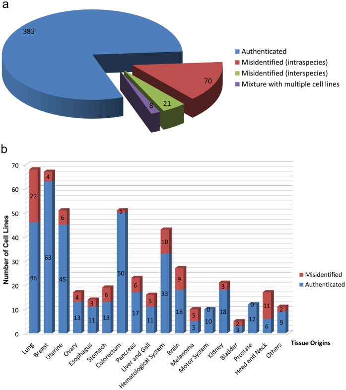
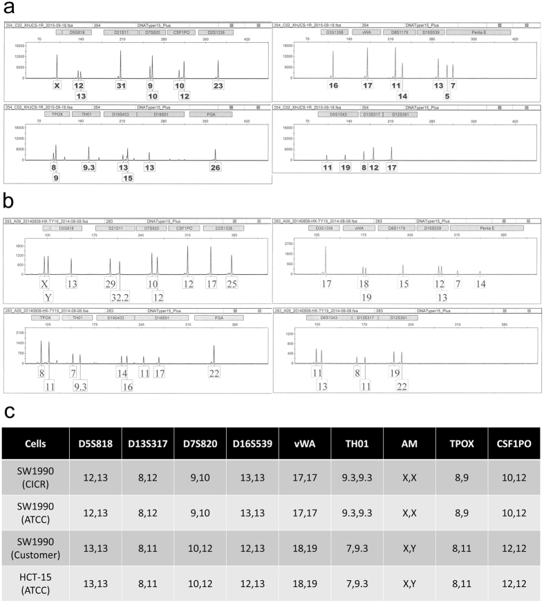
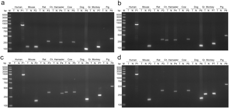
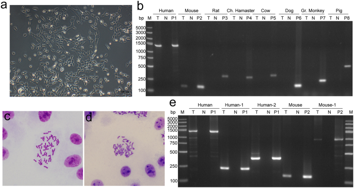
Human tumor cell lines are extremely important tools for cancer research, but a significant percentage is cross-contaminated with other cells. Short tandem repeat (STR) profiling is the prevailing standard for authenticating cell lines that originate from human tissues. Based on the analysis of 482 different human tumor cell lines used in China by STR, up to 96 cell lines were misidentified. More importantly, the study has found that STR profiling alone is insufficient to exclude inter-species cross-contamination of human cell lines. Among the 386 cell lines which had a correct STR profile, 3 of them were inter-species cross-contaminated. Careful microscopic examination may be helpful in some cases to detect changes in morphology but additional testing is needed. Additionally, species verification by PCR could easily identify the contaminants, even with a low percentage of contaminating cells. Combining STR profiling with species identification by PCR, more than 20.5% (99/482) of tumor cell lines were revealed as having been incorrectly identified, including intra-species (14.5%), inter-species (4.4%) cross-contamination and contaminating cell lines (1.7%). Therefore, quality control of cell lines is a systemic issue. Each cell line should undergo a full QA (Quality Assurance) assessment before it is used for research.


Conflict of interest statement

The authors declare that they have no competing interests.

Figures

Figure 1
Figure 1
Workflow for authentication of human tumor cell lines.
Figure 2
Figure 2
Overview of the 482 cell lines tested. (a) The pie graphs show cases of authenticated, intra- and inter-species cross-contamination of human tumor cell lines. (b) All tumor cells were divided into 18 categories according to their tissue origins and cases of authenticated and misidentified cell lines of each category are showed in the column chart. Misidentified cell lines include intra-species and inter-species cell lines.
Figure 3
Figure 3
Electropherogram of SW1990 (human pancreatic carcinoma cell line) from two laboratories. (a) STR profile of SW1990 from China Infrastructure of Cell Line Resource (CICR), which is an authentic cell line. (b) STR profile of SW1990 from a customer’s laboratory, which is cross-contaminated by HCT-8 (human colorectal carcinoma cell line). (c) Comparison of STR profiles with ATCC.
Figure 4
Figure 4
Cases of human cells fully substituted by animal cells. (ad), by PCR-based species identification, Ramos (human Burkitt’s lymphoma B cells), A549-CP-H2 (human lung cancer cells with Cas9 stable expression), A427 (human lung cancer cells) and SCaBER (human bladder cancer cells) were cross-contaminated by cells of mouse, Chinese hamster, bovine and monkey origin, respectively. T, test sample; N, deionized water used as negative control; P, cell lines of corresponding species used as positive control separately, P1, RD (human rhabdomyosarcoma cell line); P2, Hepa 1–6 (mouse hepatocarcinoma cell line); P3, PC-12 (rat phaeochromocytoma cell line); P4, CHO (Chinese Hamster ovary cells); P5, MDBK (bovine kidney cell line); P6, MDCK (dog kidney cell line); P7, VERO (African Green Monkey kidney cell line); P8, LLC-PK1(pig kidney cell line); M, DNA marker; bp, base pairs.
Figure 5
Figure 5
Electropherogram of 786–0 (human renal cell carcinoma) from two laboratories. (a) STR profile of 786–0 from China Infrastructure of Cell Line Resource (CICR), which is an authentic cell line. (b) STR profile of 786–0 from a customer’s laboratory, which is a mixture of multiple cells. (c) Comparison of STR profiles with ATCC.
Figure 6
Figure 6
Species identification of BeWo (Human choriocarcinoma cells). (a) Two distinct morphology was found in the microscopic view of BeWo(A) from A laboratory; (b) human and mouse specific bands were both found by PCR-based species identification in BeWo(A); (c) only one morphology was found in the microscopic view of BeWo(B) from B laboratory; (d) BeWo(B) was identified as human cells by PCR-based assay; (e) human chromosome was found in the metaphase spreads of BeWo(A); (f) mouse chromosome was found in the metaphase spreads of BeWo(A); (g) The result of PCR with species-specific primers targeting different DNA loci confirmed that BeWo(A) was a mixture of human and mouse cells. T, test sample; N, deionized water used as negative control; P, cell lines of corresponding species used as positive control separately, P1, RD (human rhabdomyosarcoma cell line); P2, Hepa 1–6 (mouse hepatocarcinoma cell line); P3, PC-12 (rat phaeochromocytoma cell line); P4, CHO (Chinese Hamster ovary cells); P5, MDBK (bovine kidney cell line); P6, MDCK (dog kidney cell line); P7, VERO (African Green Monkey kidney cell line); P8, LLC-PK1(pig kidney cell line); M, DNA marker; bp, base pairs.
Figure 7
Figure 7
Species identification of PG-LH7 (Human lowly metastatic lung cancer cells). (a) No distinct morphological variation was found in the microscopic view of PG-LH7; (b) human and mouse specific bands were both found by PCR-based species identification in PG-LH7. (c) Human chromosome was found in the metaphase spreads of PG-LH7; (d) mouse chromosome was found in the metaphase spreads of PG-LH7; (e) The result of PCR with species-specific primers targeting different DNA loci confirmed that PG-LH7 was a mixture of human and mouse cells. T, test sample; N, deionized water used as negative control; P, cell lines of corresponding species used as positive control separately, P1, RD (human rhabdomyosarcoma cell line); P2, Hepa 1–6 (mouse hepatocarcinoma cell line); P3, PC-12 (rat phaeochromocytoma cell line); P4, CHO (Chinese Hamster ovary cells); P5, MDBK (bovine kidney cell line); P6, MDCK (dog kidney cell line); P7, VERO (African Green Monkey kidney cell line); P8, LLC-PK1(pig kidney cell line); M, DNA marker; bp, base pairs.

Similar articles

Cited by

References

    1. Capes-Davis A, et al. Check your cultures! A list of cross-contaminated or misidentified cell lines. Int J Cancer. 2010;127:1–8. doi: 10.1002/ijc.25242. - DOI - PubMed
    1. Dirks WG, Drexler HG. STR DNA typing of human cell lines: detection of intra- and interspecies cross-contamination. Methods Mol Biol. 2013;946:27–38. doi: 10.1007/978-1-62703-128-8_3. - DOI - PubMed
    1. ASN‑0002, A. T. C. C. S. D. O. W. Cell line misidentification: the beginning of the end. Nat Rev Cancer10, 441–448 (2010). - PubMed
    1. Ayyoob K, Masoud K, Vahideh K, Jahanbakhsh A. Authentication of newly established human esophageal squamous cell carcinoma cell line (YM-1) using short tandem repeat (STR) profiling method. Tumour Biol. 2016;37:3197–3204. doi: 10.1007/s13277-015-4133-4. - DOI - PubMed
    1. Ye F, Chen CG, Qin J, Liu J, Zheng CY. Genetic profiling reveals an alarming rate of cross-contamination among human cell lines used in China. Faseb J. 2015;29:4268–4272. doi: 10.1096/fj.14-266718. - DOI - PubMed

Publication types