Delayed APC/C activation extends the first mitosis of mouse embryos
- PMID: 28851945
- PMCID: PMC5575289
- DOI: 10.1038/s41598-017-09526-1
Delayed APC/C activation extends the first mitosis of mouse embryos
Abstract
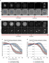
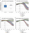
The correct temporal regulation of mitosis underpins genomic stability because it ensures the alignment of chromosomes on the mitotic spindle that is required for their proper segregation to the two daughter cells. Crucially, sister chromatid separation must be delayed until all the chromosomes have attached to the spindle; this is achieved by the Spindle Assembly Checkpoint (SAC) that inhibits the Anaphase Promoting Complex/Cyclosome (APC/C) ubiquitin ligase. In many species the first embryonic M-phase is significantly prolonged compared to the subsequent divisions, but the reason behind this has remained unclear. Here, we show that the first M-phase in the mouse embryo is significantly extended due to a delay in APC/C activation. Unlike in somatic cells, where the APC/C first targets cyclin A2 for degradation at nuclear envelope breakdown (NEBD), we find that in zygotes cyclin A2 remains stable for a significant period of time after NEBD. Our findings that the SAC prevents cyclin A2 degradation, whereas over-expressed Plk1 stimulates it, support our conclusion that the delay in cyclin A2 degradation is caused by low APC/C activity. As a consequence of delayed APC/C activation cyclin B1 stability in the first mitosis is also prolonged, leading to the unusual length of the first M-phase.
Conflict of interest statement
The authors declare that they have no competing interests.
Figures


Similar articles
-
Spatial control of the APC/C ensures the rapid degradation of cyclin B1.EMBO J. 2024 Oct;43(19):4324-4355. doi: 10.1038/s44318-024-00194-2. Epub 2024 Aug 14. EMBO J. 2024. PMID: 39143240 Free PMC article.
-
Cyclin A2 degradation during the spindle assembly checkpoint requires multiple binding modes to the APC/C.Nat Commun. 2019 Aug 27;10(1):3863. doi: 10.1038/s41467-019-11833-2. Nat Commun. 2019. PMID: 31455778 Free PMC article.
-
Mitotic phosphatase activity is required for MCC maintenance during the spindle checkpoint.Cell Cycle. 2016;15(2):225-33. doi: 10.1080/15384101.2015.1121331. Cell Cycle. 2016. PMID: 26652909 Free PMC article.
-
Cyclin A and Nek2A: APC/C-Cdc20 substrates invisible to the mitotic spindle checkpoint.Biochem Soc Trans. 2010 Feb;38(Pt 1):72-7. doi: 10.1042/BST0380072. Biochem Soc Trans. 2010. PMID: 20074038 Review.
-
The APC/C in female mammalian meiosis I.Reproduction. 2013 Jun 27;146(2):R61-71. doi: 10.1530/REP-13-0163. Print 2013 Aug. Reproduction. 2013. PMID: 23687281 Review.
Cited by
-
Delays in the final stages of fertilization are strongly associated with trichotomous cytokinesis and cleavage arrest.J Assist Reprod Genet. 2025 Jan;42(1):107-114. doi: 10.1007/s10815-024-03330-3. Epub 2024 Nov 28. J Assist Reprod Genet. 2025. PMID: 39607653
-
Single-cell quantification of ribosome occupancy in early mouse development.Nature. 2023 Jun;618(7967):1057-1064. doi: 10.1038/s41586-023-06228-9. Epub 2023 Jun 21. Nature. 2023. PMID: 37344592 Free PMC article.
-
Early onset of APC/C activity renders SAC inefficient in mouse embryos.Front Cell Dev Biol. 2024 Mar 13;12:1355979. doi: 10.3389/fcell.2024.1355979. eCollection 2024. Front Cell Dev Biol. 2024. PMID: 38544818 Free PMC article.
-
Multiple Roles of PLK1 in Mitosis and Meiosis.Cells. 2023 Jan 2;12(1):187. doi: 10.3390/cells12010187. Cells. 2023. PMID: 36611980 Free PMC article. Review.
-
Mysteries in embryonic development: How can errors arise so frequently at the beginning of mammalian life?PLoS Biol. 2019 Mar 6;17(3):e3000173. doi: 10.1371/journal.pbio.3000173. eCollection 2019 Mar. PLoS Biol. 2019. PMID: 30840627 Free PMC article.
References
Publication types
MeSH terms
Substances
Grants and funding
LinkOut - more resources
Full Text Sources
Other Literature Sources
Molecular Biology Databases
Research Materials
Miscellaneous

