. 2017 Aug 29;7(1):9794.
doi: 10.1038/s41598-017-09621-3.

AhR-deficiency as a cause of demyelinating disease and inflammation

Affiliations

AhR-deficiency as a cause of demyelinating disease and inflammation

Ludmila Juricek et al. Sci Rep. .

Abstract

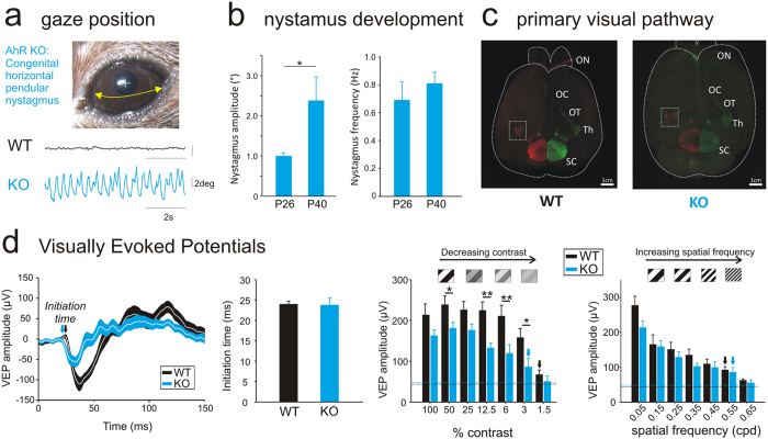
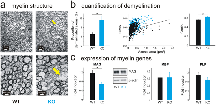
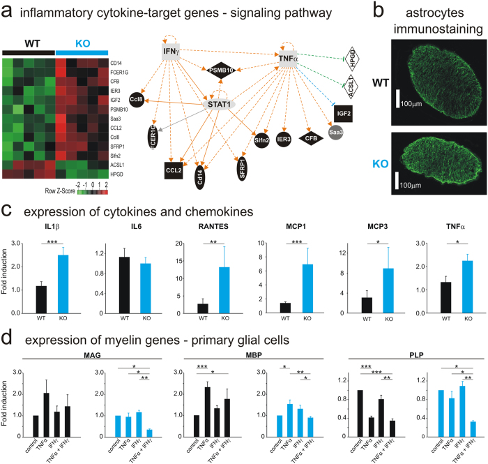
The Aryl hydrocarbon Receptor(AhR) is among the most important receptors which bind pollutants; however it also regulates signaling pathways independently of such exposure. We previously demonstrated that AhR is expressed during development of the central nervous system(CNS) and that its deletion leads to the occurrence of a congenital nystagmus. Objectives of the present study are to decipher the origin of these deficits, and to identify the role of the AhR in the development of the CNS. We show that the AhR-knockout phenotype develops during early infancy together with deficits in visual-information-processing which are associated with an altered optic nerve myelin sheath, which exhibits modifications in its lipid composition and in the expression of myelin-associated-glycoprotein(MAG), a cell adhesion molecule involved in myelin-maintenance and glia-axon interaction. In addition, we show that the expression of pro-inflammatory cytokines is increased in the impaired optic nerve and confirm that inflammation is causally related with an AhR-dependent decreased expression of MAG. Overall, our findings demonstrate the role of the AhR as a physiological regulator of myelination and inflammatory processes in the developing CNS. It identifies a mechanism by which environmental pollutants might influence CNS myelination and suggest AhR as a relevant drug target for demyelinating diseases.


Conflict of interest statement

The authors declare that they have no competing interests.

Figures

Figure 1
Figure 1
AhR knockout mice as a model of INS pathologies. (A) Raw traces of spontaneous eye movements recorded in the presence of light (8 week-old AhR-KO). (B) Nystagmus amplitude (degrees) and frequency (Hz) recorded at P26 and P40 (n = 8). (C) Example of whole brain staining of a WT and an AhR KO mouse: Optic Nerve (ON), Optic Chiasm (OC), Optic Tract (OT), Thalamus (Th) and Superior Colliculus (SC). (D) Left: Average Visually Evoked Potentials (VEP) traces recorded in WT (black line, n = 9) or AhR KO mice (blue line, n = 8) (100% contrast and 0.05 cpd stimulation). The white line represents the average and the envelope around (blue or black) represents the SEM. Middle: Initiation time of the VEPs measured on both WT (black bar) and AhR KO mice (blue bar). Right: VEP amplitudes with decreasing contrast stimulation or decreasing visual acuity (i.e. increasing spatial frequency). Dotted lines represent the noise. Arrows represent the first stimulation significantly different from the noise. (*p < 0.05; **p < 0.01).
Figure 2
Figure 2
AhR deficiency leads to myelin alterations in the optic nerve. (A) Example of transverse ultrathin sections of the optic nerve of 8 weeks-old WT (left) and AhR KO (right) mice examined by transmission electron microscopy. Scale bars: 2 µm (top) or 0,5 µm (bottom). The yellow arrows indicate zones of disorganized myelin sheath. (B) Left: percentage of low-myelinated axons in the optic nerves of WT (n = 5, black bar) and AhR-KO (n = 5, blue bar) mice. Middle: Scatter plots depicting G-ratio (ratio of axon diameter/myelinated fiber diameter) of WT (n = 352 axons, black) and AhR KO (n = 243 axons, blue) mice. Right: Quantification of the G-ratio of optic nerves for each genotype (n = 5 WT and n = 5 AhR KO). (C) Quantitative Real-Time PCR analysis of three myelin genes (MAG, MBP, PLP) in the WT (black bars) and AhR-KO (blue bars) mice optic nerves (n = 18–19/group). mRNA levels were normalized to GAPDH mRNA expression. Middle: MAG protein in whole tissue lysates of WT and AhR-KO mice optic nerves was detected by Western Blot. Total actin was used as a loading control (n = 4/group). A full-length gel is included in the supplementary file (complete western blot). MAG: myelin associated-glycoprotein; MBP: myelin basic protein; PLP: proteolipid protein; GAPDH: glyceraldehyde 3-phosphate dehydrogenase (*p < 0.05).
Figure 3
Figure 3
AhR-dependent inflammatory response controls myelin genes. (A) Left: Heatmap of the annotated genes with the highest importance in inflammatory response. Red: increased mRNAs expression; green: decreased mRNA expression (n = 5/group). Right: Connected components of the relevance network after IPA. Thirteen gene probes were analyzed by IPA software and annotated for gene network construction. Both over-expressed and under-expressed genes were mapped to the same network space. The pathway analysis revealed a central regulating role for tumor necrosis factor-alpha (TNF-α) and interferon-gamma (IFNγ). Main components of the legend: hatched line: indirect interaction; continuous line: direct interaction; arrow: acts on; line with a perpendicular line at the end: inhibits; square: cytokine (Interferon-γ and TNF-α); horizontal oval (STAT1); transcription regulator; vertical oval (SFRP1): transmembrane receptor; vertical diamond (ASCL1): enzyme; horizontal diamond (CFB): peptidase; circle (Saa3): other. (B) Expression of immunoreactive GFAP, an astrocyte marker, in the optic nerve of WT (top) and AhR KO (bottom) adult mice (Scale bar: 100 µm, n = 4/group). (C) Quantitative Real-Time PCR analysis of Interleukine-1β (IL1β), IL6, RANTES (Regulated on Activated Normal T-cell Expressed and Secreted), MCP1–3 (Monocyte Chemoattractant Protein 1–3), Tumor Necrosis Factor-α (TNF-α) in the WT (black bars) and AhR-KO (blue bars) mice optic nerves (n = 10–22/group). mRNA levels were normalized to GAPDH mRNA expression. (D) Myelin genes (MAG, MBP and PLP) mRNA expression was determined by Quantitative Real-Time PCR analysis in primary mixed glial cell cultures after 3 days-treatment with TNFα (10 ng/mL), IFNγ (10 ng/mL) or both (concentration 10ng/mL of each) or vehicle (control). The relative mRNA expression levels were represented as relative fold change compared to mRNA abundance in cells treated with vehicle. Data are expressed as the mean of four to six independent experiments. Each experiment consists of pool of 2–8 pups. IPA: Ingenuity Pathway Analysis, GFAP: Glial Fibrillary Acidic Protein (***p < 0.001, **p < 0.01; *p < 0.05).

References

    1. Barouki R, Coumoul X, Fernandez-Salguero PM. The aryl hydrocarbon receptor, more than a xenobiotic-interacting protein. FEBS Lett. 2007;581:3608–3615. doi: 10.1016/j.febslet.2007.03.046. - DOI - PubMed
    1. Choudhary, M. et al. Aryl hydrocarbon receptor knock-out exacerbates choroidal neovascularization via multiple pathogenic pathways. J. Pathol., doi:10.1002/path.4433 (2014). - PMC - PubMed
    1. Mulero-Navarro S, Fernandez-Salguero PM. New Trends in Aryl Hydrocarbon Receptor Biology. Front. Cell Dev. Biol. 2016;4 doi: 10.3389/fcell.2016.00045. - DOI - PMC - PubMed
    1. Abbott BD, Birnbaum LS, Perdew GH. Developmental expression of two members of a new class of transcription factors: I. Expression of aryl hydrocarbon receptor in the C57BL/6N mouse embryo. Dev. Dyn. Off. Publ. Am. Assoc. Anat. 1995;204:133–143. - PubMed
    1. Williamson MA, Gasiewicz TA, Opanashuk LA. Aryl hydrocarbon receptor expression and activity in cerebellar granule neuroblasts: implications for development and dioxin neurotoxicity. Toxicol. Sci. Off. J. Soc. Toxicol. 2005;83:340–348. doi: 10.1093/toxsci/kfi031. - DOI - PubMed

Publication types

MeSH terms