. 2017 Aug 29;7(1):9565.
doi: 10.1038/s41598-017-09869-9.

Linoleic acid metabolite leads to steroid resistant asthma features partially through NF-κB

Affiliations

Linoleic acid metabolite leads to steroid resistant asthma features partially through NF-κB

Lipsa Panda et al. Sci Rep. .

Abstract

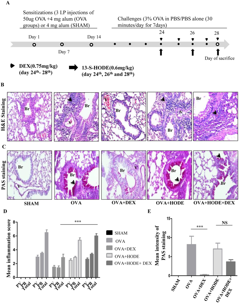
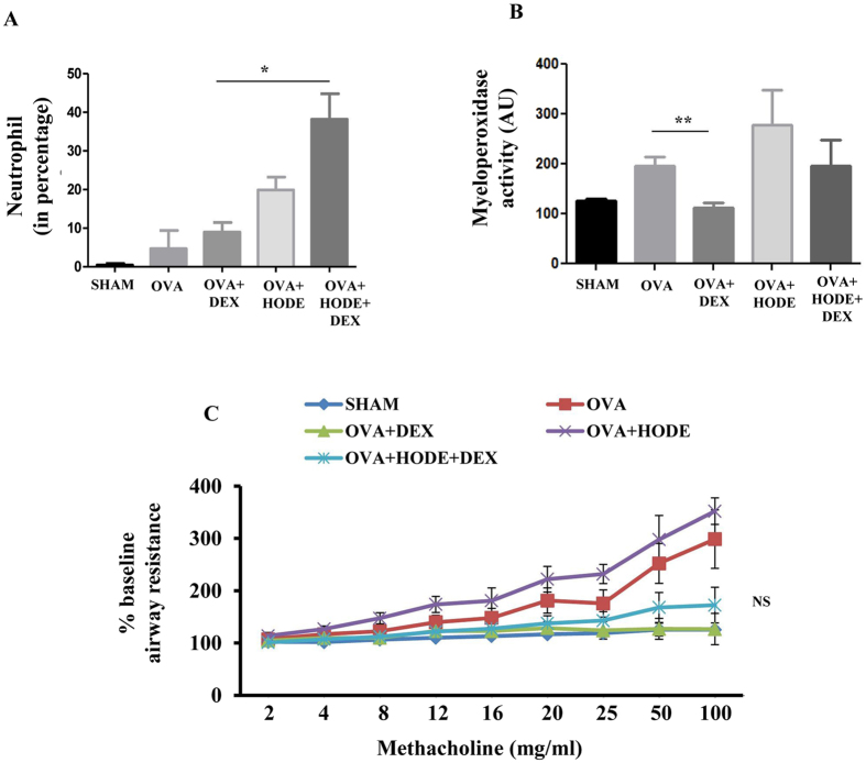
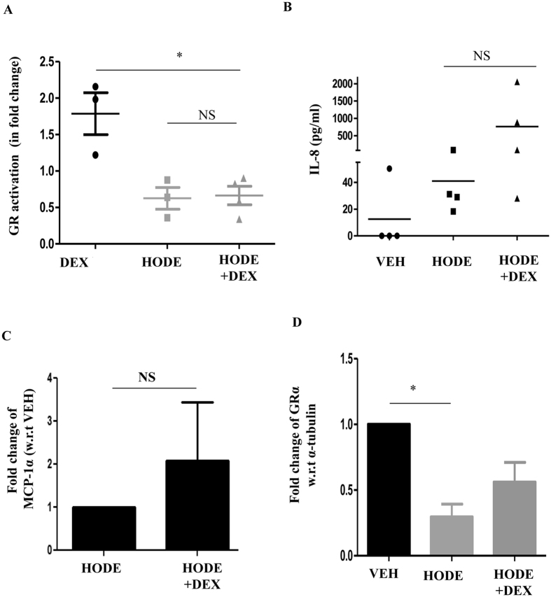
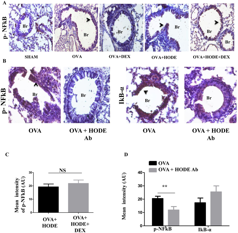
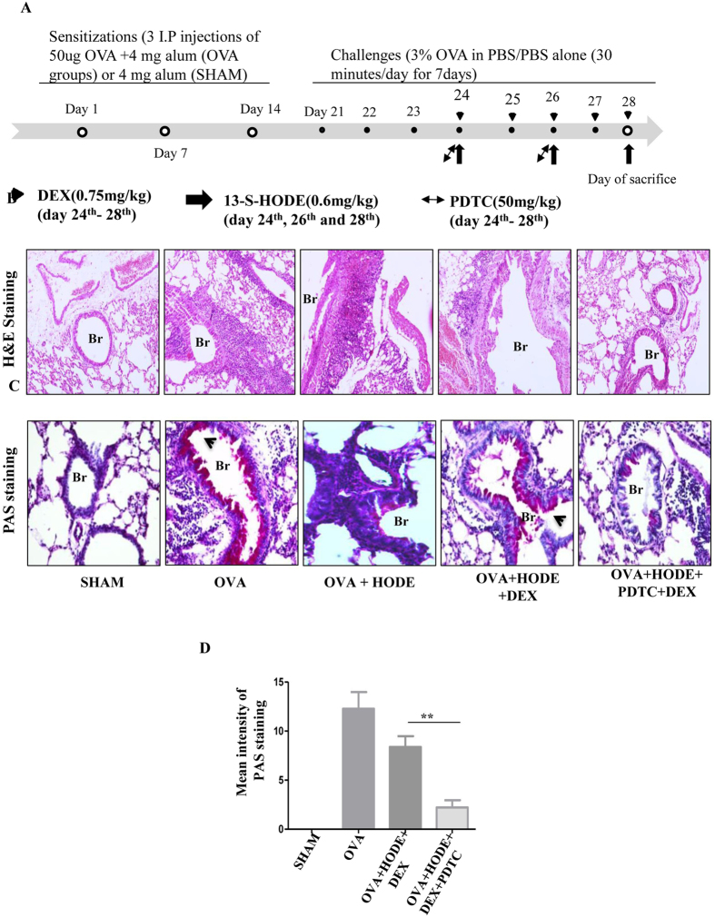
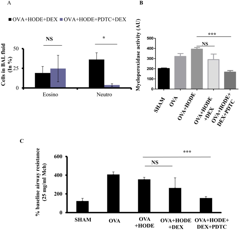
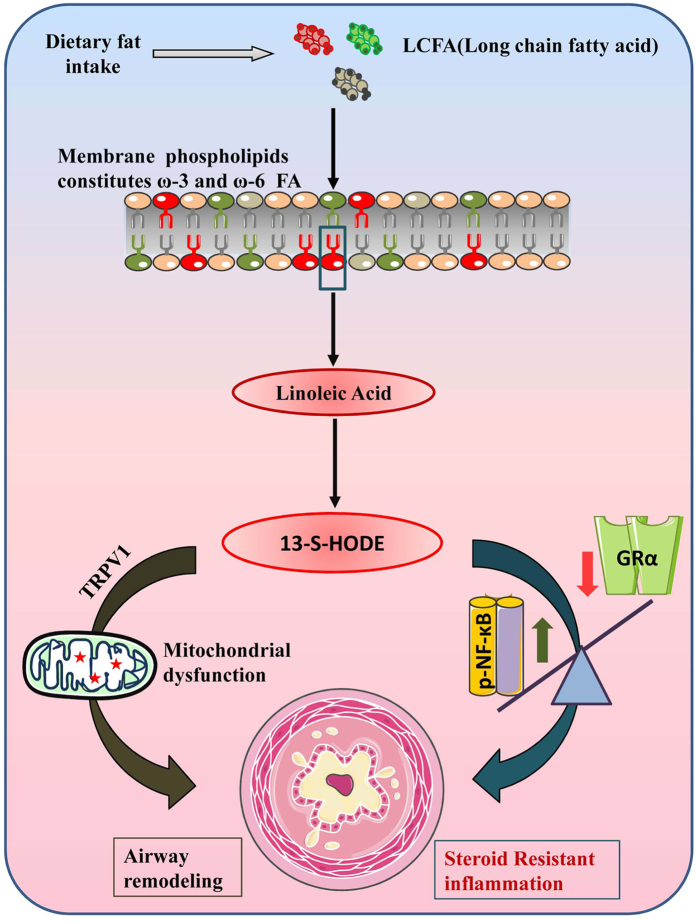
Studies have highlighted the role of nutritional and metabolic modulators in asthma pathobiology. Steroid resistance is an important clinical problem in asthma but lacks good experimental models. Linoleic acid, a polyunsaturated fatty acid, has been linked to asthma and glucocorticoid sensitivity. Its 12/15-lipoxygenase metabolite, 13-S-hydroxyoctadecadienoic acid (HODE) induces mitochondrial dysfunction, with severe airway obstruction and neutrophilic airway inflammation. Here we show that HODE administration leads to steroid unresponsiveness in an otherwise steroid responsive model of allergic airway inflammation (AAI). HODE treatment to allergic mice further increased airway hyperresponsiveness and goblet metaplasia. Treatment with dexamethasone was associated with increased neutrophilic inflammation in HODE treated allergic mice; unlike control allergic mice that showed resolution of inflammation. HODE induced loss of steroid sensitivity was associated with increased p-NFkB in mice and reduced GR-α transcript levels in cultured human bronchial epithelia. In summary, HODE modifies typical AAI to recapitulate many of the phenotypic features seen in severe steroid unresponsive asthma. We speculate that since HODE is a natural metabolite, it may be relevant to the increased asthma severity and steroid insensitivity in patients who are obese or consume high fat diets. Further characterization of HODE induced steroid insensitivity may clarify the mechanisms.


Conflict of interest statement

The authors declare that they have no competing interests.

Figures

Figure 1
Figure 1
Dexamethasone fails to attenuate airway inflammation and goblet cell metaplasia induced by HODE in allergic mice. (A) Schematic representation of experimental design/protocol as described in methods. (B and C) Representative photomicrographs (20 X magnifications) of bronchovascular regions of different groups of mice stained with haematoxylin and eosin (H & E) and periodic acid–Schiff (PAS). Arrows indicate the infiltrated inflammatory cells in (B) and goblet cell metaplasia in (C). Mean inflammation score (D) and mean intensity of mucin content (E) estimated from the images of H and E and PAS stained lung sections. Data represents mean ± SE; n = 4–6 each group; ***p < 0.001, NS, non-significant (OVA versus OVA + HODE + DEX), Br: bronchi, V: vessel.
Figure 2
Figure 2
Dexamethasone fails to alleviate HODE induced neutrophilic inflammation and AHR in allergic mice. (A and B) Neutrophil percentage and myeloperoxidase activity in BAL fluid. (C) The percentage baseline airway resistance in response to increasing concentrations of methacholine in HODE administered allergic mice. Data represents mean ± SE; n = 4–6 mice each group; *p < 0.05, **p < 0.01, NS, non-significant (OVA versus OVA + HODE + DEX).
Figure 3
Figure 3
HODE reduces GR-α and its activation in BEAS-2B cells. (A) Glucocorticoid receptor (GR) activation was estimated in nuclear extract of HODE induced Beas2B cells (details in methods). (B and C) Levels of IL-8 and MCP1-α in the supernatants of cultured human bronchial epithelia, induced with HODE and DEX. (D) Transcript levels of GR-α normalized to α-tubulin. Data represents mean ± SE; n = 3–5; *p < 0.05, NS: non-significant.
Figure 4
Figure 4
HODE administration increases p-NFκB in AAI mice. (A and C) Representative IHC images (20X magnifications) and quantification of the expression of p-NFκB in HODE induced steroid resistance model. (B and D) Representative IHC images (20X magnifications) and quantification of p-NFκB and IκB-α in the lung sections of HODE neutralized allergic mice. Data represents mean ± SE; n = 3–6; **p < 0.01, NS: non-significant. Br: Bronchi. Arrows indicate the positive expression
Figure 5
Figure 5
NFκB inhibitor treatment increases steroid sensitivity, reduces airway inflammation and goblet cell metaplasia in HODE induced steroid resistant mice. (A) Schematic representation of experimental design. PDTC (50 mg/kg) was administered intraperitoneally on day 24th and day 26th, 2 hrs and 4 hrs after the administration to HODE and DEX respectively (details explained in material and methods). (B and C) The representative photomicrographs of H and E (20x magnifications) and PAS, respectively. (D) Mean intensity of PAS calculated by Image J software. Data represents mean ± SE; n = 3–6; **p < 0.01, NS: non-significant. Br: bronchi, V: vessel, Arrows indicate the goblet cell metaplasia.
Figure 6
Figure 6
NFκB inhibitor treatment alleviates neutrophilic inflammation and airway hyperresponsiveness in HODE induced steroid resistant mice. (A) The counts of neutrophils and eosinophils in BAL fluid of steroid resistant mice administered with PDTC. (B) The myeloperoxidase activity in mouse BAL fluid supernatants. (C) The percentage baseline airway resistance in response to 25 mg/ml methacholine dose. Data represents mean ± SE; n = 3–6 each group; *p < 0.05,***p < 0.001, NS: non-significant.
Figure 7
Figure 7
13-S-HODE mediates steroid resistance via NF-κB and GR-α. Dietary lipids, absorbed through intestine are converted to long chain fatty acids which serve as a precursor for the formation of phospholipids. While, ω-6 fatty acids (red color phospholipid) are oxidized into pro-inflammatory lipid mediators, ω-3 is (green color phospholipid) oxidized to anti-inflammatory. 13-S-HODE is a lipid metabolite of linoleic acid (ω-6 fatty acid), oxidized by 15-lipoxygenase. 13-S-HODE, causes airway remodeling and goblet cell metaplasia by mitochondrial dysfunction via TRPV1 channels. In another independent pathway, it increases p-NFκB and reduces GR-α leading to steroid resistant asthma.

References

    1. Raphael W, Sordillo LM. Dietary polyunsaturated fatty acids and inflammation: The role of phospholipid biosynthesis. Int. J. Mol. Sci. 2013;14:21167–21188. doi: 10.3390/ijms141021167. - DOI - PMC - PubMed
    1. Arvaniti F, et al. Salty-snack eating, television or video-game viewing, and asthma symptoms among 10- to 12-year-old children: the PANACEA study. J. Am. Diet. Assoc. 2011;111:251–257. doi: 10.1016/j.jada.2010.10.051. - DOI - PubMed
    1. Black PN, Sharpe S. Dietary fat and asthma: Is there a connection? Eur. Respir. J. 1997;10:6–12. doi: 10.1183/09031936.97.10010006. - DOI - PubMed
    1. Ellwood P, et al. Do fast foods cause asthma, rhinoconjunctivitis and eczema? Global findings from the International Study of Asthma and Allergies in Childhood (ISAAC) phase three. Thorax. 2013;68:351–360. doi: 10.1136/thoraxjnl-2012-202285. - DOI - PubMed
    1. Mickleborough TD, Rundell KW. Dietary polyunsaturated fatty acids in asthma- and exercise-induced bronchoconstriction. Eur. J. Clin. Nutr. 2005;59:1335–1346. doi: 10.1038/sj.ejcn.1602250. - DOI - PubMed

Publication types

MeSH terms