. 2017 Aug 29;7(1):9801.
doi: 10.1038/s41598-017-09875-x.

New insights into tenocyte-immune cell interplay in an in vitro model of inflammation

Affiliations

New insights into tenocyte-immune cell interplay in an in vitro model of inflammation

Meaghan Stolk et al. Sci Rep. .

Abstract

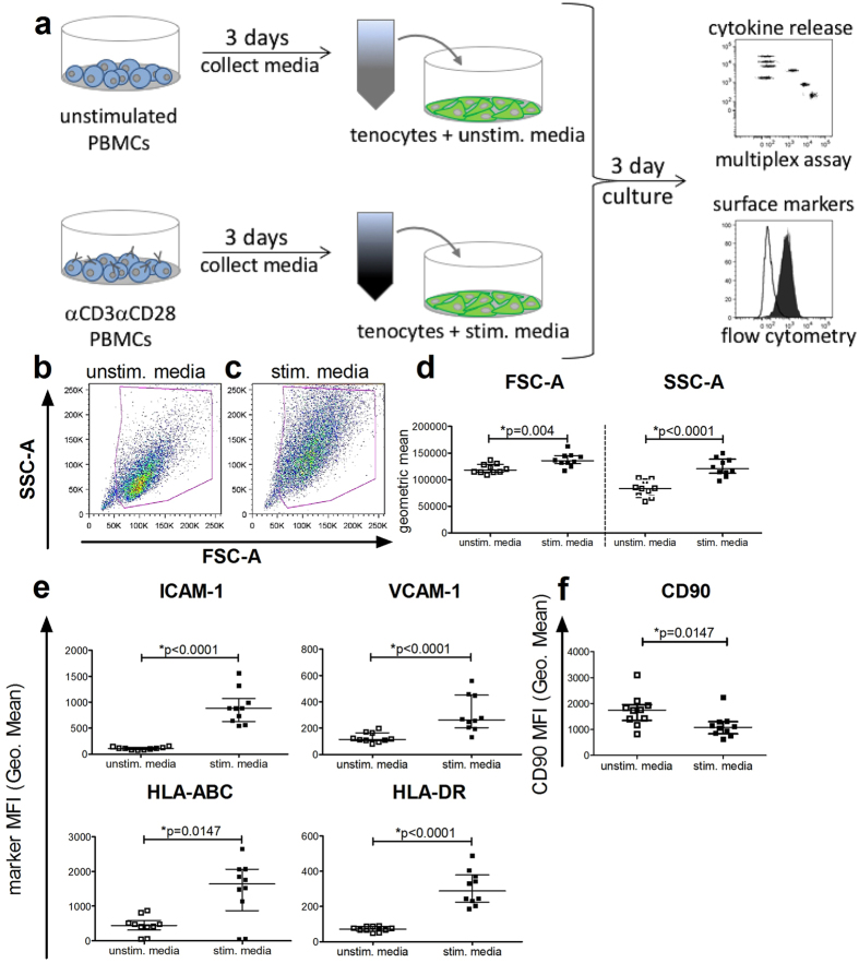
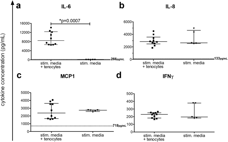
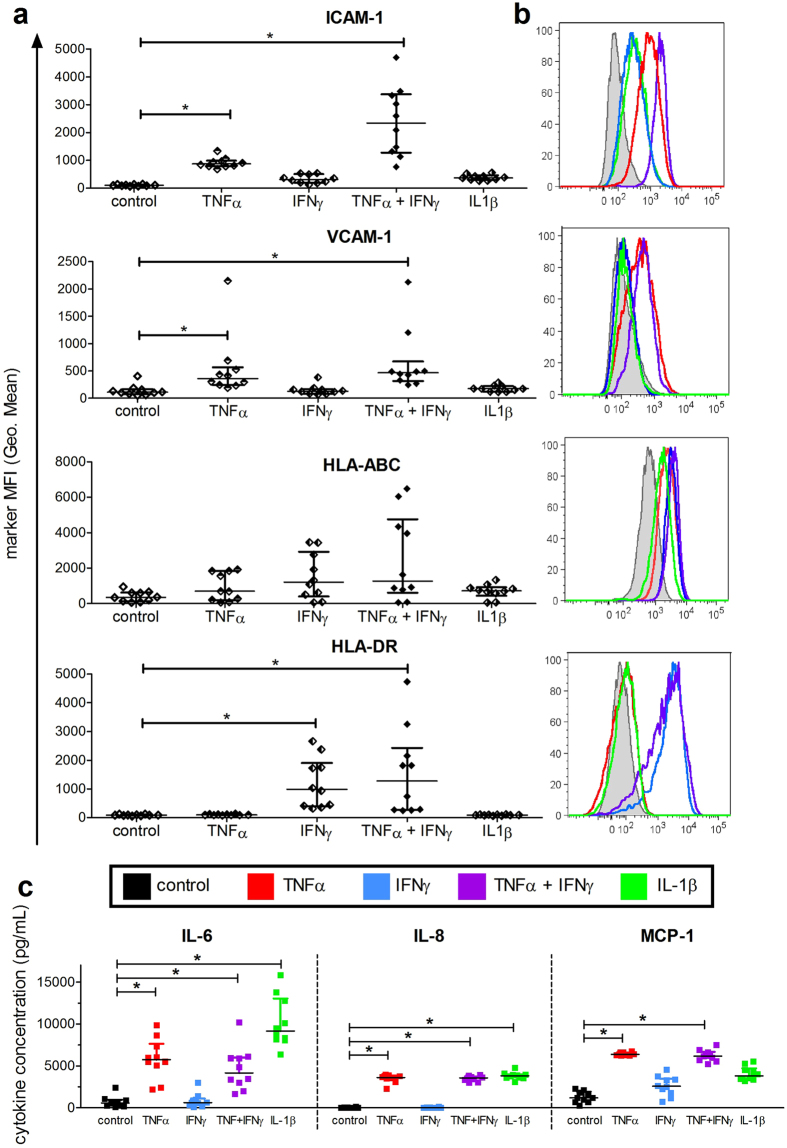
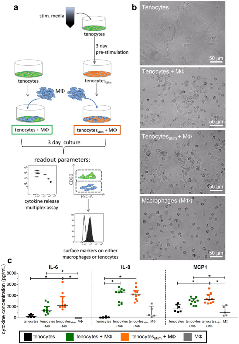
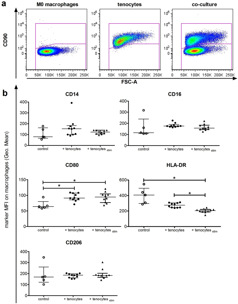
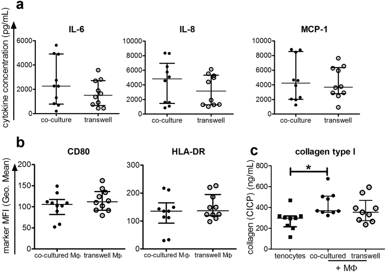
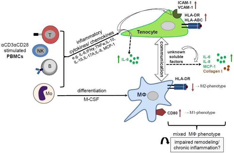
Inflammation plays an important role in the development and resolution of tendon diseases, but underlying mechanisms are poorly understood. We therefore aimed to analyze the response of human tenocytes to inflammatory stimuli and to uncover their interplay with macrophages in vitro. Tenocytes from human ruptured supraspinatus tendons (n = 10) were treated for three days with a stimulation mixture derived from activated mononuclear cells isolated from healthy human peripheral blood. Significantly increased expression levels of selected adhesion- and human leukocyte antigen (HLA)-molecules, and enhanced interleukin (IL)-6 release were detected by flow cytometry. Tenocyte stimulation with the pro-inflammatory cytokines interferon gamma, tumor necrosis factor alpha and IL-1ß triggered similar changes in surface markers and enhanced the release of IL-6, IL-8 and monocyte chemoattractant protein 1 (MCP-1). In co-cultures of macrophages with pre-stimulated tenocytes, macrophages significantly increased CD80 expression, but simultaneously decreased HLA-DR-expression, which are both typical pro-inflammatory polarization markers. Co-cultures also released more IL-6, IL-8, MCP-1 than tenocyte-cultures alone. We demonstrate that tenocytes respond to inflammatory environments in vitro with altered surface marker and cytokine profiles and influence macrophage polarization. Importantly, all changes detected in direct co-cultures were also present in a transwell setting, implicating that communication between the cells involves soluble factors.


Conflict of interest statement

The authors declare that they have no competing interests.

Figures

Figure 1
Figure 1
Pro-inflammatory stimulation media increases the size and granularity of tenocytes, and changes their surface marker expression. Following incubation for 3 days with either the control media from unstimulated PBMC (unstim. media) or the pro-inflammatory stimulation media from αCD3αCD28 stimulated PBMCs (stim. media) (a), supernatants were collected for later cytokine evaluation before the tenocytes were stained with a panel of human-specific antibodies to characteristic surface markers and analyzed by flow cytometry. Tenocytes included in the viable gate with unstim. media (b) or with the stim. media (c) are shown here with representative flow cytometry density plots. The mean fluorescence intensity (MFI) of forward scatter area (FSC-A) and sidewards scatter area (SSC-A) indicating cell size and granularity are shown (d). The expression of the adhesion molecules ICAM-1 (CD54) and VCAM-1 (CD106), as well as the HLA class I molecule HLA-ABC and the HLA class II molecule HLA-DR (e) and CD90 (Thy-1) (f) are also shown after incubation with unstimulated and stimulated media. Data are presented as the median of n = 10 with the interquartile range and are considered significantly different when *p < 0.05 with the Mann-Whitney non-parametric t-test.
Figure 2
Figure 2
Tenocytes produce IL-6 upon stimulation. Cytokines were measured in media from αCD3αCD28 stimulated PBMCs (stim. media) (see Fig. 1a) using the LegendplexTM human inflammation panel. After tenocytes were cultured for 3 days with either the control media from unstimulated PBMC (unstim. media + tenocytes; dotted line) or stimulation media (stim. media + tenocytes), cytokines were measured in the supernatants. The levels of IL-6 (a), IL-8 (b), MCP-1 (c) and IFNγ (d) are shown for the tenocytes cultured with stimulated media compared to the stimulated media without tenocytes present. The median levels for tenocytes incubated with the unstimulated media are shown next to the dotted line at that level on a–c, and IFNγ was undetectable. Samples were treated following the manufacturer’s instructions and measured by flow cytometry. Analysis was done using Legendplex software. Data are presented as the median with the interquartile range with an n = 5 for stimulated media alone and n = 10 for stimulated media + tenocytes, and are considered significantly different when *p < 0.05 with the Mann-Whitney non-parametric t-test.
Figure 3
Figure 3
Increased marker expression and cytokine release after stimulation of tenocytes with single or combined pro-inflammatory recombinant cytokines. Tenocytes were seeded overnight and then recombinant cytokines were added for a final concentration of 10 ng/mL. Following incubation for 3 days, supernatants were collected before tenocytes were evaluated by flow cytometry for various markers. The mean fluorescence intensity (MFI) of ICAM-1 (CD54), VCAM-1 (CD106), HLA-ABC and HLA-DR was evaluated (a). Representative histogram overlays for each marker following stimulation with TNFα (red), IFNγ (blue), TNFα + IFNγ (purple) and IL-1ß (green) compared to the unstimulated control (black) are shown in (b). Supernatants from the cultures were evaluated with the LegendplexTM human inflammation panel. Shown are the summarized data for IL-6, IL-8 and MCP-1 levels in tenocyte cultures after stimulation with the cytokines in comparsion to the unstimulated control (c). All data are presented as the median of n = 10 with the interquartile range and are considered significantly different when *p < 0.05 with the Kruskal Wallis non-parametric ANOVA with Dunn’s post-test.
Figure 4
Figure 4
Co-cultures of tenocytes with macrophages increases the release of cytokines. A schematic representation of tenocyte and macrophage direct co-cultures is presented in (a). Tenocytes were either pre-stimulated with a mix of 50% media and 50% stimulation media for 3 days (tenocytesstim), or directly seeded into a 6 well plate. One day later, M0 macrophages (MΦ) derived from CD14+ monocytes using 50 ng/mL M-CSF for 6 days were added in a ratio of 1 tenocyte:5 macrophages to obtain co-cultures (tenocytes + MΦ) and pre-stimulated co-cultures (tenocytesstim + MΦ). Additional wells of MΦ or tenocytes alone were seeded as controls. After 3 days of culture, supernatants were collected for later cytokine analysis before photos were taken of the cell cultures (b). Supernatants were evaluated using the LegendplexTM human inflammation panel and the levels of IL-6, IL-8, and MCP-1 are shown (c). Data are presented as the median with the interquartile range of n = 5–6 for either tenocytes or macrophages alone and n = 10 for tenocyte/macrophage co-cultures and are considered significantly different when *p < 0.05 with the Kruskal Wallis non-parametric ANOVA with Dunn’s post-test.
Figure 5
Figure 5
Co-culture with tenocytes changes the surface marker expression on macrophages. M0 macrophages were co-cultured together with tenocytes (+ tenocytes) or pre-stimulated tenocytes (+ tenocytesstim) in 6 well plates for 3 days. Additional wells of M0 macrophages alone (control) were cultured under the same conditions. After supernatants and culture photographs were taken, the cells were stained with a panel of human-specific antibodies to macrophage surface markers. Flow cytometry analysis of markers on the macrophages was achieved by first gating on the CD90 negative population to identify macrophages from the direct co-cultures (a). Shown is the mean fluorescence intensity (MFI) of CD14, CD16, CD80, HLA-DR and CD206 on macrophages from co-cultures with unstimulated or pre-stimulated tenocytes in comparison to control M0 macrophages (b). Data are presented as the median with interquartile range for an n = 5 for macrophages alone or n = 10 for tenocyte/macrophage co-cultures and are considered significantly different when *p < 0.05 with the Kruskal Wallis non-parametric ANOVA with Dunn’s post-test.
Figure 6
Figure 6
Soluble factors released in co-cultures were similar when performed in transwell assays. Supernatants were evaluated using the LegendplexTM human inflammation panel and the levels of IL-6, IL-8, and MCP-1 are shown for either direct co-culture or transwell assays where tenocytes were cultured in inserts above adherent macrophages (a). The mean fluorescence intensity (MFI) is shown for CD80 and HLA-DR on macrophages following direct co culture (co-cultured MΦ) or transwell co-culture (transwell MΦ) with tenocytes (b). Supernatants were tested for CICP (C-terminal of type I collagen) using an EIA Kit from tenocytes alone and tenocytes co-cultured with macrophages directly or in transwell assays (c). Data are presented as the median with the interquartile range of n = 10 for (a) and (b) and n = 9 for (c), and are considered significantly different when *p < 0.05 with the Kruskal Wallis non-parametric ANOVA with Dunn’s post-test.
Figure 7
Figure 7
Potential role of cross-talk between tenocytes and macrophages in an inflammatory milieu in tendinopathy. The scheme illustrates the effects of inflammatory conditions on human tenocytes alone and during their interplay with human macrophages on surface marker expression and cytokine release patterns. A mixed cytokine stimulation originating from αCD3αCD28 activated human PBMCs including all subsets (T cells, B cells, NK cells and monocytes (Mo)) induces the up-regulation of HLA-molecules (HLA-DR, HLA-ABC) and adhesion molecules (ICAM-1, VCAM-1) on tenocytes and enhances their IL-6 secretion. After co-culturing monocyte-derived macrophages (Mϕ) with pre-stimulated tenocytes, the macrophages display a mixed M1/M2 macrophage phenotype with enhanced expression of CD80 (characteristic for M1-Mϕ), but reduced HLA-DR (characteristic for M2-Mϕ). As yet unknown soluble factors lead to higher levels of IL-6, IL-8 and MCP-1 secretion. We hypothesize, that this interaction might contribute to a disturbed resolution of the immune response after tendon injury leading to chronic tendinopathies.

Similar articles

Cited by

References

    1. Boileau P, et al. Arthroscopic repair of full-thickness tears of the supraspinatus: does the tendon really heal? The Journal of bone and joint surgery. American volume. 2005;87:1229–1240. - PubMed
    1. Kelly BA, Proffen BL, Haslauer CM, Murray MM. Platelets and plasma stimulate sheep rotator cuff tendon tenocytes when cultured in an extracellular matrix scaffold. Journal of orthopaedic research: official publication of the Orthopaedic Research Society. 2016;34:623–629. doi: 10.1002/jor.23058. - DOI - PMC - PubMed
    1. Abate M, et al. Pathogenesis of tendinopathies: inflammation or degeneration? Arthritis research & therapy. 2009;11 doi: 10.1186/ar2723. - DOI - PMC - PubMed
    1. Rees JD, Stride M, Scott A. Tendons–time to revisit inflammation. British journal of sports medicine. 2014;48:1553–1557. doi: 10.1136/bjsports-2012-091957. - DOI - PMC - PubMed
    1. Dakin SG, et al. Inflammation activation and resolution in human tendon disease. Science translational medicine. 2015;7 doi: 10.1126/scitranslmed.aac4269. - DOI - PMC - PubMed

Publication types