. 2017 Aug 29;7(1):9906.
doi: 10.1038/s41598-017-09972-x.

Retrograde trafficking of β-dystroglycan from the plasma membrane to the nucleus

Affiliations

Retrograde trafficking of β-dystroglycan from the plasma membrane to the nucleus

Viridiana Gracida-Jiménez et al. Sci Rep. .

Erratum in

Abstract

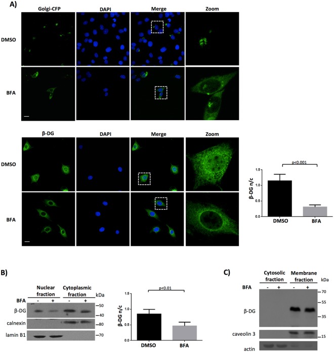
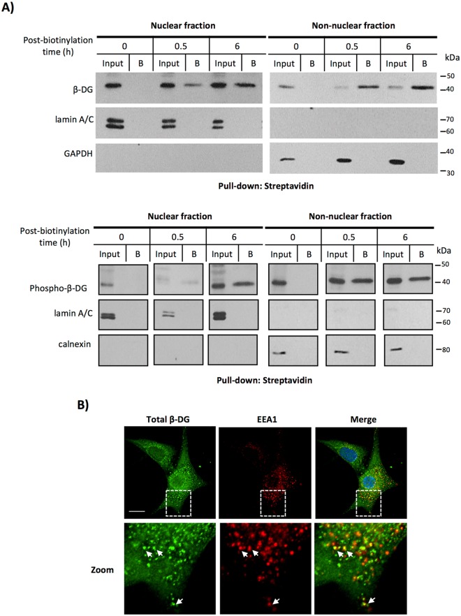
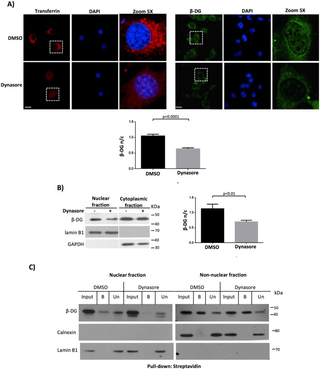
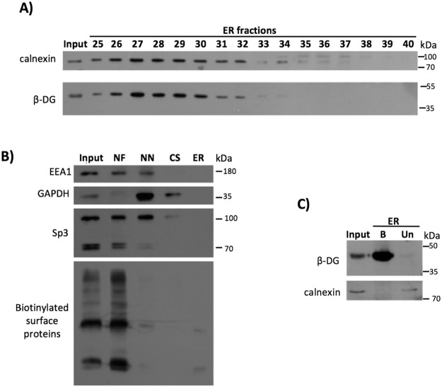
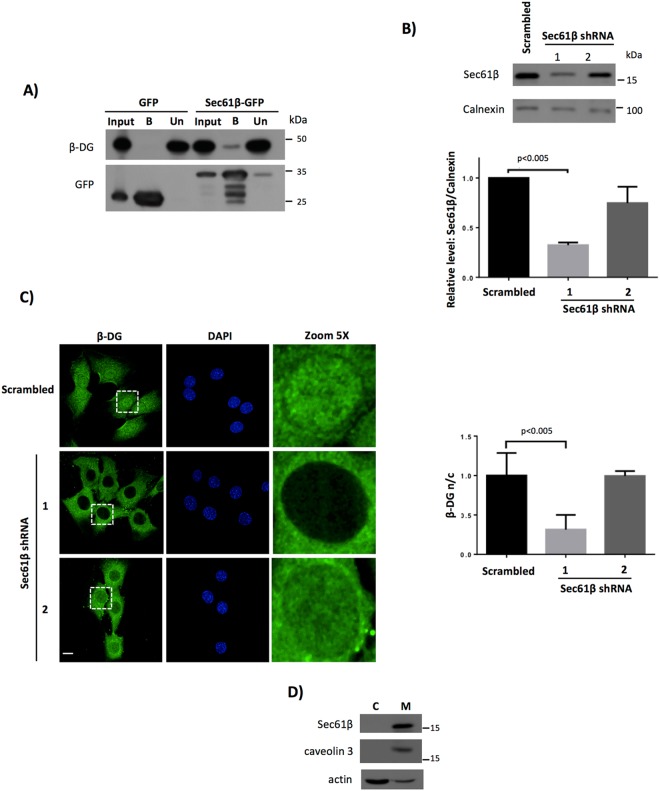
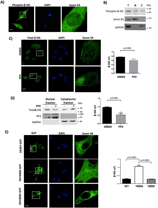
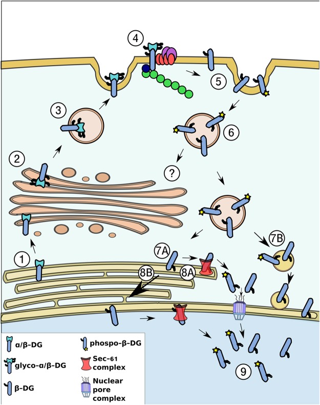
β-Dystroglycan (β-DG) is a transmembrane protein with critical roles in cell adhesion, cytoskeleton remodeling and nuclear architecture. This functional diversity is attributed to the ability of β-DG to target to, and conform specific protein assemblies at the plasma membrane (PM) and nuclear envelope (NE). Although a classical NLS and importin α/β mediated nuclear import pathway has already been described for β-DG, the intracellular trafficking route by which β-DG reaches the nucleus is unknown. In this study, we demonstrated that β-DG undergoes retrograde intracellular trafficking from the PM to the nucleus via the endosome-ER network. Furthermore, we provided evidence indicating that the translocon complex Sec61 mediates the release of β-DG from the ER membrane, making it accessible for importins and nuclear import. Finally, we show that phosphorylation of β-DG at Tyr890 is a key stimulus for β-DG nuclear translocation. Collectively our data describe the retrograde intracellular trafficking route that β-DG follows from PM to the nucleus. This dual role for a cell adhesion receptor permits the cell to functionally connect the PM with the nucleus and represents to our knowledge the first example of a cell adhesion receptor exhibiting retrograde nuclear trafficking and having dual roles in PM and NE.


Conflict of interest statement

The authors declare that they have no competing interests.

Figures

Figure 1
Figure 1
β-DG traffics from ER to Golgi prior to its nuclear localization. (A) C2C12 cells cultured on glass coverslips were transiently transfected to express the galactosyl-transferase-CFP fusion protein (Golgi-CFP) and 16 h post-transfection they were treated with BFA or vehicle alone (DMSO; see Methods). Afterwards, cells were fixed, stained with DAPI and further analyzed by CLSM (upper panel). C2C12 cells seeded on coverslips were treated with vehicle or BFA, fixed, immunolabeled for non-phosphorylated β-DG and counterstained with DAPI for nuclei visualization prior to analysis by CLSM (lower panel). Maximum intensity projections are shown. The fluorescence intensity in the nucleus and cytoplasm (F n/c ratio) was quantified to estimate β-DG nuclear accumulation (see Methods). Data shown in the graph (right) represent the mean +/− SD from three separate experiments (n = 30 cells), with p value denoting statistical significance (Student t-test). Scale bar 20 µm. (B) Control and BFA-treated cells were fractionated into cytoplasmic and nuclear extracts and further subjected to SDS-PAGE/Western blotting analysis using anti-β-DG antibodies for the immunodetetection of total β-DG. Membranes were striped and reprobed for calnexin (cytoplasmic marker) and lamin B1 (nuclear marker). Densitometric analysis of immunoblot autoradiograms was carried out and the relative levels of β-DG in the nucleus and cytoplasmic (n/c) were obtained. Results represent the mean +/− SD of 4 separate experiments, with p values indicating significant differences (Student t-test). (C) Vehicle- or BFA-treated cells were fractionated into cytosolic and total membrane extracts prior to being subjected to SDS-PAGE/Western blotting analysis, using anti-β-DG antibodies for total β-DG. Stripped membranes were reprobed for caveolin (membrane protein) and actin (cytosolic marker).
Figure 2
Figure 2
Nuclear β-DG derives from the PM. (A) C2C12 cells were incubated with biotin for the indicated time intervals to label cell surface proteins (see Methods). Cells were then subjected to subcellular fractionation to isolate nuclear and non-nuclear fractions and biotinylated proteins were pulled-down using streptavidin-agarose beads and analyzed by SDS-PAGE/Western blotting using primary antibodies for non-phosphorylated-β-DG (upper panel) and phosphorylated β-DG (lower panel). Lower panel blot was reorganized so that the time points order matched the ones shown in upper panel, original blot is shown in Supp. Figure 1. Input: immunoblotting analysis of cellular fractions prior to streptavidin-mediated pull-down. B: Bound/precipitated fraction. Membranes were stripped and reprobed for lamin A/C and GAPDH/calnexin as purity controls for nuclear and non-nuclear fractions respectively. (B) C2C12 cells cultured on glass coverslips were double-immunostained for total β-DG and the early endosomal marker EEA1. Nuclei were counterstained with DAPI prior to CLSM analysis. A typical single Z-section from three independent experiments is shown. Scale bar 20 µm.
Figure 3
Figure 3
Inhibition of dynamin-dependent endocytosis reduces nuclear localization of β-DG. (A, left panel) Serum-starved C2C12 cells, seeded on coverslips, were treated with 40 µM dynasore (endocytosis inhibitor) or 0.05% DMSO (vehicle) for 30 min at 37 °C and then incubated for 5 min at 20 °C with Alexa594-transferrin (red). After 15 min at 37 °C, cells were fixed, stained with DAPI (nuclei) and transferrin uptake was monitored by CLSM analysis, with typical images shown (scale bar is 20 µm). (A, right panel) Cells treated with dynasore or DMSO, as above, were immunolabeled for β-DG and counterstained with DAPI prior to be imaged by CLSM, with typical Z-sections shown (scale bar is 20 µm). (A, lower panel). Nuclear accumulation of β-DG (F n/c) was estimated as described in Methods. Data correspond to mean +/− SD from a series of three separate experiments (n = 30 cells). (B) Cytoplasmic and nuclear extracts obtained from dynasore- or DMSO-treated cells were analyzed by Western blotting using anti-β-DG antibodies. Stripped membranes were reprobed for lamin A/C and GAPDH as purity and loading controls for nuclear and cytoplasmic extracts respectively. The nuclear/cytoplasmic ratio (n/c) of β-DG was estimated by densitometry analysis. Results represent the mean +/− SD for 3 separate experiments, with significant differences denoted by p values (Student t-test). (C) DMSO- and dynasore-treated cells were subjected to biotinylation assays as in Fig. 2. At 1 h post-biotinyation time, cells were fractionated into nuclear and non-nuclear fractions and pulled-down using streptavidin-agarose beads. Recovered and unbound proteins were analyzed by SDS-PAGE/Western blotting using primary antibodies for total β-DG. Membranes were stripped and reprobed for lamin A/C and calnexin; markers for nuclear and non-nuclear fractions respectively. Input: immunoblotting analysis of cellular fractions prior to pull-down. B, bound/precipitated fraction. Un, unbound fraction.
Figure 4
Figure 4
Retrograde trafficking of β-DG from the PM to the ER. (A) ER was purified using density gradient techniques (OptiPrep) and then ER fractions were immunoblotted for the ER marker calnexin or β-DG on the same membrane. (B) Verification of the purity of ER fractions: Aliquots from each step of the ER purification were analyzed by Western blotting using primary antibodies against EEA1 (early endosomal marker), GAPDH (cytosolic marker) and Sp3 (nuclear marker). As a PM marker, ER was isolated from biotinylated cells at 4 °C, the lysates were pulldown using streptavidin-agarose beads and then blotted with HRP-streptavidin. NF: Nuclear fraction; NN: Non-nuclear fraction; CS: Cytosolic fraction; ER: Endoplasmic reticulum fraction. (C) Cells were subjected to cell surface biotinylation and subsequently to ER purification using the OptiPrep gradient. The ER fractions were combined and biotinylated proteins were precipitated using streptavidin-agarose beads and then analyzed by SDS-PAGE/Western blotting with antibodies against β-DG and calnexin.
Figure 5
Figure 5
Nuclear translocation of β-DG is dependent on the Sec61 translocon. (A) C2C12 cells were transiently transfected to express Sec61β-GFP or GFP alone. The transfected cells were lysed 8 h post-transfection and immunoprecipitated using the GFP-Trap system; the precipitated proteins were analyzed by Western blot using antibodies against β-DG. Input corresponds to 5% of protein extract prior to immunoprecipitation; Un, unbound proteins; B, bound proteins. (B) Lysates from C2C12 cells stably transfected with vectors encoding shRNAs directed against mouse Sec61β mRNA (Sec61β shRNA1 and 2) or a scrambled shRNA (control) were analyzed by western blotting using antibodies against Sec61β and calnexin (loading control). (C) C2C12 cells expressing the scrambled shRNA or the Sec61β shRNAs (1 or 2) were cultured on glass coverslips, fixed, immunostained for total β-DG and counterstained with DAPI for nuclei visualization, prior to being analyzed by CLSM, with typical single Z-sections shown (scale bar is 20 µm). Quantitative analysis of the levels of β-DG nuclear accumulation (Fn/c ratio) was performed (right panel) and results represent the mean +/− SD for three separate experiments (n ≥ 50), with significant differences denoted by the p values (Student’s t-test). (D) Cells were fractionated to obtain cytosolic and membrane extracts. Distribution of Sec61β was analyzed by SDS-PAGE/Western blotting analysis using anti-Sec61β antibodies. Purity of cell fractions was analyzed by using primary antibodies against caveolin (membrane marker) and actin (cytosolic marker). C, cytosolic fraction; M, membrane fraction.
Figure 6
Figure 6
Src-dependent Tyrosine890 phosphorylation of β-DG drives its nuclear targeting. (A) C2C12 cells cultured on glass coverslips were treated with sodium orthovanadate (OV) for 3 h, fixed and immunostained using a phospho-specific antibody that recognizes Tyr890 phosphorylated β-DG. Nuclei were stained with DAPI (blue color) prior to CLMS analysis, with single optical Z-sections. Scale bar = 20 μm. (B) Cultures from OV-treated cells were fractionated into total (T), cytoplasmic (C) and nuclear (N) extracts and these extracts were separated by SDS-PAGE and subjected to Western blot analysis using phospho-β-DG antibodies. Nuclear (lamin B1) and cytoplasmic (GAPDH) protein markers were analyzed in parallel to validate the purity of the fractions. (C) C2C12 cells grown on glass coverslips were treated with PP2 or vehicle, fixed and immunolabeled for total β-DG and counterstaining with DAPI to visualize nuclei. Cells were imaged by CLMS and typical single Z-sections are shown (scale bar is 20 µm). Right panel. Quantitative analysis of CLMS images was carried out to obtain the nuclear to cytoplasmic fluorescence intensity of total β-DG (F n/c, see Methods). Results represent mean +/− SD for 3 separate experiments, with significant differences denoted by the p value (Student t-test). (D) Nuclear and cytoplasmic fractions obtained from control and PP2-treated cells were analyzed by SDS-PAGE/Western blotting using specific antibodies against total β-DG. Sp3 and GAPDH were used as loading controls for nuclear and cytoplasmic fractions respectively. Right panel. Nuclear to cytoplasmic ratio (n/c) of total β-DG was measured by densitometry analysis and data correspond to mean +/− SD for 3 independent experiments, with significant differences indicated by the p value (Student t-test). (E) C2C12 cells grown on glass coverslips were transiently transfected to express DGWT-GFP, DGY890E-GFP or DGY890F-GFP proteins. At 24 h post-transfection, cells were fixed, stained with DAPI and subjected to CLMS, with typical single optical Z-sections shown. Scale bar is 20 µM. Right panel. The F n/c ratio for DGWT-GFP, DGY890E-GFP and DGY890F-GFP was obtained and plotted. Results correspond to mean +/− SD for 3 separate experiments (n = 50 cells), with significant differences indicated by the p value (Student t-test).
Figure 7
Figure 7
Schematic diagram of the retrograde trafficking of β-DG from the cell surface to the nucleus. (1) DG is synthesized in the ER as a precursor that undergoes a proteolytic cleavage to generate two subunits: α- and β-DG. (2) α-DG and β-DG maintain a non-covalent interaction and are transported from the ER to the Golgi apparatus, where both proteins are glycosylated (3) and then transported to the PM, where they interact with the DAPC (4). (5) β-DG is endocytosed from the PM, a process positively modulated by its phosphorylation on Tyr890. (7) β-DG is further translocated from the PM to the ER, and based on the β-DG-Sec61β interaction, it is likely that the Sec61 translocon releases β-DG from the ER membrane, prior to be recognized by the importin system to enter the nucleus through the NPC (8A-9). As both β-DG and Sec61 have been found in the NE, an alternative possibility is that β-DG moves from the ER to the NE by lateral diffusion to interact there with Sec61 to be directly delivered to the nucleoplasm (8B-9). Another alternative route for nuclear translocation is that β-DG-containing endosomes fuse directly to the nuclear membrane to discharge their contents in the nuclear envelope so that β-DG gets further translocated into the nucleoplasm (7B).

Similar articles

Cited by

References

    1. Ervasti JM, Campbell KP. A role for the dystrophin-glycoprotein complex as a transmembrane linker between laminin and actin. J Cell Biol. 1993;122:809–823. doi: 10.1083/jcb.122.4.809. - DOI - PMC - PubMed
    1. Ibraghimov-Beskrovnaya O, Sheffield VC, Campbell KP. Single base polymorphism in the DAG1 gene detected by DGGE and mismatch PCR. Human molecular genetics. 1993;2:1983. doi: 10.1093/hmg/2.11.1983-a. - DOI - PubMed
    1. Henry MD, Campbell KP. Dystroglycan inside and out. Curr Opin Cell Biol. 1999;11:602–607. doi: 10.1016/S0955-0674(99)00024-1. - DOI - PubMed
    1. Moore CJ, Winder SJ. Dystroglycan versatility in cell adhesion: a tale of multiple motifs. Cell Commun Signal. 2010;8:3. doi: 10.1186/1478-811X-8-3. - DOI - PMC - PubMed
    1. Durbeej M, Ekblom P. Dystroglycan and laminins: glycoconjugates involved in branching epithelial morphogenesis. Exp Lung Res. 1997;23:109–118. doi: 10.3109/01902149709074024. - DOI - PubMed

Publication types

LinkOut - more resources