

Localization and Orientation of Xanthophylls in a Lipid Bilayer
- PMID: 28852075
- PMCID: PMC5575131
- DOI: 10.1038/s41598-017-10183-7
Localization and Orientation of Xanthophylls in a Lipid Bilayer
Abstract
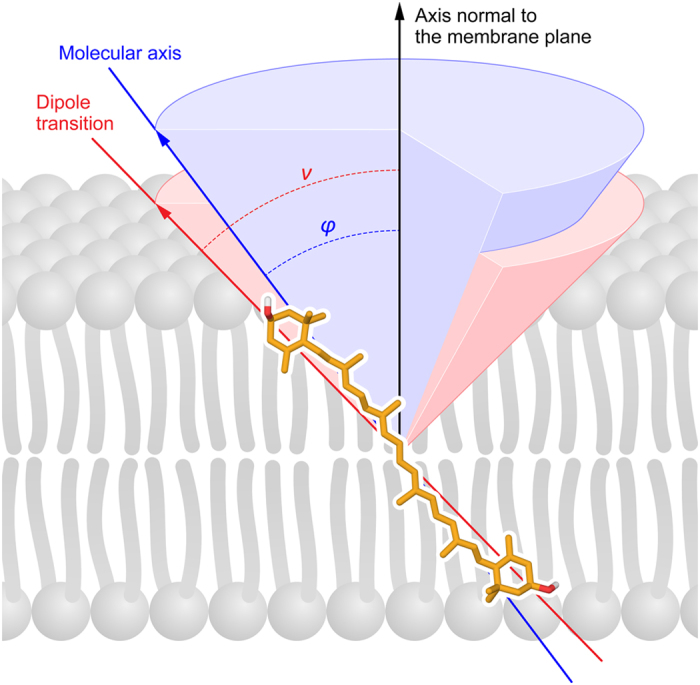
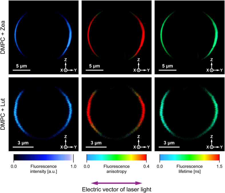
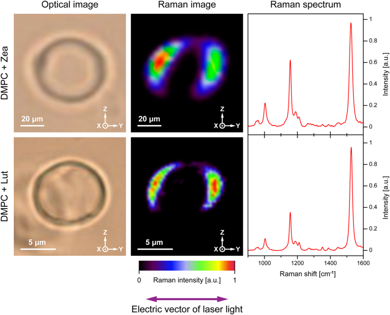
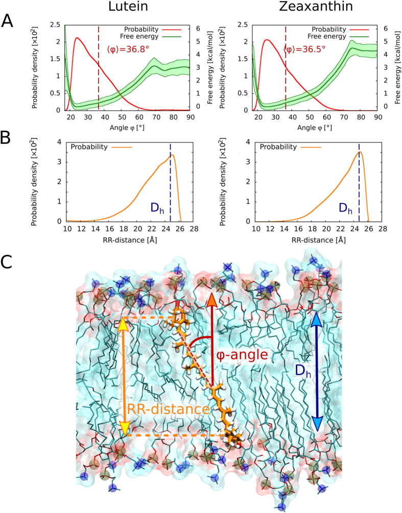
Xanthophylls (polar carotenoids) play diverse biological roles, among which are modulation of the physical properties of lipid membranes and protection of biomembranes against oxidative damage. Molecular mechanisms underlying these functions are intimately related to the localization and orientation of xanthophyll molecules in lipid membranes. In the present work, we address the problem of localization and orientation of two xanthophylls present in the photosynthetic apparatus of plants and in the retina of the human eye, zeaxanthin and lutein, in a single lipid bilayer membrane formed with dimyristoylphosphatidylcholine. By using fluorescence microscopic analysis and Raman imaging of giant unilamellar vesicles, as well as molecular dynamics simulations, we show that lutein and zeaxanthin adopt a very similar transmembrane orientation within a lipid membrane. In experimental and computational approach, the average tilt angle of xanthophylls relative to the membrane normal is independently found to be ~40 deg, and results from hydrophobic mismatch between the membrane thickness and the distance between the terminal hydroxyl groups of the xanthophylls. Consequences of such a localization and orientation for biological activity of xanthophylls are discussed.
Conflict of interest statement
The authors declare that they have no competing interests.
Figures
Similar articles
-
Xanthophyll pigments lutein and zeaxanthin in lipid multibilayers formed with dimyristoylphosphatidylcholine.J Photochem Photobiol B. 2002 Aug;68(1):39-44. doi: 10.1016/s1011-1344(02)00330-5. J Photochem Photobiol B. 2002. PMID: 12208035
-
Organisation of xanthophyll pigments lutein and zeaxanthin in lipid membranes formed with dipalmitoylphosphatidylcholine.Biochim Biophys Acta. 2000 Dec 20;1509(1-2):255-63. doi: 10.1016/s0005-2736(00)00299-6. Biochim Biophys Acta. 2000. PMID: 11118537
-
Can Xanthophyll-Membrane Interactions Explain Their Selective Presence in the Retina and Brain?Foods. 2016 Mar;5(1):7. doi: 10.3390/foods5010007. Epub 2016 Jan 12. Foods. 2016. PMID: 27030822 Free PMC article.
-
Factors Differentiating the Antioxidant Activity of Macular Xanthophylls in the Human Eye Retina.Antioxidants (Basel). 2021 Apr 14;10(4):601. doi: 10.3390/antiox10040601. Antioxidants (Basel). 2021. PMID: 33919673 Free PMC article. Review.
-
The role of the xanthophyll cycle and of lutein in photoprotection of photosystem II.Biochim Biophys Acta. 2012 Jan;1817(1):182-93. doi: 10.1016/j.bbabio.2011.04.012. Epub 2011 May 1. Biochim Biophys Acta. 2012. PMID: 21565154 Review.
Cited by
-
Antioxidant Interactions between Citrus Fruit Carotenoids and Ascorbic Acid in New Models of Animal Cell Membranes.Antioxidants (Basel). 2023 Sep 7;12(9):1733. doi: 10.3390/antiox12091733. Antioxidants (Basel). 2023. PMID: 37760036 Free PMC article.
-
Inflammation/bioenergetics-associated neurodegenerative pathologies and concomitant diseases: a role of mitochondria targeted catalase and xanthophylls.Neural Regen Res. 2021 Feb;16(2):223-233. doi: 10.4103/1673-5374.290878. Neural Regen Res. 2021. PMID: 32859768 Free PMC article. Review.
-
The impact of selected xanthophylls on oil hydrolysis by pancreatic lipase: in silico and in vitro studies.Sci Rep. 2024 Feb 1;14(1):2731. doi: 10.1038/s41598-024-53312-9. Sci Rep. 2024. PMID: 38302772 Free PMC article.
-
An Overview of Lutein in the Lipid Membrane.Int J Mol Sci. 2023 Aug 18;24(16):12948. doi: 10.3390/ijms241612948. Int J Mol Sci. 2023. PMID: 37629129 Free PMC article. Review.
-
Origin and Evolution of Polycyclic Triterpene Synthesis.Mol Biol Evol. 2020 Jul 1;37(7):1925-1941. doi: 10.1093/molbev/msaa054. Mol Biol Evol. 2020. PMID: 32125435 Free PMC article.
References
-
- Britton, G. Functions of intact carotenoids. in Carotenoids. Natural Functions, Vol. 4 (eds Britton, G., Liaaen-Jensen, S. & Pfander, H.) (Birkhauser, Basel, Boston, Berlin, 2008).
-
- Kaczor, A. & Baranska, M. (eds). Carotenoids. Nutrition, Analysis and Technology, (Wiley Oxford, 2016).
-
- Gruszecki, W.I. Carotenoid orientation: Role in membrane stabilization. in Carotenoids in health and disease (eds Krinsky, N. I., Mayne, S. T. & Sies, H.) 151–163 (Marcel Dekker, New York, 2004).
-
- Gruszecki, W.I. Carotenoids in lipid membranes. in Carotenoids. Physical, Chemical and Biological Functions and Properties (ed. Landrum, J. T.) 19–30 (CRC Press, London, New York, Boca Raton FL, 2010).
Publication types
LinkOut - more resources
Full Text Sources
Other Literature Sources
