. 2017 Aug 29;7(1):9755.
doi: 10.1038/s41598-017-10122-6.

Mechanisms of stem cell based cardiac repair-gap junctional signaling promotes the cardiac lineage specification of mesenchymal stem cells

Affiliations

Mechanisms of stem cell based cardiac repair-gap junctional signaling promotes the cardiac lineage specification of mesenchymal stem cells

Heiko Lemcke et al. Sci Rep. .

Abstract

Different subtypes of bone marrow-derived stem cells are characterized by varying functionality and activity after transplantation into the infarcted heart. Improvement of stem cell therapeutics requires deep knowledge about the mechanisms that mediate the benefits of stem cell treatment. Here, we demonstrated that co-transplantation of mesenchymal stem cells (MSCs) and hematopoietic stem cells (HSCs) led to enhanced synergistic effects on cardiac remodeling. While HSCs were associated with blood vessel formation, MSCs were found to possess transdifferentiation capacity. This cardiomyogenic plasticity of MSCs was strongly promoted by a gap junction-dependent crosstalk between myocytes and stem cells. The inhibition of cell-cell coupling significantly reduced the expression of the cardiac specific transcription factors NKX2.5 and GATA4. Interestingly, we observed that small non-coding RNAs are exchanged between MSCs and cardiomyocytes in a GJ-dependent manner that might contribute to the transdifferentiation process of MSCs within a cardiac environment. Our results suggest that the predominant mechanism of HSCs contribution to cardiac regeneration is based on their ability to regulate angiogenesis. In contrast, transplanted MSCs have the capability for intercellular communication with surrounding cardiomyocytes, which triggers the intrinsic program of cardiogenic lineage specification of MSCs by providing cardiomyocyte-derived cues.


Conflict of interest statement

The authors declare that they have no competing interests.

Figures

Figure 1
Figure 1
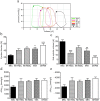
Flow cytometric characterization of CD271+ and CD133+ stem cell populations in human BM. (a) Population overlap of CD271+/CD133+ cells was determined in total BM, CD271+ and CD133+ subpopulations, respectively. Boxes enclose 25% - 75% values with median line, each outlier is displayed, n = 6. (b) Cell purity and viability of MACS-isolated CD271+ (n = 44) and CD133 + cell fractions, n = 75. (c) Analysis of CD45 expression of purified CD271+ cells revealed distinct subpopulations. The high expression of CD45dim demonstrates that mesenchymal stem cells represent the major cell population within the CD271+ fraction, n = 40. (d) CFU-F assay of mononuclear cells, purified CD271+ cells and CD271 fraction confirmed that all mesenchymal progenitors originated from CD271+ cells, n = 11. Graphs are shown as mean ± SEM. Statistical differences were analyzed using one-way ANOVA, followed by Tukey’s post hoc test (***P < 0.0001).
Figure 2
Figure 2
Therapeutic effect of MSCs and HSCs treatment on cardiac functions. (a) Representative pressure volume (PV)-loop graph used to evaluate cardiac performance. (b,c) Compared to untreated MI (MIC), ejection fraction and end-systolic volume were significantly improved when MSC and HSC populations were applied, both as single injection (MI HSC, MI MSC) and co-administration (MIX), n = 7. (d,e) Likewise, PV-loop analysis showed that stem cell treated hearts demonstrated an improved velocity of pressure rise (max/min), compared to the untreated MIC group, n = 7. Graphs are shown as mean ± SEM. Statistical differences were analyzed using one-way ANOVA, followed by Tukey’s post hoc test (*P < 0.05, **P < 0.01, P < 0.001), vs. MIC.
Figure 3
Figure 3
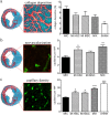
Impact of MSCs and HSCs on MI-induced cardiac remodeling and neovascularization defined for co-transplanted cells and single populations. (a) Stem cell treatment (MSC, HSC, MIX) decreased collagen deposition at the infarction border in comparison to untreated MI (MIC), n = 7. (b) Effect of stem cell transplantation on neovascularization determined within the infarcted area. Infiltration of the infarcted scar with new blood vessels was improved in stem cell treated hearts compared to MIC, n = 7. (c) The capillary density in the border zone was significantly higher following single injection or co-administration of MSCs and HSCs, in contrast to untreated MIC, n = 7. Graphs are shown as mean ± SEM. Statistical differences were analyzed using one-way ANOVA, followed by Tukey’s post hoc test (*P < 0.05, **P < 0.01, P < 0.001), vs. MIC.
Figure 4
Figure 4
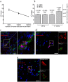
Retention, engraftment and therapeutic activity of MSCs and HSCs in vivo. (a) Sensitivity test of qRT-PCR with stem cell treated cryosectioned hearts revealed a detection threshold of 1 × 103 cells using human GAPDH, n = 6. (b) While the number of retained cells per murine heart was similar 48 h after injection, HSCs demonstrated an increased retention rate three weeks post infarction compared to MSCs, n = 6. (c) Immunostaining of HSCs in stem cell-treated cryosectioned hearts gave evidence for the co-localization of HSCs with tomato-lectin labeled blood vessels. No co-localization with vessels was found for MSCs. (d,e) Following transplantation into mice hearts, MSCs demonstrate cardiac specific lineage specification as shown by labeling of the cardiac specific transcription factor NKX2.5 and GATA-4. Scale bars 20 µm. Graphs are shown as mean ± SEM.
Figure 5
Figure 5
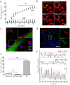
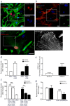
Establishment of functional GJs between MSCs and CMs. (a) MSCs and HSCs were co-cultured with cardiomyocytes, loaded with calcein and subjected to FRAP microscopy. While MSCs demonstrated strong fluorescence recovery, no recovery was observed for HSCs, n = 3, 38 cells. (b) Representative FRAP images demonstrated the influx of calcein from surrounding CMs into the bleached MSC (CFDA labeled), indicating the presence of functional GJs (c) Immunolabeling of MSC-CM co-cultures revealed the presence of Cx43 plaques (*) at cell borders between GFP-labeled MSC and CMs. (d) A strong cytoplasmic expression of Cx43 in MSCs was also confirmed in vivo, following injection into mice heart. (e) Gap junctional coupling with CMs induced Ca2+ transients in MSCs. Changes of the intracellular Ca2+ level were visualized using X-Rhod-1. Compared to co-culture with non-CMs, co-cultivation with CMs induced significant stronger changes of the intracellular Ca2+ level in MSCs. As expected, the alterations of intracellular Ca2+ were more pronounced in CMs, n = 3. (f) Representative plots of Ca2+ level changes. The amplitude of the oscillating signal of X-Rhod-1 in MSCs is higher when cells are co-cultured with CMs, compared co-culture with non-CMs. However, these Ca2+ transients are less distinct and demonstrated a more irregular pattern than Ca2+ transients found in CMs. Scale Bars 20 µm, Graphs are shown as mean ± SEM. FRAP curves were statistically analyzed by two-way ANOVA (*P < 0.05, **P < 0.01, ***P ≤ 0.001), followed by Bonferroni’s post-hoc test. Statistical significance between Ca2+ levels was analyzed using Statistical differences were analyzed using one-way ANOVA, followed by Dunn’s post hoc test (***P < 0.001).
Figure 6
Figure 6
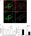
Gap junctional coupling with CMs promotes the cardiogenic differentiation of MSCs. (a,b) Immunofluorescence labeling of cardiac specific markers revealed the transdifferentiation of MSCs into a cardiac-like phenotype. MSCs co-cultured with CMs showed expression of GATA-4 and NKX2.5, two early cardiac specific transcription factors. (c) MSCs were found to express α-actinin, indicating the formation of pre-sarcomeric structures. (d) Quantitative assessment of NKX2.5 and GATA-4 expression by fluorescence microscopy demonstrated a marked increase when MSCs were co-cultivated with CMs, n = 3, 175 cells, note the logarithmic scale. (e) qRT-PCR confirmed the pronounced up-regulation of NKX2.5 and GATA-4 induced by co-culture with CMs, compared to MSC mono-culture, n = 3. (f) The establishment of cell-cell contacts between MSCs and CMs resulted in a significant higher protein level of GATA-4 and NKX2.5 compared to MSCs that share the same medium but lack direct cell-cell interaction with CMs, n = 3, 148 cells, note the logarithmic scale. (g) Downregulation of Cx43 in co-cultured MSCs reduced the expression levels of both transcription factors, indicating a promoting role of GJs in the transdifferentiation process of MSCs induced by adjacent CMs, n = 5, 409 cells. Scale Bars 20 µm, Graphs are shown as mean ± SEM. Statistical significance was analyzed using two-tailed Student’s t-test (*P ≤ 0.05, **P ≤ 0.01, ***P ≤ 0.001).
Figure 7
Figure 7
GJ-dependent delivery of small RNA from CM influences the transdifferentiation of MSCs. (a) EGFP-labeled MSCs were co-cultured with CMs transfected with DY-547 labeled miRNA. 24 h after co-cultivation, miRNA was also found in MSCs (asterisk), indicating the exchange of small RNAs between these two cell types, Scale bar 20 µm. (b) CM-derived small RNAs induce gene silencing in MSCs. MSCs were transfected with a plasmid coding for EGFP and co-cultured with CMs transfected with anti-EGFP siRNA. Following co-cultivation for 2 days, EGFP expression in MSCs was decreased by 20% when co-cultured with CMs containing anti-EGFP-siRNA. Downregulation of Cx43 in MSCs diminishes the EGFP-reducing effect of CMs. Efficiency of the EGFP/siRNA reporter construct was evaluated by double transfection of MSCs with EGFP and siRNA, resulting in a reduced EGFP intensity of ~50%. n = 3. (c) To verify the impact of CM-derived small RNAs on the transdifferentiation of MSCs, Dicer was down-regulated in CMs. Resulting disruption of the miRNA machinery in CMs impaired the transdifferentiation process in co-cultured MSCs. While NKX2.5 was significantly decreased in MSCs, no effect was observed on the expression level of GATA-4 upon co-cultivation with dicer knockdown CMs, n = 3, 255 cells. Graphs are shown as mean ± SEM. Statistical significance between EGFP fluorescence intensities was analyzed using one-way ANOVA, followed by Dunn’s post hoc test (**P < 0.01, ***P < 0.001). Statistical significance for GATA-4 and NKX2.5 levels was analyzed using two-tailed Student’s t-test (*P ≤ 0.05, ***P ≤ 0.001).

Similar articles

Cited by

References

    1. Tongers J, Losordo DW, Landmesser U. Stem and progenitor cell-based therapy in ischaemic heart disease: Promise, uncertainties, and challenges. European Heart Journal. 2011;32:1197–1206. doi: 10.1093/eurheartj/ehr018. - DOI - PMC - PubMed
    1. Ni NC, et al. The promise and challenges of cardiac stem cell therapy. Semin. Thorac. Cardiovasc. Surg. 2014;26:44–52. doi: 10.1053/j.semtcvs.2014.03.001. - DOI - PubMed
    1. Marban E, Malliaras K. Mixed results for bone marrow – derived cell therapy for ischemic heart disease. J. Am. Med. Assoc. 2012;308:2405–2406. doi: 10.1001/jama.2012.64751. - DOI - PubMed
    1. Chavakis E, Koyanagi M, Dimmeler S. Enhancing the outcome of cell therapy for cardiac repair: Progress from bench to bedside and back. Circulation. 2010;121:325–335. doi: 10.1161/CIRCULATIONAHA.109.901405. - DOI - PubMed
    1. Boyle AJ, Schulman SP, Hare JM. Stem cell therapy for cardiac repair: Ready for the next step. Circulation. 2006;114:339–352. doi: 10.1161/CIRCULATIONAHA.105.590653. - DOI - PubMed

Publication types