. 2017 Aug 29;7(1):9816.
doi: 10.1038/s41598-017-10358-2.

A novel function of cIAP1 as a mediator of CHIP-driven eIF4E regulation

Affiliations

A novel function of cIAP1 as a mediator of CHIP-driven eIF4E regulation

Tae Woong Seo et al. Sci Rep. .

Abstract

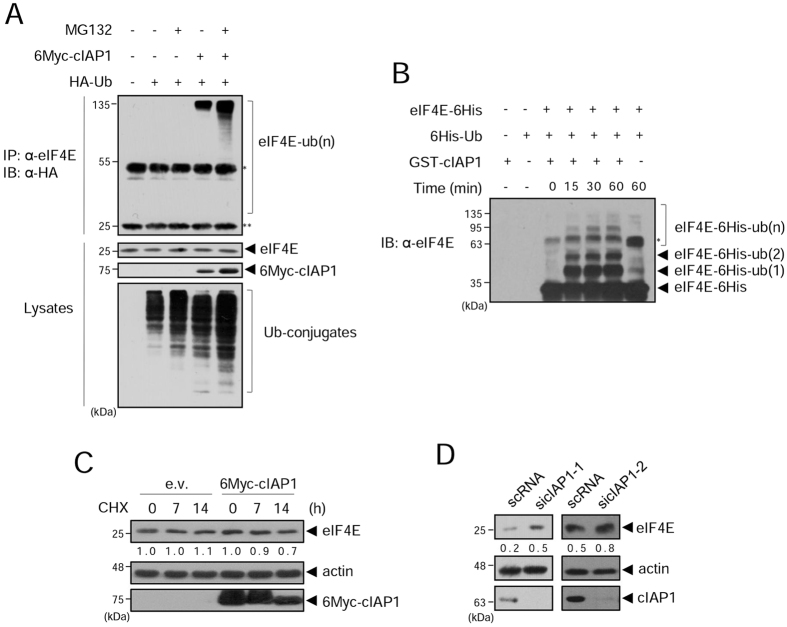
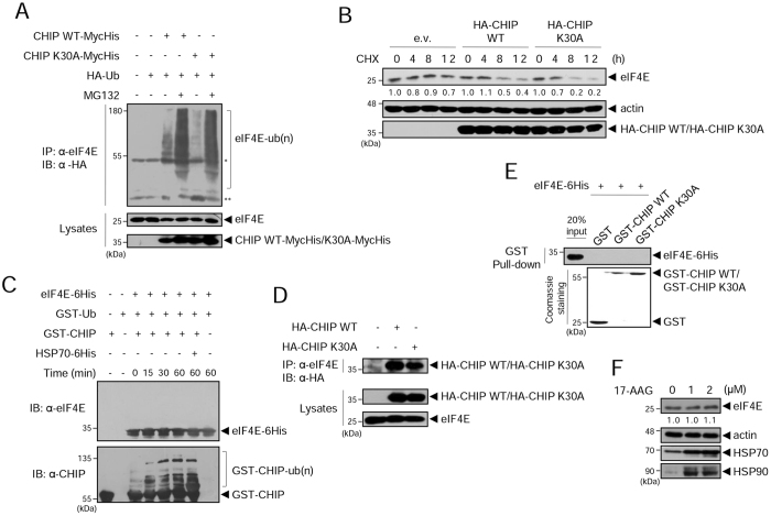
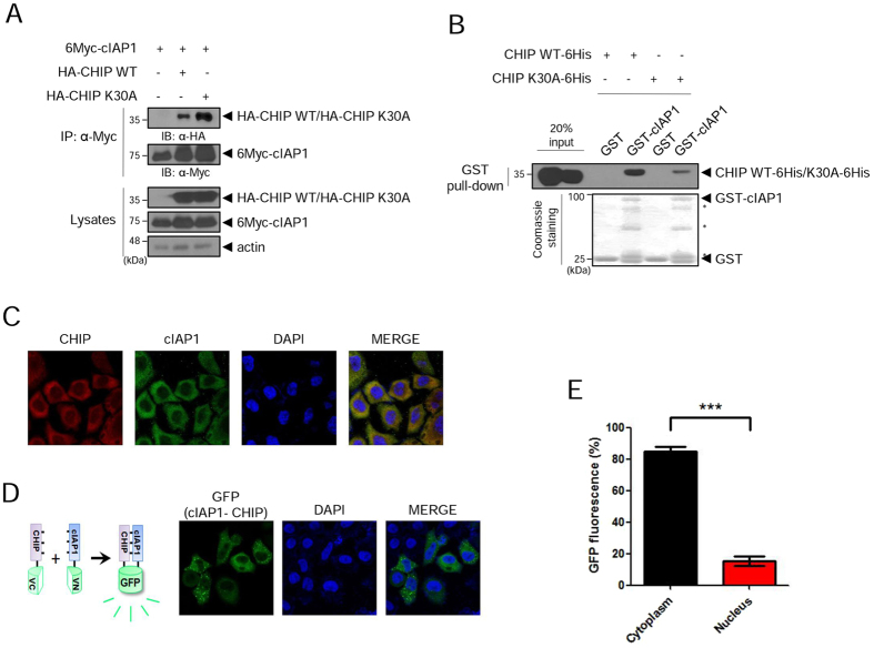
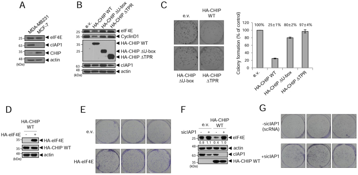
eIF4E is an initiator protein in cap-dependent translation. Its overexpression is linked to tumorigenesis in various human cancers, suggesting that the levels of eIF4E must be under tight control in normal cells. Although several eIF4E regulatory mechanisms have been demonstrated, the intracellular mechanisms controlling eIF4E protein levels remain poorly understood. Here, we report that eIF4E is efficiently regulated by dual mechanisms, both involving human inhibitor of apoptosis family protein cIAP1. cIAP1 itself ubiquitinates eIF4E as an E3 ligase, and interestingly, cIAP1 also functions as a mediator to present eIF4E to another E3 ligase, CHIP. This collaborative activity of cIAP1 and CHIP directs eIF4E toward degradation, controlling its levels and suppressing tumorigenesis. Our results provide the first evidence for a mediator function of cIAP1 and collaborative activity of cIAP1 and CHIP, suggesting that maintaining balanced levels of these E3 ligases might be beneficial for normal cell growth.


Conflict of interest statement

The authors declare that they have no competing interests.

Figures

Figure 1
Figure 1
Among human IAPs, cIAP1 specifically interacts with eIF4E. (A) HEK 293 cells were co-transfected with 6Myc-ML-IAP, 6Myc-XIAP, 6Myc-cIAP1, 6Myc-cIAP2, or eIF4E-MycHis. After 24 h, whole cell lysates (WCL) were co-immunoprecipitated with anti-His antibody analyzed by western blot (WB) with anti-Myc antibody (top). Expression of IAPs and eIF4E was determined by WB using anti- His or anti-Myc (bottom) antibodies. *IgG heavy chain. (B) eIF4E-6His protein was incubated with GST or GST-cIAP1 proteins on glutathione beads. Precipitated eIF4E-6His protein was subjected to WB with anti-His antibody. *Non-specific or GST-cIAP1 fragment band. (C) HeLa cells were immunostained with anti-eIF4E (red) and anti-cIAP1 (green) antibodies. DAPI (blue) was used to counterstain nuclear DNA. WB images are cropped from the original blots shown in supplementary information.
Figure 2
Figure 2
The ubiquitination and levels of eIF4E protein are regulated by cIAP1. (A) HEK 293 T cells were co-transfected with 6Myc-cIAP1 and HA-ubiquitin. After 24 h, cells were treated with 20 μM of MG132 for 6 h, and WCL were co-immunoprecipitated with anti-eIF4E antibody and analyzed by WB with anti-HA antibody. *IgG heavy chain, **IgG light chain. (B) Purified eIF4E-6His and GST-cIAP1 proteins were subjected to the in vitro ubiquitination assay. After incubation, samples were analyzed by WB with anti-eIF4E antibody. *Non-specific background band after 1 h incubation without GST-cIAP1. (C) HeLa cells were transfected with empty vector (e.v.) or 6Myc-cIAP1. After 48 h, cells were treated with 200 μg/ml cycloheximide (CHX) and harvested at the indicated times. WCL were analyzed by WB with anti-eIF4E and anti-Myc antibodies. Actin was used as a quantitative loading control. The numbers indicate relative eIF4E expression to actin within the indicated vector-expressing cells. (D) HeLa cells were transfected with control scRNA (scRNA) or cIAP1-targeted siRNAs (sicIAP1-1, sicIAP1-2). WCL were analyzed by WB with anti-eIF4E, anti-cIAP1, and anti-actin antibodies at two days after transfection. The numbers indicate expression of eIF4E relative to actin. WB images are cropped from the original blots shown in supplementary information.
Figure 3
Figure 3
eIF4E is regulated by CHIP but not via direct binding. (A) HEK 293 T cells were co-transfected with HA-ubiquitin and CHIP WT-MycHis or CHIP K30A-MycHis. After 24 h, cells were treated with 20 μM MG132 for 6 h, and WCL were co-immunoprecipitated with anti-eIF4E antibody, followed by WB with anti-HA antibody. *IgG heavy chain, **IgG light chain. (B) HeLa cells were transfected with HA-CHIP WT, HA-CHIP K30A, or empty vector (e.v.). After 48 h, cells were treated with 200 μg/ml CHX and harvested at the indicated times. WCL were analyzed by WB with anti-eIF4E and anti-HA antibodies. The numbers indicate relative eIF4E expression to actin within the indicated vector-expressing cells. (C) The in vitro ubiquitination assay using purified eIF4E-6His, GST-CHIP, and Hsp70-6His was analyzed by WB with anti-eIF4E antibody (top) and with anti-CHIP antibody (bottom). (D) HEK 293 cells were transfected with HA-CHIP WT or HA-CHIP K30A. After 24 h, WCL were co-immunoprecipitated with anti-eIF4E antibody and WB with anti-HA antibody. (E) eIF4E-6His protein was incubated with GST, GST-CHIP WT, or GST-CHIP K30A proteins on glutathione beads. Precipitated eIF4E-6His protein was subjected to SDS-PAGE and WB with anti-His antibody. (F) MDA-MB231 cells were treated with 17-(Allylamino)geldanamycin (17-AAG) for 24 h and WCL were analyzed by WB with the indicated antibodies. The numbers indicate relative eIF4E expression to actin. WB images are cropped from the original blots shown in supplementary information.
Figure 4
Figure 4
cIAP1 and CHIP interact in vitro and in vivo. (A) HEK 293 cells were co-transfected with 6Myc-cIAP1 and HA-CHIP WT or HA-CHIP K30A. After 24 h, WCL were co-immunoprecipitated with anti-Myc antibody and analyzed by WB with anti-HA antibody. (B) Purified GST and GST-cIAP1 proteins on glutathione beads were incubated with purified CHIP WT-6His or CHIP K30A-6His proteins. After incubation, the beads were analyzed by WB using anti-His antibody. *GST-cIAP1 fragments or non-specific bands. (C) HeLa cells were immunostained with anti-cIAP1 antibody (green) and anti-CHIP antibody (red). DAPI (blue) was used to counterstain nuclear DNA. (D) Hela cells were co-transfected with cIAP1-VN and CHIP-VC for BiFC assay. After fixation and permeabilization, cells were stained with DAPI to visualize the nucleus. The interaction of the two proteins was visualized by GFP fluorescence. (E) Quantification of GFP fluorescence per cell in Hela cells using Image J program. Values represent means ± s.e.m from triplicate independent experiments (***p < 0.0001, unpaired t-test). WB images are cropped from the original blots shown in supplementary information.
Figure 5
Figure 5
Interaction of eIF4E with CHIP is dependent on cIAP1. (A) Purified GST, GST-CHIP, GST-cIAP1, and eIF4E-6His proteins were used in the His pull-down assay. After incubation, the beads were analyzed by WB using anti-GST antibody. (B) His pull-down assay with increased concentration of GST-cIAP1 proteins. Complexes of eIF4E-6His, GST-cIAP1, and GST-CHIP were analyzed by SDS-PAGE and WB using anti-GST antibody. (C) HeLa cells were transfected with scRNA or sicIAP1. After 24 h transfection, WCL were immunoprecipitated with anti-CHIP antibody, and analyzed by WB with the indicated antibodies. The numbers indicate levels of co-immunoprecipitated eIF4E relative to immunoprecipitated CHIP. (D) HeLa cells were co-transfected with cIAP1-VN and CHIP-VC. After 24 h transfection, cells were immunostained with anti-eIF4E antibody (red) and DAPI to visualize the nucleus (blue). GFP fluorescence indicating an interaction between cIAP1 and CHIP was merged with the endogenous eIF4E staining (red) in the cytoplasm. WB images are cropped from the original blots shown in supplementary information.
Figure 6
Figure 6
The BIR1 domain of cIAP1 is responsible for eIF4E binding while BIR3 is responsible for CHIP binding. (A) Schematic representation of the cIAP1 WT, RING domain mutant (H588A/C592A), and various truncation constructs used in the study. (B) HEK 293Tcells were co-transfected with the various 6Myc-cIAP1 constructs (A) and HA-eIF4E. After 24 h, WCL were co-immunoprecipitated with anti-HA antibody and analyzed by WB with anti-Myc antibody. *IgG heavy chain. (C) HEK 293 T cells were co-transfected with the various 6Myc-cIAP1 constructs (A) and HA-CHIP. After 24 h, WCL were co-immunoprecipitated with anti-HA antibody and analyzed by WB with anti-Myc antibody. *IgG heavy chain. (D) Schematic representation of the CHIP WT, K30A, and various truncation constructs. (E) HEK 293 T cells were co-transfected with 6Myc-cIAP1 and the various HA-CHIP constructs (D). After 24 h, WCL were co-immunoprecipitated with anti-HA antibody and analyzed by WB with anti-Myc antibody. WB images are cropped from the original blots shown in supplementary information.
Figure 7
Figure 7
The collaborative activity of cIAP1 and CHIP regulates eIF4E. (A) HEK 293 T cells were transfected with sicIAP1 (top) or siCHIP (bottom) for 24 h, then co-transfected with HA-ubiquitin and CHIP-MycHis (top) or 6Myc-cIAP1 (bottom). After 24 h, cells were treated with 20 μM MG132 for 6 h, and WCL were co-immunoprecipitated with anti-eIF4E antibody, followed by WB with anti-HA antibody. *IgG heavy chain. (B) HeLa cells were transfected with scRNA or sicIAP1. After 24 h, cells were transfected with HA-CHIP for 24 h and half-life was measured at the indicated times. WCL were analyzed by WB with the indicated antibodies. The numbers indicate relative eIF4E expression to actin within the indicated condition. (C) HEK 293 T cells were transfected with scRNA or siCHIP. After 24 h, cells were co-transfected with various 6Myc-cIAP1 constructs (C1, C4, C5 and C7 in Fig. 6A) and HA-ubiquitin for 24 h. After 24 h, 20 μM of MG132 was treated for 6 h. Co-immunoprecipitations were performed using anti-eIF4E antibody and analyzed by WB with anti-HA antibody to reveal ubiquitinated eIF4E. (D) HEK 293 T cells were co-transfected with 6Myc-cIAP1, HA-CHIP, or both, and HA-ubiquitin. After 24 h, cells were treated with 20 μM MG132 for 6 h and WCL were co-immunoprecipitated with anti-eIF4E antibody, followed by WB with anti-HA antibody. *IgG heavy chain. (E) Measurement of eIF4E half-life in HeLa cells expressing 6Myc-cIAP1 alone or 6Myc-cIAP1 and HA-CHIP together (top), and in HeLa cells transfected with 6Myc-cIAP1 and scRNA or siCHIP (bottom). The numbers indicate relative eIF4E expression to actin within the indicated condition. (F) HeLa cells were depleted with either sicIAP1 (sicIAP1-1), siCHIP or both. After 48 h, WCL were analyzed by WB with the indicated antibodies (top). The numbers indicate expression of eIF4E relative to actin. Means ± SEM from three independent experiments, **p < 0.01, one-way ANOVA (bottom). WB images are cropped from the original blots shown in supplementary information.
Figure 8
Figure 8
Depletion of cIAP1 reverses the reduced growth of CHIP WT-overexpressing MDA-MB231 cells. (A) The protein levels of endogenous eIF4E, cIAP1, and CHIP in MDA-MB231 and MCF7 cells. (B) MDA-MB231 cell lines stably expressing empty vector, HA-CHIP WT, HA-CHIP ∆U-box, and HA-CHIP ∆TPR were generated. WCL were analyzed by WB with the indicated antibodies. (C) The stable cell lines were grown in medium containing G418 for 2 weeks. Colonies were stained with crystal violet and photographed (left panel) and visible colonies were counted (right panel). Results are depicted as mean ± S.D. from three independent experiments. (D and E) MDA-MB231 cell lines stably expressing CHIP WT were transfected with HA-eIF4E. WCL were analyzed by WB with anti-HA antibody (D) and the colony formation assay was carried out for 2 weeks (E). (F and G) MDA-MB231 cell lines stably expressing empty vector or CHIP WT were transfected with scRNA or sicIAP1. WCL were analyzed by WB with indicated antibodies (F) and colonies were stained using crystal violet (G). The numbers indicate expression of eIF4E relative to actin. WB images are cropped from the original blots shown in supplementary information.
Figure 9
Figure 9
The RING activity of cIAP1 is not absolutely required for its mediator function. (A) MCF-7 cell lines stably expressing empty vector, 6Myc-cIAP1 WT, or 6Myc-cIAP1 RM were generated. WCL were analyzed by WB with indicated antibodies. (B) Cell proliferation was measured using the MTT assay for a total of 4 days in MCF-7 cell lines stably expressing indicated cIAP1 constructs. (C) Colony formation assay was carried out in medium containing G418 for 2 weeks. Colonies were stained with crystal violet and photographed (left panel) and visible colonies were counted (right panel). Results are depicted as mean ± S.D. from three independent experiments.

References

    1. Vaux DL, Silke J. IAPs, RINGs and ubiquitylation. Nat. Rev. Mol. Cell Biol. 2005;6:287–297. doi: 10.1038/nrm1621. - DOI - PubMed
    1. Shi Y. Mechanisms of caspase activation and inhibition during apoptosis. Mol. Cell. 2002;9:459–470. doi: 10.1016/S1097-2765(02)00482-3. - DOI - PubMed
    1. Yang Y, Fang S, Jensen JP, Weissman AM, Ashwell JD. Ubiquitination protein ligase activity of IAPs and their degradation in proteasomes in response to apoptotic stimuli. Science. 2000;288:874–877. doi: 10.1126/science.288.5467.874. - DOI - PubMed
    1. Cartier J, et al. Cellular inhibitor of apoptosis protein-1 (cIAP1) can regulate E2F1 transcription factor-mediated control of cyclin transcription. J. Biol. Chem. 2011;286:26406–26417. doi: 10.1074/jbc.M110.191239. - DOI - PMC - PubMed
    1. Xu L, et al. c-IAP1 cooperates with Myc by acting as a ubiquitin ligase for Mad1. Mol. Cell. 2007;28:914–922. doi: 10.1016/j.molcel.2007.10.027. - DOI - PubMed

Publication types

MeSH terms

Substances

LinkOut - more resources