. 2017 Aug 29;7(1):9842.
doi: 10.1038/s41598-017-10555-z.

Phosphatidylinositol-3 Kinase Inhibitors, Buparlisib and Alpelisib, Sensitize Estrogen Receptor-positive Breast Cancer Cells to Tamoxifen

Affiliations

Phosphatidylinositol-3 Kinase Inhibitors, Buparlisib and Alpelisib, Sensitize Estrogen Receptor-positive Breast Cancer Cells to Tamoxifen

I-Chun Chen et al. Sci Rep. .

Abstract

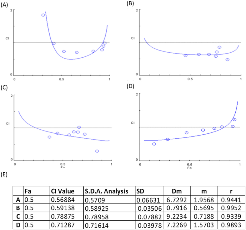
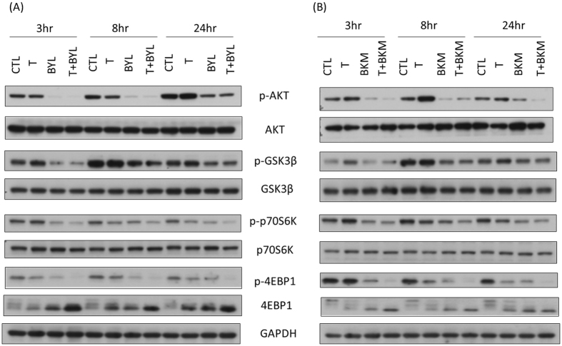
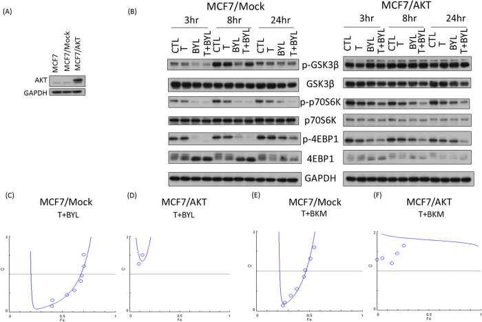
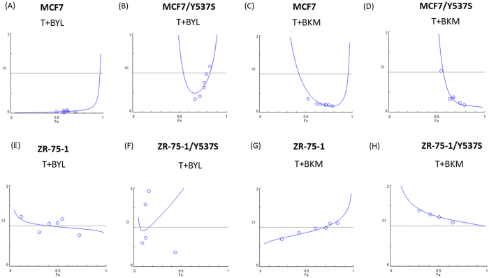
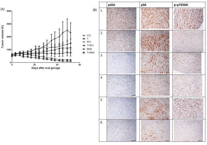
Tamoxifen is the standard first-line hormonal therapy for premenopausal women with estrogen receptor (ER)-positive metastatic breast cancer (BC). One of the crucial mechanisms underlying hormonal therapy resistance is the collateral activation of the phosphatidylinositol-3 kinase (PI3K)/AKT pathway. We explored whether PI3K inhibitors, buparlisib and alpelisib, enhance the efficacy of tamoxifen against ER-positive BC cells. We have observed a synergism between alpelisib or buparlisib and tamoxifen in the treatment for ER-positive BC cell lines harboring different PI3K alterations. Immunoblotting analysis showed alpelisib, buparlisib, or either drug in combination with tamoxifen downregulated the PI3K downstream targets in the MCF-7 and ZR75-1 cells. In the MCF-7 cells transfected with a constitutive active (myristoylated) AKT1 construct or mutant ER, the synergistic effect between alpelisib and tamoxifen was markedly attenuated, indicating that synergism depends on AKT inhibition or normally functioning ER. Combining alpelisib or buparlisib with tamoxifen also attenuated MCF-7 tumor growth in Balb/c nude mice. Our data suggest that additional PI3K blockade might be effective in enhancing the therapeutic effect of tamoxifen in ER-positive BC and support the rationale combination in clinical trials.


Conflict of interest statement

The authors declare that they have no competing interests.

Figures

Figure 1
Figure 1
Combination Index for Tamoxifen and Alpelisib in Different ER-Positive Breast Cancer Cell Lines. (A) MCF-7 (PIK3CA mutation), (B) T47D (PIK3CA mutation), (C) ZR75-1 (PIK3CA wild type, PTEN loss), (D) HCC1500 (PIK3CA wild type, PTEN intact) and (E) Calculated combination index. These ER-positive breast cancer cells were treated with tamoxifen and alpelisib at different concentrations for 6 days. Combination indices were calculated to determine the relationship between drug synergism and genetic alterations in the PI3K pathway.
Figure 2
Figure 2
Combining Tamoxifen with (A) Alpelisib or (B) Buparlisib Attenuates Signal Transduction Downstream of PI3K in ZR75-1 Cells (tamoxifen: 8 µM; alpelisib: 5 µM; and buparlisib: 5 µM). Cells treated with a combination of tamoxifen with alpelisib or buparlisib had lower pAKT, pGSK3β, p-p70S6K, and p4EBP1 expression levels at 3, 8, and 24 hours after treatment, compared with tamoxifen-treated or control cells.
Figure 3
Figure 3
Tamoxifen Combined with Alpelisib or Buparlisib Promotes Apoptosis. (A) Cell cycle stage was determined through flow cytometry after the treatment of MCF-7 cells with control(CTL), tamoxifen (8 µM, T), buparlisib (5 µM, BKM), alpelisib (5 µM, BYL), tamoxifen (8 µM) + buparlisib (5 µM) (T + BKM), and tamoxifen (8 µM) + alpelisib (5 µM) (T + BYL) for 24, 48, and 72 hours. (B) SubG1 proportion was quantified and depicted in a bar graph. (C) MCF-7 cells were stained for PI and annexin V after treatment for 72 hours. Flow cytometry was used to quantify each population and the results depicted as a dot plot. (D) Proportions of PI and annexin V double-positive cells. (*p < 0.05) Representative figures from the three experiments are depicted in Fig. 3.
Figure 4
Figure 4
Constitutively Active AKT Abolishes the Synergistic Effect of Treatment with Tamoxifen and PI3K inhibitor. (A) AKT expression level was measured by Western blot analysis in MCF-7 cells without transfection (parental), cells transfected with empty vector (MCF-7/mock), and cells transfected with myristoylated AKT (MCF-7/AKT). (B) Comparison of p-GSK3b, pp-70S6K, and p-4EBP1 expression levels between MCF-7/mock and MCF-7/AKT cells treated with control (CTL), tamoxifen (T, 8 µM), alpelisib (BYL, 5 µM), or tamoxifen and alpelisib (T + BYL, 8 µM + 5 µM) at 3, 8, and 24 hours. Fa plots of combination indices for treatment with T + BYL in MCF-7/mock (C) or MCF-7/AKT (D) cells and treatment with T + BKM in MCF-7/mock (E) or MCF-7/AKT (F) cells.
Figure 5
Figure 5
Mutant ER Results in Loss of Synergism Between Tamoxifen and PI3K Inhibitor. Y537S mutant was transfected into MCF-7 (MCF-7/Y537S) or ZR 75-1 (ZR75-1/Y537S) by lipofectamine 2000. Combination indices for T + BYL in (A) MCF-7, (B) MCF-7/Y537S, (E) ZR 75-1, (F) ZR 75-1/Y537S or tamoxifen(T) + buparisib(BKM), in (C) MCF-7, (D) MCF-7/Y537S, (G) ZR 75-1, (H) ZR 75-1/Y537S after 72 hours of treatment.
Figure 6
Figure 6
Treatment with Tamoxifen and a PI3K Inhibitor Controls MCF-7 Tumor Growth In Vivo. Female Balb/c nude mice were inoculated with MCF-7 cells at the flank and randomized into six different treatment groups when the tumor volume reached 500 mm3. (A) Tumor volumes measured using a caliper for treatments with CTL (solid circles; PBS), T (open circles; tamoxifen, 50 mg/kg), BYL (solid triangles; alpelisib, 35 mg/kg), T + BYL (open triangles; tamoxifen + alpelisib, 50 mg/kg + 35 mg/kg), BKM (solid squares; 25 mg/kg), and T + BKM (open squares; tamoxifen + buparlisib, 50 mg/kg + 25 mg/kg). The figure is representative of two independent experiments (N = 5 and 7 in each treatment group). (B) Representative figures of paraffin-embedded MCF-7 tumors from (1) CTL, (2) T, (3) BYL, (4) T + BYL, (5) BKM, and (6) T + BKM treatments were stained for pAKT, pS6, and p-p70S6K. All images were obtained using a 10x eyepiece and 40x objective lens (scale bar = 50 µm).

Similar articles

Cited by

References

    1. Cole MP, Jones CT, Todd ID. A new anti-oestrogenic agent in late breast cancer. An early clinical appraisal of ICI46474. British journal of cancer. 1971;25:270–275. - PMC - PubMed
    1. Jordan VC. Third annual William L. McGuire Memorial Lecture. “Studies on the estrogen receptor in breast cancer”–20 years as a target for the treatment and prevention of cancer. Breast cancer research and treatment. 1995;36:267–285. doi: 10.1007/BF00713399. - DOI - PubMed
    1. Sunderland MC, Osborne CK. Tamoxifen in premenopausal patients with metastatic breast cancer: a review. Journal of clinical oncology: official journal of the American Society of Clinical Oncology. 1991;9:1283–1297. doi: 10.1200/JCO.1991.9.7.1283. - DOI - PubMed
    1. Kaufmann M, et al. Goserelin, a depot gonadotrophin-releasing hormone agonist in the treatment of premenopausal patients with metastatic breast cancer. German Zoladex Trial Group. Journal of clinical oncology: official journal of the American Society of Clinical Oncology. 1989;7:1113–1119. doi: 10.1200/JCO.1989.7.8.1113. - DOI - PubMed
    1. Klijn JG, et al. Combined tamoxifen and luteinizing hormone-releasing hormone (LHRH) agonist versus LHRH agonist alone in premenopausal advanced breast cancer: a meta-analysis of four randomized trials. Journal of clinical oncology: official journal of the American Society of Clinical Oncology. 2001;19:343–353. doi: 10.1200/JCO.2001.19.2.343. - DOI - PubMed

Publication types

MeSH terms

LinkOut - more resources